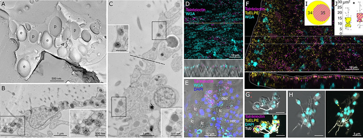
Fig 5. Main secretory cells in the dorsal epithelium.
(A) Freeze fracture replica at the apex of a dorsal epithelial cell (DEC) imaged in TEM reveals e- or p-faces of numerous ~500 nm diameter secretory granules. (B, C) TEM of ultrathin sections labelled with nanogold conjugated WGA. Transverse section in the dorsal epithelium (B) shows multiple WGA-stained granules in ciliated DECs; insets show enlarged view of boxed regions. Transverse section at the transition region between dorsal and ventral epithelia (C, the border is demarcated by dotted line) shows that DEC granules bind more WGA than do morphologically similar granules in VEC. (D-H) Confocal images of wholemounts (D, F) and dissociated cell preparations (E, G, H). Many DEC express Ta Intelectin 60661 (D, whole mount; E, dissociated cells) as evident from co-labeling with WGA. Mucocytes (m) label intensely with WGA, but do not express Ta Intelectin 60661. Other cell types (VEC, lipophil (lc) and fiber (fc) cells) are not labeled. (F) Ta ELPE is expressed in nearly all DEC and a few VEC (see xz inset); (G, I) About half of Ta ELPE+ co-expresses Ta Intelectin 60661. (H) Some VEC, identified based on their small sizes and cylindrical shapes express Ta ELPE but not Ta Intelectin. (J) Cells co-expressing Ta ELPE and Ta Intelectin are larger than those expressing only Ta ELPE. e – e face; fc – fiber cells; lc – lipophil cells; m – mucocytes; p – p face.

