



Abstract
Objectives
Due to Albendazole's relatively low efficacy and bioavailability, Echinococcosis has proven a challenge to manage successfully, with several studies investigating ways to improve the outcome, mainly showing mixed results. We, therefore, aimed to evaluate whether Sulfonated Graphene Oxide (S-GO), as nanocarriers, could improve the mentioned outcome.
Methods
Echinococcus protoscoleces were divided into four groups based on the agent they received, which comprised control, S-GO, Albendazole, and Albendazole-loaded S-GO (S-GO-Albendazole). Then, the Bax and Bcl-2 gene expression levels and the number of surviving protoscoleces in each group were determined.
Results
Bax gene expression increased by 121% in the 50 μg/ml concentration of the S-GO-Albendazole, while Bcl-2 gene expression decreased by 64%. Moreover, S-GO-Albendazole was approximately 18% more effective at neutralizing protoscoleces than Albendazole and 14% and 31% more effective at improving the expression of the mentioned genes, respectively (p < 0.05). In addition, the number of surviving protoscoleces after exposure to the mentioned concentration reduced by approximately 99%.
Conclusions
S-GO, despite not having significant lethality on protoscoleces, significantly increased the lethality of Albendazole and, therefore, is a suitable nanocarrier. However, we recommend conducting in vivo and clinical studies to more accurately determine this nanocomplex's potential and side effects.
1. Introduction
Echinococcosis, a zoonotic infection caused by the Taeniidea cestode family, has affected over 3 million individuals worldwide [1]. Moreover, the process of treating the disorder has not only been arduous but costly, too, amounting to more than 763 million dollars and 100,0006,62 disability-adjusted life years (DALYs) annually [2].
Besides, even though the mortality rate of Cystic Echinococcosis (CE) is estimated to be around 2–4%, the figure can go even higher if the infection is not adequately taken care of [1]. In this regard, several management techniques have been introduced during the past few decades, consisting of classic surgical removal, percutaneous sterilization or Puncture, Aspiration, Injection, and Re-aspiration (PAIR), plain follow-up of the cysts (the watch and wait method), and isolated medical therapy with anthelminthic agents [1, 3]. However, each technique has merits and shortcomings, leading to several newly introduced but not well-investigated methods [1, 3].
In addition, the conservative treatment, consisting of Albendazole (ABZ), which is a benzimidazole (BMZ), is only effective in a few inoperable cases (i.e., cases where multiple cysts have affected more than two organs), cases with peritoneal infiltrations, and before and after cyst operation (to prevent potential complications following spillages). Besides, their efficacy is thought to be overestimated, with recent studies showing full recovery in 18% to just over 50% of cases [4–7]. Furthermore, the current Albendazole (ABZ) formulations facing significant limitations. These include poor bioavailability, low water solubility, limited intestinal absorption (5%), and inadequate cyst-to-serum concentration ratios (25%). These challenges hinder the effective delivery of therapeutic ABZ concentrations to target tissues, compromising treatment efficacy. Consequently, there is an urgent need for innovative strategies to enhance the pharmaceutical properties and therapeutic performance of ABZ [1, 3–12].
Subsequently, numerous studies have been carried out to improve the mentioned low ABZ bioavailability, reporting various degrees of improvement. These methods include (a) Ingesting ABZ with fat-rich meals [13], (b) utilizing ABZ-containing sodium salts [14], (c) oil surfactant suspensions [15], and (d) incorporating ABZ or its active metabolite, ABZ sulfoxide (ABZ-SO), into nanoparticles or nanocapsules. Moreover, the most commonly utilized nanoparticles include metal nanoparticles (e.g., gold, zinc, copper, silver, and selenium), metal oxide nanoparticles (e.g., Zinc Oxide, Titanium Oxide, Cerium Oxide, and Zirconium Oxide), Chitosan and its derivatives, Liposomes, Solid lipid nanocapsules (SLN), ABZ-poly-lactic-co-glycolic-acid (ABZ-PLGA) nanoparticles, and Poloxamer 188 [11, 16–23]. Nevertheless, there is still much to be known, and some believe that other not-yet-utilized nanoparticles can achieve even higher feats.
One such group is the Graphene family of nanoparticles (GFN), a carbon-based, up-and-coming, yet cost-effective family of nanomaterials with relatively low environmental hazards [24, 25]. There are several members in the family, with the most notable being Graphene Oxide (GO), Reduced Graphene Oxide (rGO), Graphene quantum dots (GQD), and Graphene Nanoribbons (GNR) [25]. In addition, their characteristics, including their sizable surface area, high loading capacity, highly desirable thermal and mechanical properties, pH responsiveness, and capable enhanced permeability and retention (EPR) effects, have been well described in the literature [25–27]. Their toxic effects have also been investigated on C. elegans worms and Zebrafish, but the results were not as straightforward, with significant toxicity shown to the former but no adverse effects on the latter [28, 29]. In addition, their effects on human cells are not yet fully comprehended, with studies reporting negligible to significant toxic effects based on the utilized nanocomponent's size, coating, and structure [26].
Nevertheless, the positive effects of GFNs have been widely studied, which include their interesting potential in drug and gene delivery systems, tissue engineering and regeneration, and antimicrobial regimens [30–34]. For instance, the usefulness of the GFN compounds in drug delivery has been demonstrated on anticancer agents Rituxan and Doxorubicin (DOX) and antibiotics such as gentamicin, ciprofloxacin, and benzylpenicillin [35–40]. However, anthelminthic agents have still not been the subject of such studies. Therefore, based on the promise this agent could provide, we evaluated the effects of the graphene-based nanocomposites (namely, sulfonated GO or S-GO) on the efficacy of ABZ on Echinococcus protoscoleces (PSCs).
2. Materials and Methods
2.1. Design
The present in vitro investigation was conducted on viable protoscoleces (PSCs) of the sheep strain of E. granulosus, subsequent to obtaining protocol approval from the local Institutional Review Board (IRB). Additionally, efforts were made to minimize the number of animals utilized, ensuring only the necessary quantity to achieve the required PSC counts. An illustration of the study design is provided below. (see Figure 1).
Figure 1.
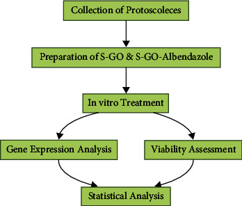
Illustration of the study design.
2.2. PSC Collection
We collected the livers and lungs of the infected sheep (kept under appropriate and ethically approved minimally harmful conditions) from a local industrial slaughterhouse and transferred them to the parasitology laboratory under sterile and thermally isolated conditions. After disinfecting the cysts' surfaces with 70% ethyl alcohol, 10–50 ml of the cyst fluid was aspirated using a sterile syringe and transferred to a 50 ml falcon tube, while the germinal layer was also isolated and added to the tube with the mix left to rest for 5 minutes. Then, the supernatant fluid was removed, and the obtained PSCs, which settled down at the bottom, were washed five times with a mixture of Phosphate-Buffer Saline (PBS, pH = 7.1) and Penicillin-Streptomycin (Biosera, USA). Then, samples with >90% viability (determined via the 0.1% eosin stain produced by dissolving 1 g of eosin powder in 1000 mL of distilled water) were deemed suitable [41]. These PSCs (5000 PSCs per 100 microliter of fluid) were then cultured in a medium comprising 10 ml of RPMI 1640, 10% Fetal Bovine Serum (FBS; Gibco, Germany), and 1% Penicillin-Streptomycin in aseptic conditions [42–45].
2.3. Preparation of GO
GO was synthesized using the modified Hummer's method and the graphite powder as raw material [46]. First, 2 g of the powder and 1.5 g of sodium nitrate were slowly added to a flask containing 70 ml of concentrated sulfuric acid (H2SO4), which was placed in an ice bath, and the mixture was stirred at a temperature of 0–5°C. After homogenization, 8 g of potassium permanganate (KMnO4) was added gradually for 2 hours. Then, the temperature of the reaction mixture was increased to 35°C and stirred for one hour to obtain a light brown viscose mixture. Next, 100 ml of distilled water was slowly added while stirring the solution, with the temperature increased to 90°C for one hour. The reaction was then stopped by adding 20 ml of 30% hydrogen peroxide (H2O2), and the mixture was stirred for 24 hours at room temperature. Then, the resulting suspension was washed with 3% hydrochloric acid (HCl) and distilled water until a pH of seven was achieved. Finally, the neutralized solution was sonicated using an ultrasonic homogenizer with an output power of 40% for two hours. The resulting brown solution was centrifuged to remove unlaminated graphite oxide sheets. The final step was drying the graphene oxide solution using a freeze-dryer to obtain graphene oxide powder with laminated plates.
2.4. Preparation of S-GO
To synthesize S-GO, 1 g of GO was added to a flask containing 350 mL of distilled water and then ultrasonicated for 30 minutes. Afterward, 5 g of sodium nitrite (NaNO2), dissolved in 75 ml of distilled water, was added to the mix and placed in an ice water bath. Additionally, 4 g of sulfanilic acid (C6H7NO3S) was dissolved in 50 ml of HCl (1 M) and transferred to a burette. In the next step, to synthesize S-GO through diazonium salt formation, the solution was drop-by-drop added to the flask containing GO and NaNO2 (within a temperature of 0 to 5°C) and stirred for 24 hours. Then, the resulting mixture was washed, centrifuged with distilled water to reach a neutral pH, and dried by a freeze-dryer.
2.5. Synthesis of ABZ-Loaded S-GO Nanocomposite
For this purpose, 5 mg ABZ and 2.5 mg S-GO (2 : 1 ratio) were added to a flask to synthesize a 50 μg/ml ABZ-loaded S-GO (S-GO-ABZ) solution, with distilled water added until the solution reached 100 ml. The solution was then exposed to ultrasonic waves for 30 minutes to disperse completely. Finally, the ABZ-loaded S-GO solution was stored in the refrigerator at 5°C and in dark conditions. Furthermore, other concentrations were prepared similarly, with an ABZ-to-S-GO ratio of 2 : 1.
2.6. Nanocomposite Characterization
Fourier-transform infrared spectroscopy (FT-IR) was used to identify functional groups on the S-GO samples. X-ray diffraction (XRD) analysis using a Panalytica X'Pert PRO powder diffractometer with Cu Kα radiation (λ = 1.5404 Å) was employed to study the crystallinity of the materials.
2.7. RNA Isolation and Complementary DNA (cDNA) Synthesis
After 60 min, we isolated the cellular RNA from the study groups on the desired timescales using an RNX-plus kit (CinnaGen, Iran) and determined the quantity and quality using the NanoDrop 2000c spectrophotometer (Thermo Scientific, USA). The resulting RNA was then reverse-transcribed into cDNA using a reverse transcriptase kit (Thermo Scientific, USA) and taken into account as the PCR amplification template.
2.8. Primer Design and Quantitative Real-Time Polymerase Chain Reaction (qRT-PCR)
We considered B-cell lymphoma 2 (Bcl-2) and BCL2 Associated X, Apoptosis Regulator (Bax) genes as the determinants of PSC apoptosis. Initially, we checked the Deoxyribonucleic Acid (DNA) and Ribonucleic acid (RNA) sequences of the genes on the genome databases of the National Center for Biotechnology Information (NCBI; available at https://www.ncbi.nlm.nih.gov) and European Bioinformatics Institute (Ensembl; available at https://www.ensembl.org). Then, after identifying different exons of each gene, we designed the appropriate primers (Isogen, Netherlands), provided in Table 1, using the Primer3 tool (available at https://frodo.wi.mit.edu/cgibin/primer3/primer3_www.cgi), while also double checking their integrity using the 3.5th edition of the Gene Runner tool (Hasting Software, Inc., 1994). We then assessed the specificity of the primers via the Basic Local Alignment Search Tool (BLAST).
Table 1.
The sequences of the designed primers.
| Gene | Primer sequence |
|---|---|
| Bcl-2 | F: AAGCATCAACCAATCTGTCAGAG R: ACTATCGAAGGAGGCATAAATGTC |
|
| |
| Bax | F: TCCTCCTCGGATGAAGCAGA R: CTGATGTACCGGTCTCTCGC |
|
| |
| 5.8S | F: GTCGATGAAGAGTGCAGCCAAC |
Ultimately, we determined the expression levels of Bcl-2 and Bax (at baseline and four weeks after cell differentiation) via performing the qRT-PCR in 15 µl reaction volume containing 1 μl of cDNA, 4.5 μl of sterile deionized water, 7.5 μl of FastStart SYBR Green Master ROX solution (Roche), and 2 μl of the mentioned primers in the FAM/SYBR and ROX channels of the ROTOR GENE 3000 (Corbett) device. The resulting data from the former channel were normalized with the latter channel and transferred to the LinRegPCR tool (available at https://www.gene-quantification.de/). These data from the band intensities were then normalized against their primary internal control gene concentrations to determine their expression levels objectively.
2.9. In Vitro Treatment and Scolicidal Assay
To minimize bias in allocating treatment, PSCs were randomly divided into four main groups based on the agent and dosage they received: (1) negative untreated control, (2) ABZ, (3) S-GO, and (4) S-GO-ABZ. These groups also comprised three subgroups with 10 µg/ml, 20 µg/ml, and 50 µg/ml concentrations, respectively. Finally, the agents' scolicidal effects were assessed after 15, 30, and 60 minutes. Blinding was used when assessing the results of PSC viability and gene expression to further reduce potential bias.
2.10. Statistical Analysis
We used the 27th version of SPSS statistical software (IBM Corp. Released 2020. IBM SPSS Statistics for Windows, Version 27.0. Armonk, NY: IBM Corp) to analyze the obtained data. Moreover, the differences between the means of the PSC viability in the active and control groups were analyzed using the One-way analysis of variance (ANOVA) with the Student–Newman–Keuls (SNK) method as the post hoc analysis. Moreover, p < 0.05 were considered statistically significant.
3. Results
3.1. FT-IR Spectra
In Figure 2, we compared the spectra of (a) GO and (b) S-GO. In Figure 2(a), the broad peak at 3448 cm−1 is related to the O–H stretching vibration caused by functional hydroxyl and carboxyl groups. Moreover, the peaks that appeared at 2922 and 2848 cm−1 are assigned to the C-H stretching vibrations, while the strong peak at 1382 cm−1 is assigned to the C-H bending vibrations. The peak at 1735 cm−1 is also attributed to the stretching vibration of the carbonyl group (C=O), while the peak at 1640 cm−1 is attributed to the stretching vibration of aromatic rings (C=C). The 1221 and 1068 cm−1 peaks also correspond to OC (due to C-O-C) and OC (due to C-OH of alcohol groups) stretching vibrations, respectively. Finally, according to the peaks showing oxygen presence, the graphite oxidation reaction can be confirmed [47].
Figure 2.
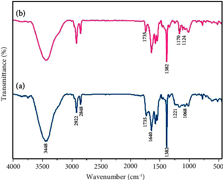
FT-IR spectra of GO (a) and S-GO (b).
The peaks corresponding to S-GO are shown in Figure 2(b). The peaks at 1170 and 1124 cm−1 are related to the stretching vibrations of two S-O groups and confirm the presence of sulfonic acid. It is also observed that after the sulfonation process, the peaks of 1735 and 1382 cm−1 have weakened [48, 49] (Figure 2).
3.2. XRD Pattern of S-GO
The pattern was recorded for the GO sample in the range of 5° to 90° (2θ) at room temperature, at a temperature increase speed of 0.02°/s is depicted in Figure 3. The structural information about GO shows two characteristic peaks at 11.5° and 42.8° angles due to the reflections of (002) and (101) planes [50] (Figure 3).
Figure 3.
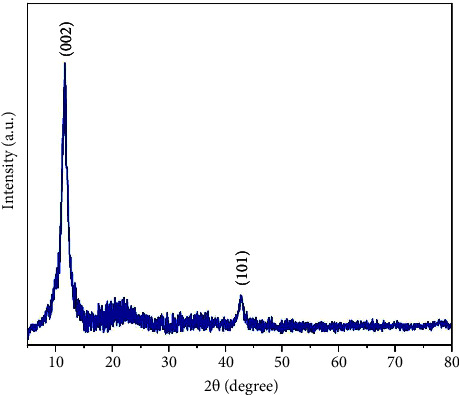
XRD patterns of S-GO.
3.2.1. Gene Expression
In the S-GO group, the Bax gene expression increased by 12% in the 10 μg/dl concentration, 14% in the 20 μg/dl concentration, and 32% in the 50 μg/dl concentration, respectively. Moreover, the expression of the Bcl-2 gene decreased by 7%, 9%, and 13% in the mentioned concentrations, respectively.
In the ABZ group, the Bax gene expression increased by 10%, 30%, and 90% in the 10 μg/dl, 20 μg/dl, and 50 μg/dl concentrations, respectively, while Bcl-2 expression decreased by 9%, 18%, and 49% in the mentioned concentrations.
In the S-GO-ABZ group, the Bax gene expression increased by 18%, 50%, and 121% in the 10 μg/dl, 20 μg/dl, and 50 μg/dl concentrations, respectively, while Bcl-2 expression decreased by 18%, 38%, and 64%, respectively. The analyses showed, however, that only the changes in the S-GO-ABZ group's 50 μg/ml concentration were statistically significant (p < 0.05) (Figures 4 and 5).
Figure 4.
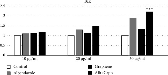
Bax gene expression in the S-GO-ABZ, ABZ, S-GO, and control groups. However, only the increase in the S-GO-ABZ group was significant. Bars represent mean ± standard deviation (SD) from duplicate experiments. (∗P < 0.05, ∗∗P < 0.01, ∗∗∗P < 0.001) (Alb, Albendazole; Grph, S-GO).
Figure 5.
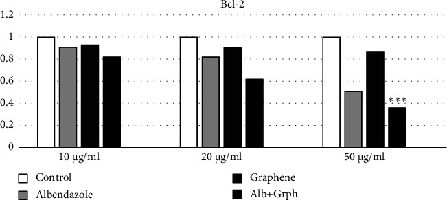
Bcl-2 gene expression in the S-GO-ABZ, ABZ, S-GO, and control groups. However, only the decrease in the GFN-ABZ was significant. (∗∗∗p < 0.05) (Alb, Albendazole; Grph, S-GO).
3.3. PSC Viability
As shown in Figure 5(a), dead PSCs were stained red after exposure to eosin, while in Figure 5(b), viable ones were unstained with flame cell activity. Moreover, the effects of various agents (i.e., ABZ, S-GO, and S-GO-ABZ) on PSCs at different intervals and concentrations are shown in Figure 6.
Figure 6.
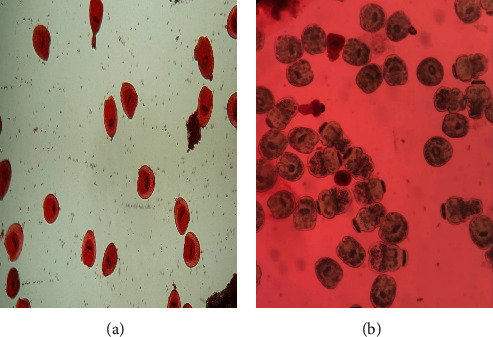
(a) The dead and (b) viable protoscoleces (PSCs) after treatment with S-GO-ABZ.
According to the observed results, the number of viable PSCs decreased by 2% when the 1 μg/ml concentration of S-GO was applied, by 7% when the 10 μg/ml concentration was applied, by 14% when the 20 μg/ml concentration was applied, and by 18% when the 50 μg/ml concentration was applied. Furthermore, following the application of the mentioned concentrations, the decreases in the PSC counts of the ABZ group were 3%, 34%, 69%, and 81%, respectively. In the S-GO-ABZ group, the mentioned decreases were 24%, 49%, 93%, and 99%, respectively. However, such decreases were more significantly pronounced in the S-GO-ABZ group compared to the ABZ group (p < 0.05) (Figures 6 and 7).
Figure 7.
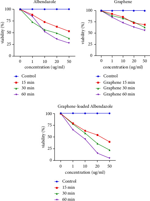
Number of viable protoscoleces (PSCs) after applying different concentrations of each agent in the S-GO-ABZ, ABZ, S-GO, and control groups. However, only the decreases in the S-GO-ABZ and ABZ groups were significant (p < 0.05).
4. Discussion
One of the reasons for the difficulty in controlling and managing Echinococcosis is the low effectiveness of Albendazole, for which no suitable replacement has yet been discovered. However, the findings of our study indicate that at a specific concentration (50 μg/ml), S-GO-ABZ is approximately 18% more effective than ABZ in neutralizing protoscoleces and 14% and 31% more effective in altering the expression of Bax and Bcl-2 apoptosis regulator genes, respectively (i.e., increase in Bax expression and decrease in Bcl-2 expression).
However, based on our literature review, this study was the first to investigate the effectiveness of S-GO as a nanocarrier on the toxicity of ABZ on Echinococcus PSCs. Therefore, we could only attempt to compare our findings with previous remotely similar studies to achieve a relative in vitro efficacy profile. One study investigated the combined effects of gold nanoparticles incorporation and lasers on Echinococcus PSCs, with an 89.3% toxicity rate after 120 minutes [51]. In another study, the PSC toxicity of copper, iron, silver, silicon, and zinc nanoparticles was investigated after 60 minutes of exposure, demonstrating that zinc nanoparticles possessed the highest toxicity (80%), followed by silicon (52%) [52]. Another similar study investigated the alterations in caspase-3 gene expressions and DNA fragmentation rates after exposure to ABZ-PLGA, which, even though ABZ and ABZ-PLGA both demonstrated significant toxicities, the differences between them were not statistically significant [53].
Moreover, another study assessed the PSC toxic effects of ABZ-loaded copper nanoparticles (with a concentration of 750 mg/ml) along with alterations in caspase-3 gene expression, demonstrating that even though ABZ-loaded copper nanoparticles neutralized 100% of the PSCs after 60 minutes and decreased the caspase-3 expression by 36%, the nanoparticle alone killed 73%, therefore, not showing how copper nanoparticles improved ABZ drug delivery and obscuring the results. Nevertheless, even though our study achieved a similar toxicity profile, the PSC toxicity of S-GO alone was negligible, objectively showing its capabilities in improving the effectiveness of ABZ purely as a nanocarrier.
Ultimately, we acknowledge that the investigation of the nanocarrier profile of GFNs for ABZ is in its first steps, and several steps need to be taken for them to be considered. Therefore, this in vitro study may not completely mimic the intricate in vivo surroundings. Further in vivo studies are needed to confirm the effectiveness and safety of S-GO-ABZ in animal models before it can be used clinically, as the interactions among the nanocomposite, the host immune system, and the parasite in the hydatid cyst may impact treatment results. Furthermore, the research did not examine the extended-term durability and distribution timing of Albendazole from the S-GO nanocarrier. Future research should determine the drug release characteristics of S-GO-ABZ and improve the formulation for extended drug release.
Additionally, while our study focused on protoscolex viability as a measure of antiparasitic activity, future research could explore other aspects of E. granulosus biology, such as the effects of ALB-GO and S-GO on cyst development, parasite migration, and host immune responses. Understanding the broader impact of these nanomaterials on the parasite life cycle could provide valuable insights into their therapeutic potential.
5. Conclusions
Despite not having significant toxicity on Echonicoccus PSCs, S-GO substantially increased ABZ's toxicity. Therefore, we believe that S-GO can be considered a suitable nanocarrier for improving the therapeutic efficacy of ABZ in Echinococcosis. By elucidating their mechanisms of action and addressing current limitations, further research can significantly advance the development of effective and sustainable therapies for this neglected tropical disease.
Contributor Information
Mohammad Shafie'ei, Email: ms6584@gmail.com.
Ali Derakhshani, Email: derakhshani.sci@gmail.com.
Data Availability
The dataset supporting the conclusions of this manuscript is available from the corresponding author upon reasonable request.
Ethical Approval
This study was approved by the Kerman University of Medical Sciences Institutional Review Board under the license “IR.KMU.AH.REC.1398.110”.
Conflicts of Interest
The authors declare that they have no conflicts of interest.
Authors' Contributions
MRL, MS, and FK prepared the first draft of the manuscript. MS, ZA, and AD collected the protoscoleces. GS and HD prepared GO and S-GO. AD, GS, and HD synthesized the Albendazole-loaded S-GO nanocomposite and the nanocomposite characterization. AD, MS, and ZA performed the RNA isolation, cDNA synthesis, primer design, qRT-PCR, in vitro treatment, and scolicidal assay. All authors participated in the review and critical revision of the manuscript. MRL and MS contributed equally to this work and should be considered as co-first authors. All named authors meet the International Committee of Medical Journal Editors (ICMJE) criteria for authorship for this article, take responsibility for the integrity of the work, and approve this version to be published.
References
- 1.McManus D. P., Gray D. J., Zhang W., Yang Y. Diagnosis, treatment, and management of Echinococcosis. BMJ . 2012;344(jun11 1):p. e3866. doi: 10.1136/bmj.e3866. [DOI] [PubMed] [Google Scholar]
- 2.Budke C. M., Deplazes P., Torgerson P. R. Global socioeconomic impact of cystic Echinococcosis. Emerging Infectious Diseases . 2006;12(2):296–303. doi: 10.3201/eid1202.050499. [DOI] [PMC free article] [PubMed] [Google Scholar]
- 3.Brunetti E., Kern P., Vuitton D. A. Expert consensus for the diagnosis and treatment of cystic and alveolar Echinococcosis in humans. Acta Tropica . 2010;114(1):1–16. doi: 10.1016/j.actatropica.2009.11.001. [DOI] [PubMed] [Google Scholar]
- 4.Bloom A. K., Ryan E. T. Albendazole. In: Magill A. J., Hill D. R., Solomon T., Ryan E. T., editors. Hunter’s Tropical Medicine and Emerging Infectious Disease . London, UK: W.B. Saunders; 2013. pp. 1088–1089. [Google Scholar]
- 5.Cherry J., Demmler-Harrison G. J., Kaplan S. L., Steinbach W. J., Hotez P. J. Feigin and Cherry’s Textbook of Pediatric Infectious Diseases E-Book: 2-Volume Set . Amsterdam, Netherlands: Elsevier Health Sciences; 2013. [Google Scholar]
- 6.Davis A., Dixon H., Pawlowski Z. S. Multicentre clinical trials of benzimidazole-carbamates in human cystic echinococcosis (phase 2) Bulletin of the World Health Organization . 1989;67(5):503–508. [PMC free article] [PubMed] [Google Scholar]
- 7.Horton R. J. Albendazole in treatment of human cystic Echinococcosis: 12 years of experience. Acta Tropica . 1997;64(1-2):79–93. doi: 10.1016/s0001-706x(96)00640-7. [DOI] [PubMed] [Google Scholar]
- 8.Dehkordi A. B., Sanei B., Yousefi M., et al. Albendazole and treatment of hydatid cyst: review of the literature. Infectious Disorders: Drug Targets . 2019;19(2):101–104. doi: 10.2174/1871526518666180629134511. [DOI] [PubMed] [Google Scholar]
- 9.Bakhtiar N. M., Akbarzadeh A., Casulli A., et al. Therapeutic efficacy of nanocompounds in the treatment of cystic and alveolar echinococcoses: challenges and future prospects. Parasitology Research . 2019;118(9):2455–2466. doi: 10.1007/s00436-019-06416-5. [DOI] [PubMed] [Google Scholar]
- 10.Wen H., Vuitton L., Tuxun T., et al. Echinococcosis: Advances in the 21st century. Clinical Microbiology Reviews . 2019;32(2) doi: 10.1128/cmr.00075-18. [DOI] [PMC free article] [PubMed] [Google Scholar]
- 11.Pensel P. E., Castro S., Allemandi D., Bruni S. S., Palma S. D., Elissondo M. C. Enhanced chemoprophylactic and clinical efficacy of Albendazole formulated as solid dispersions in experimental cystic Echinococcosis. Veterinary Parasitology . 2014;203(1-2):80–86. doi: 10.1016/j.vetpar.2014.01.027. [DOI] [PubMed] [Google Scholar]
- 12.Kapan S., Turhan A. N., Kalayci M. U., Alis H., Aygun E. Albendazole is not effective for primary treatment of hepatic hydatid cysts. Journal of Gastrointestinal Surgery . 2008;12(5):867–871. doi: 10.1007/s11605-007-0458-7. [DOI] [PubMed] [Google Scholar]
- 13.Lange H., Eggers R., Bircher J. Increased systemic availability of Albendazole when taken with a fatty meal. European Journal of Clinical Pharmacology . 1988;34(3):315–317. doi: 10.1007/bf00540964. [DOI] [PubMed] [Google Scholar]
- 14.Vural G., Yardimci M., Kocak M., et al. Efficacy of novel albendazole salt formulations against secondary cystic Echinococcosis in experimentally infected mice. Parasitology . 2020;147(13):1425–1432. doi: 10.1017/s0031182020001225. [DOI] [PMC free article] [PubMed] [Google Scholar]
- 15.Rigter I. M., Schipper H. G., Koopmans R. P., et al. Relative bioavailability of three newly developed albendazole formulations: a randomized crossover study with healthy volunteers. Antimicrobial Agents and Chemotherapy . 2004;48(3):1051–1054. doi: 10.1128/aac.48.3.1051-1054.2004. [DOI] [PMC free article] [PubMed] [Google Scholar]
- 16.Albalawi A. E., Alanazi A. D., Baharvand P., Sepahvand M., Mahmoudvand H. High potency of organic and inorganic nanoparticles to treat cystic echinococcosis: an evidence-based review. Nanomaterials . 2020;10(12):p. 2538. doi: 10.3390/nano10122538. [DOI] [PMC free article] [PubMed] [Google Scholar]
- 17.Nassef N. E., Saad A. G. E., Harba N. M., et al. Evaluation of the therapeutic efficacy of albendazole-loaded silver nanoparticles against Echinococcus granulosus infection in experimental mice. Journal of Parasitic Diseases . 2019;43(4):658–671. doi: 10.1007/s12639-019-01145-z. [DOI] [PMC free article] [PubMed] [Google Scholar]
- 18.Wen H., New R. R., Muhmut M., et al. Pharmacology and efficacy of liposome-entrapped albendazole in experimental secondary alveolar echinococcosis and effect of co-administration with cimetidine. Parasitology . 1996;113(2):111–121. doi: 10.1017/s003118200006635x. [DOI] [PubMed] [Google Scholar]
- 19.Ahmadnia S., Moazeni M., Mohammadi-Samani S., Oryan A. In vivo evaluation of the efficacy of albendazole sulfoxide and albendazole sulfoxide loaded solid lipid nanoparticles against hydatid cyst. Experimental Parasitology . 2013;135(2):314–319. doi: 10.1016/j.exppara.2013.07.017. [DOI] [PubMed] [Google Scholar]
- 20.Aminpour S., Rafiei A., Jelowdar A., Kouchak M. Evaluation of the protoscolicidal effects of albendazole and albendazole loaded solid lipid nanoparticles. Iranian Journal of Parasitology . 2019;14(1):127–135. doi: 10.18502/ijpa.v14i1.726. [DOI] [PMC free article] [PubMed] [Google Scholar]
- 21.Pensel P. E., Ullio Gamboa G., Fabbri J., et al. Cystic echinococcosis therapy: albendazole-loaded lipid nanocapsules enhance the oral bioavailability and efficacy in experimentally infected mice. Acta Tropica . 2015;152:185–194. doi: 10.1016/j.actatropica.2015.09.016. [DOI] [PubMed] [Google Scholar]
- 22.Ullio Gamboa G. V., Pensel P. E., Elissondo M. C., et al. Albendazole-lipid nanocapsules: optimization, characterization and chemoprophylactic efficacy in mice infected with Echinococcus granulosus. Experimental Parasitology . 2019;198:79–86. doi: 10.1016/j.exppara.2019.02.002. [DOI] [PubMed] [Google Scholar]
- 23.Darvishi M. M., Moazeni M., Alizadeh M., Abedi M., Tamaddon A. M. Evaluation of the efficacy of albendazole sulfoxide (ABZ-SO)-loaded chitosan-PLGA nanoparticles in the treatment of cystic Echinococcosis in laboratory mice. Parasitology Research . 2020;119(12):4233–4241. doi: 10.1007/s00436-020-06901-2. [DOI] [PubMed] [Google Scholar]
- 24.Novoselov K. S., Geim A. K., Morozov S. V., et al. Electric field effect in atomically thin carbon films. Science . 2004;306(5696):666–669. doi: 10.1126/science.1102896. [DOI] [PubMed] [Google Scholar]
- 25.Joshi K., Mazumder B., Chattopadhyay P., Bora N. S., Goyary D., Karmakar S. Graphene family of nanomaterials: Reviewing advanced applications in drug delivery and medicine. Current Drug Delivery . 2019;16(3):195–214. doi: 10.2174/1567201815666181031162208. [DOI] [PubMed] [Google Scholar]
- 26.Lu N., Wang L., Lv M., Tang Z., Fan C. Graphene-based nanomaterials in biosystems. Nano Research . 2019;12(2):247–264. doi: 10.1007/s12274-018-2209-3. [DOI] [PMC free article] [PubMed] [Google Scholar]
- 27.Zhao X., Wei Z., Zhao Z., et al. Design and development of graphene oxide nanoparticle/chitosan hybrids showing pH-sensitive surface charge-reversible ability for efficient intracellular Doxorubicin delivery. ACS Applied Materials and Interfaces . 2018;10(7):6608–6617. doi: 10.1021/acsami.7b16910. [DOI] [PubMed] [Google Scholar]
- 28.Zhang W., Wang C., Li Z., et al. Unraveling stress-induced toxicity properties of graphene oxide and the underlying mechanism. Advanced Materials . 2012;24(39):5391–5397. doi: 10.1002/adma.201202678. [DOI] [PubMed] [Google Scholar]
- 29.Gollavelli G., Ling Y. C. Multi-functional graphene as an in vitro and in vivo imaging probe. Biomaterials . 2012;33(8):2532–2545. doi: 10.1016/j.biomaterials.2011.12.010. [DOI] [PubMed] [Google Scholar]
- 30.Li R., Wang Y., Du J., et al. Graphene oxide loaded with tumor-targeted peptide and anti-cancer drugs for cancer target therapy. Scientific Reports . 2021;11(1):p. 1725. doi: 10.1038/s41598-021-81218-3. [DOI] [PMC free article] [PubMed] [Google Scholar]
- 31.Chen B., Liu M., Zhang L., Huang J., Yao J., Zhang Z. Polyethylenimine-functionalized graphene oxide as an efficient gene delivery vector. Journal of Materials Chemistry . 2011;21(21):7736–7741. doi: 10.1039/c1jm10341e. [DOI] [Google Scholar]
- 32.Yao C., Tu Y., Ding L., et al. Tumor cell-specific nuclear targeting of functionalized graphene quantum dots in vivo. Bioconjugate Chemistry . 2017;28(10):2608–2619. doi: 10.1021/acs.bioconjchem.7b00466. [DOI] [PubMed] [Google Scholar]
- 33.Lu B., Li T., Zhao H., et al. Graphene-based composite materials beneficial to wound healing. Nanoscale . 2012;4(9):2978–2982. doi: 10.1039/c2nr11958g. [DOI] [PubMed] [Google Scholar]
- 34.Karahan H. E., Wiraja C., Xu C., et al. Graphene materials in antimicrobial nanomedicine: current status and future perspectives. Advanced Healthcare Materials . 2018;7(13) doi: 10.1002/adhm.201701406.e1701406 [DOI] [PubMed] [Google Scholar]
- 35.Sun X., Liu Z., Welsher K., et al. Nano-graphene oxide for cellular imaging and drug delivery. Nano Research . 2008;1(3):203–212. doi: 10.1007/s12274-008-8021-8. [DOI] [PMC free article] [PubMed] [Google Scholar]
- 36.Ding H., Zhang F., Zhao C., et al. Beyond a carrier: graphene quantum dots as a probe for programmatically monitoring anti-cancer drug delivery, release, and response. ACS Applied Materials and Interfaces . 2017;9(33):27396–27401. doi: 10.1021/acsami.7b08824. [DOI] [PubMed] [Google Scholar]
- 37.Pandey H., Parashar V., Parashar R., Prakash R., Ramteke P. W., Pandey A. C. Controlled drug release characteristics and enhanced antibacterial effect of graphene nanosheets containing gentamicin sulfate. Nanoscale . 2011;3(10):4104–4108. doi: 10.1039/c1nr10661a. [DOI] [PubMed] [Google Scholar]
- 38.Huang T., Zhang L., Chen H., Gao C. A cross-linking graphene oxide–polyethyleneimine hybrid film containing ciprofloxacin: one-step preparation, controlled drug release and antibacterial performance. Journal of Materials Chemistry B . 2015;3(8):1605–1611. doi: 10.1039/c4tb01896f. [DOI] [PubMed] [Google Scholar]
- 39.Matulewicz K., Kaźmierski Ł, Wiśniewski M., et al. Ciprofloxacin and graphene oxide combination-new face of a known drug. Materials . 2020;13(19):p. 4224. doi: 10.3390/ma13194224. [DOI] [PMC free article] [PubMed] [Google Scholar]
- 40.Wang Y., Zhang D., Bao Q., Wu J., Wan Y. Controlled drug release characteristics and enhanced antibacterial effect of graphene oxide–drug intercalated layered double hydroxide hybrid films. Journal of Materials Chemistry . 2012;22(43):23106–23113. doi: 10.1039/c2jm35144g. [DOI] [Google Scholar]
- 41.Smyth J., Barrett N. Procedures for testing the viability of human hydatid cysts following surgical removal, especially after chemotherapy. Transactions of the Royal Society of Tropical Medicine and Hygiene . 1980;74(5):649–652. doi: 10.1016/0035-9203(80)90157-1. [DOI] [PubMed] [Google Scholar]
- 42.Mohammadzadeh T., Sadjjadi S., Rahimi H., Shams S. Establishment of a modified in vitro cultivation of protoscoleces to adult Echinococcus granulosus; an important way for new investigations on hydatidosis. Iranian Journal of Parasitology . 2012;7(1):59–66. [PMC free article] [PubMed] [Google Scholar]
- 43.Smyth J., Davies Z. In vitro culture of the strobilar stage of Echinococcus granulosus (sheep strain): a review of basic problems and results. International Journal for Parasitology . 1974;4(6):631–644. doi: 10.1016/0020-7519(74)90028-9. [DOI] [PubMed] [Google Scholar]
- 44.Smyth J., Miller H. J., Howkins A. Further analysis of the factors controlling strobilization, differentiation, and maturation of Echinococcus granulosus in vitro. Experimental Parasitology . 1967;21(1):31–41. doi: 10.1016/0014-4894(67)90064-1. [DOI] [PubMed] [Google Scholar]
- 45.Zou F., Wang X., Han X., et al. Expression and function of tetraspanins and their interacting partners in B cells. Frontiers in Immunology . 2018;9:p. 1606. doi: 10.3389/fimmu.2018.01606. [DOI] [PMC free article] [PubMed] [Google Scholar]
- 46.Hummers W. S., Offeman R. E., Offeman R. E. Preparation of graphitic oxide. Journal of the American Chemical Society . 1958;80(6):p. 1339. doi: 10.1021/ja01539a017. [DOI] [Google Scholar]
- 47.Daneshafruz H., Barani H., Sheibani H. Palladium nanoparticles-decorated β-cyclodextrin–cyanoguanidine modified graphene oxide: a heterogeneous nanocatalyst for suzuki–miyaura coupling and reduction of 4-nitrophenol reactions in aqueous media. Journal of Inorganic and Organometallic Polymers and Materials . 2022;32(3):791–802. doi: 10.1007/s10904-021-02218-4. [DOI] [Google Scholar]
- 48.Si Y., Samulski E. T. Synthesis of water soluble graphene. Nano Letters . 2008;8(6):1679–1682. doi: 10.1021/nl080604h. [DOI] [PubMed] [Google Scholar]
- 49.Zhang L., Shi T., Wu S., Zhou H. Sulfonated graphene oxide: the new and effective material for synthesis of polystyrene-based nanocomposites. Colloid and Polymer Science . 2013;291(9):2061–2068. doi: 10.1007/s00396-013-2943-8. [DOI] [Google Scholar]
- 50.Vinodha G., Shima P., Cindrella L. Mesoporous magnetite nanoparticle-decorated graphene oxide nanosheets for efficient electrochemical detection of hydrazine. Journal of Materials Science . 2019;54(5):4073–4088. doi: 10.1007/s10853-018-3145-z. [DOI] [Google Scholar]
- 51.Çolak B., Aksoy F., Yavuz S., Emin Demircili M. Investigating the effect of gold nanoparticles on hydatid cyst protoscolices under low-power green laser irradiation. Turkish Journal of Surgery . 2019;35(4):314–320. doi: 10.5578/turkjsurg.4354. [DOI] [PMC free article] [PubMed] [Google Scholar]
- 52.Norouzi R., Ataei A., Hejazy M., Noreddin A., El Zowalaty M. E. Scolicidal effects of nanoparticles against hydatid cyst protoscolices in vitro. International Journal of Nanomedicine . 2020;15:1095–1100. doi: 10.2147/ijn.s228538. [DOI] [PMC free article] [PubMed] [Google Scholar]
- 53.Naseri M., Akbarzadeh A., Spotin A., Akbari N. A., Mahami-Oskouei M., Ahmadpour E. Scolicidal and apoptotic activities of albendazole sulfoxide and Albendazole sulfoxide-loaded PLGA-PEG as a novel nanopolymeric particle against Echinococcus granulosus protoscoleces. Parasitology Research . 2016;115(12):4595–4603. doi: 10.1007/s00436-016-5250-8. [DOI] [PubMed] [Google Scholar]
Associated Data
This section collects any data citations, data availability statements, or supplementary materials included in this article.
Data Availability Statement
The dataset supporting the conclusions of this manuscript is available from the corresponding author upon reasonable request.