Abstract
Introduction
Oral squamous cell carcinoma (OSCC) is a formidable malignancy in the Indian subcontinent, characterized by high morbidity and mortality, with a dismal 5-year survival rate of 40–50%. The tumor’s histopathological heterogeneity is well documented, particularly in its differentiation status, which ranges from well-differentiated lesions with prominent keratin pearls to poorly differentiated forms lacking such structures.
Objectives
Existing literature has elucidated the role of neutrophils and macrophages in the degradation of keratin pearls, the involvement of multinucleated giant cells (MNGCs) in this process remains cryptic.
Case report
This study reports a novel case of a 49-year-old male with moderately differentiated OSCC, characterized by ulcerative growth in the left buccal mucosa. Histopathological analysis revealed neoplastic cell infiltration, keratinization, and abnormal mitoses, alongside the degradation of keratin pearls by large foreign body and Langhans MNGCs. This intricate keratin pearl degradation by MNGCs in OSCC highlights tumor heterogeneity and aggressiveness, offering profound insights into surgical, radiotherapy, and chemotherapy strategies. Surgeons must meticulously consider this process as a marker of aggressive behavior, warranting precise surgical planning and a multidisciplinary approach for optimal outcomes.
Conclusion
This case emphasizes the critical role of foreign body and Langhans MNGCs in the degradation of keratin pearls within OSCC, revealing a hitherto unrecognized facet of tumor biology. This discovery holds profound implications for understanding OSCC progression, prognosis, and therapeutic responsiveness, warranting further investigation into the molecular mechanisms underpinning this process.
Keywords: Aggressiveness, Foreign body multinucleated giant cells, Keratin pearl degradation, Langhans multinucleated giant cells, Oral squamous cell carcinoma
Background
Oral squamous cell carcinoma (OSCC) ranks as the sixth most prevalent malignancy in the Indian subcontinent, characterized by substantial morbidity and mortality, and an overall 5-year survival rate of 40–50% [1, 2]. This malignancy exhibits notable heterogeneity at cellular and molecular levels and is histopathologically classified into well-differentiated, moderately differentiated, and poorly differentiated lesions [2, 3]. The well-differentiated variant is distinguished by the presence of abundant keratin pearls within epithelial islands, sequestered from stromal tissues [4]. Comprehensive literature, including findings by Esaa AA et al., elucidates that the degradation of keratin pearls in OSCC is a meticulously orchestrated process initiated by neutrophils and completed by macrophages [5].
Researches Perspectives
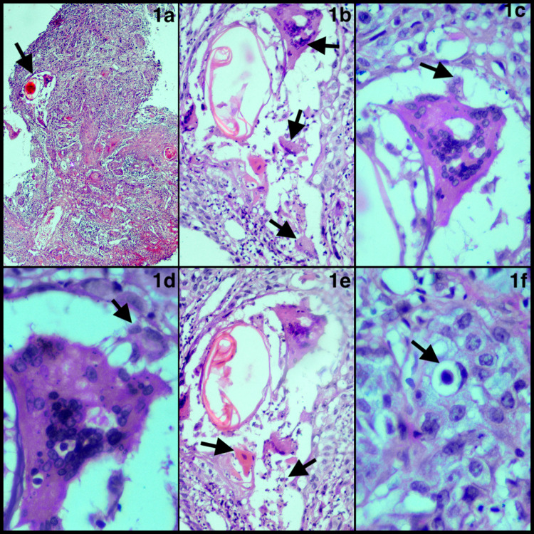
Building upon this background knowledge, we present a novel case of a 49-year-old male with an ulcerative growth in the left buccal mucosa. Histopathology revealed ulcerated dysplastic epithelium and atypical epithelial cells with moderate pleomorphism infiltrating the connective tissue stroma in sheets, strands, and small islands. Notable findings included individual cell keratinization, abnormal mitoses, and stromal multinucleated giant cells (MNGCs). The host response was moderate, with an average of 4 cannibalistic cells and 5 mitotic figures per 10 high-power fields. The evaluation confirmed moderately differentiated OSCC. Notably, keratin pearls within tumor islands were surrounded and degraded by large foreign body MNGCs (50–70 nuclei) and Langhans MNGCs (5–7 nuclei). Neoplastic cells infiltrated or integrated into these giant cells, and a few inflammatory and dyskeratotic cells indicated ongoing degradation mechanisms (Fig. 1).
Fig. 1.
a Keratin pearl degradation by multinucleated giant cells within the tumor island (4X). b Presence of foreign body and Langhans multinucleated giant cells (20X). c and d Neoplastic cells from the surrounding tumor island appeared to infiltrate the cytoplasm or integrate into the foreign body giant cells (40X). e Presence of dyskeratotic and inflammatory cells (20X). f Presence of cellular cannibalism within the tumor island (60X)
This novel documentation of keratin pearl degradation by foreign body MNGCs in OSCC reveals a sophisticated, orchestrated process involving keratin engulfment and degradation [6]. MNGCs, through complex signaling and molecular interactions, actively participate in this degradation within tumor islands [6, 7]. Existing literature highlights the primary role of neutrophils and monocytes in keratin pearl degradation [5], but we propose that the large keratin material presents a phagocytic challenge for macrophages and neutrophils. Consequently, tumor cells might acquire a macrophage-like phenotype, [8] facilitating MNGCs formation through tumor cell fusion. These MNGCs, genetically and immunologically programmed, remove keratin from the tumor island [9], indicating a shift toward poorer differentiation [10]. The degradation process induces intra-tumoral heterogeneity, impacting the tumor’s biological behavior and demonstrating a pro-tumorigenic effect in OSCC [10].
In OSCC, the presence of giant cells is a noteworthy phenomenon and holds significant pathological and diagnostic importance, even though their specific functions remain unclear. This case introduces novel perspectives on the formation of foreign body/Langhans MNGCs through the fusion of macrophage phenotype of tumor cells in response to keratin pearl formation and is involved in the engulfment and degradation of keratin pearl within the tumor island. Intriguingly, the presented case, classified as moderately differentiated OSCC, exhibited heightened cannibalistic activity. This suggests that the degradation of keratin pearls by foreign body/Langhans MNGCs could be indicative of a more aggressive tumor phenotype in OSCC. The presence of keratin pearls in OSCC indicates well-differentiated tumors. Their degradation by MNGCs might signal an inflammatory response, influencing tumor progression and behavior, possibly leading to a more aggressive phenotype. MNGCs activity within the tumor might correlate with worse outcomes, depending on the immune reaction’s context and extent [9, 10].
The degradation of keratin pearls by foreign body/Langhans MNGCs in OSCC has key implications for treatment: (a) Surgical Management: Understanding keratin pearl degradation helps evaluate tumor margins and differentiation, aiding in precise resection planning [5–8, 10]. (b) Radiotherapy: The tumor’s microenvironment, including MNGCs and inflammation, can affect radiotherapy efficacy. A hypoxic environment created by MNGCs may reduce effectiveness [7, 8]. (c) Chemotherapy: Signs of active cellular turnover and immune response, such as keratin pearl degradation and MNGCs, may enhance the effectiveness of chemotherapy and immunotherapy [6–10].
The disintegration of keratin pearls by foreign body/Langhans MNGCs in OSCC underscores a pivotal consideration for the attending surgeon: (A) Surgical Planning: Pathological reports on keratin pearl degradation help surgeons understand tumor differentiation and immune response, guiding resection decisions. (B) Prognosis: The presence of MNGCs informs prognosis and potential tumor aggressiveness, influencing treatment strategies. (C) Multidisciplinary Approach: Awareness of these features allows for better collaboration among surgeons, oncologists, and radiologists to optimize patient outcomes.
In conclusion, the degradation of keratin pearls by foreign body/Langhans MNGCs in OSCC underscores intricate tumor microenvironment interactions, with profound implications for treatment planning and prognosis. Surgeons should meticulously recognize these degradations, as they signify a more aggressive phenotype, necessitating judicious decisions on surgical extent and adjunctive therapies.
Acknowledgements
None.
Funding
No funds, grants, or other support were received. The authors declare they have no financial interests.
Declarations
Conflict of interest
The authors have no relevant financial or nonfinancial interests to disclose.
Ethical Approval
This article does not contain any studies with human participants performed by the author.
Informed Consent
Informed consent is not mandatory for this sort of study.
Consent for Publication
Consent for publication is not mandatory for this sort of study.
Footnotes
Publisher's Note
Springer Nature remains neutral with regard to jurisdictional claims in published maps and institutional affiliations.
References
- 1.Majumdar B, Patil S, Sarode SC, Sarode GS, Rao RS (2017) Clinico-pathological prognosticators in oral squamous cell carcinoma: an update. Transl Res Oral Oncol. 2:2057178X17738912 [Google Scholar]
- 2.Jadhav KB, Gupta N (2013) Clinicopathological prognostic implicators of oral squamous cell carcinoma: need to understand and revise. N Am J Med Sci 5:671 [DOI] [PMC free article] [PubMed] [Google Scholar]
- 3.Bhargava A, Saigal S, Chalishazar M (2010) Histopathological grading systems in oral squamous cell carcinoma: a review. J Int Oral Health 2(4):1 [Google Scholar]
- 4.Patil S, Rao RS, Ganavi BS (2013) A foreigner in squamous cell carcinoma! J Int Oral Health 5(5):147 [PMC free article] [PubMed] [Google Scholar]
- 5.Essa AA, Yamazaki M, Maruyama S, Abé T, Babkair H, Cheng J, Saku T (2014) Keratin pearl degradation in oral squamous cell carcinoma: reciprocal roles of neutrophils and macrophages. J Oral Pathol Med 43(10):778–784 [DOI] [PubMed] [Google Scholar]
- 6.Ahmadzadeh K, Vanoppen M, Rose CD, Matthys P, Wouters CH (2022) Multinucleated giant cells: current insights in phenotype, biological activities, and mechanism of formation. Front Cell Dev Biol 10:873226 [DOI] [PMC free article] [PubMed] [Google Scholar]
- 7.Smetana K Jr (1987) Multinucleate foreign-body giant cell formation. Exp Mol Pathol 46(3):258–265 [DOI] [PubMed] [Google Scholar]
- 8.Sarode SC, Sarode GS (2014) Neutrophil-tumor cell cannibalism in oral squamous cell carcinoma. J Oral Pathol Med 43(6):454–458 [DOI] [PubMed] [Google Scholar]
- 9.Sánchez-Romero C, Carlos R, DantasSoares C, Paes de Almeida O (2018) Unusual multinucleated giant cell reaction in a tongue squamous cell carcinoma: histopathological and immunohistochemical features. Head Neck Pathol 12:580–586 [DOI] [PMC free article] [PubMed] [Google Scholar]
- 10.Zhong K, Song X, Zhao XK, Hu JF, Zhou FY, Li JL, Ren JL, Li XM, Wang XZ, Huang GR, Ku JW (2022) The prognostic value of keratin pearls in patients with esophageal squamous cell carcinoma. Am J Transl Res 14(12):8947 [PMC free article] [PubMed] [Google Scholar]


