Abstract
This review aims to systematically evaluate the incidence, management strategies, and clinical outcomes of iatrogenic durotomy (ID) in endoscopic spine surgery and to propose a management flowchart based on the tear size and associated complications. A comprehensive literature search was conducted, focusing on studies involving endoscopic spinal procedures and incidental durotomy. The selected studies were analyzed for management techniques and outcomes, particularly in relation to the size of the dural tear and the presence of nerve root herniation. Based on these findings, a flowchart for intraoperative management was developed. A total of 14 studies were included, encompassing 68,546 patients. Varying incidences of ID, with management strategies largely dependent on the size of the dural tear, were found. Small tears (less than 5 mm) were often left untreated or managed with absorbable hemostatic agents, while medium (5–10 mm) and large tears (greater than 10 mm) required more complex approaches like endoscopic patch repair or open surgery. The presence of nerve root herniation necessitated immediate action, often influencing the decision to convert to open repair. Effective management of ID in endoscopic spine surgery requires a nuanced approach tailored to the size of the tear and specific intraoperative challenges, such as nerve root herniation. The proposed flowchart offers a structured approach to these complexities, potentially enhancing clinical outcomes and reducing complication rates. Future research with more rigorous methodologies is necessary to refine these management strategies further and broaden the applications of endoscopic spine surgery.
Keywords: Iatrogenic durotomy, Dural injury, Dura tear, Complication, Management, Surgical technique, Systematic review
INTRODUCTION
Endoscopic spine surgery (ESS), first introduced in the early 1990s specifically for discectomy procedures, has since evolved significantly. Its applications now extend to a broader spectrum of spinal interventions, including decompression and fusion techniques. Over the past decade, ESS has emerged as a globally recognized trend in spinal surgery, evidenced by a marked increase in related scholarly studies [1,2]. This evolution in practice is mirrored in the classification of ESS into 2 primary categories: full ESS (uniportal system) and unilateral biportal ESS (UBE, or the biportal system). The full-endoscopic approach involves simultaneous viewing and operative intervention through a single portal channel, whereas the UBE technique distinguishes itself by utilizing separate channels for viewing and operational purposes. The latter has gained increasing acceptance due to its compatibility with standard surgical instruments and a comparatively shorter learning curve for practitioners [3].
Despite its minimally invasive nature and associated benefits, such as reduced intraoperative blood loss and shorter hospital stays, ESS is not without its challenges. A significant concern is iatrogenic durotomy (ID), a prevalent intraoperative complication. This complication is particularly challenging to address due to the limited working space available in ESS procedures. ID can lead to a range of adverse outcomes, from pseudomeningocele and persistent headaches to more severe complications like infection, persistent radiculopathy, dysesthesia, and cerebrospinal fluid (CSF) leakage, affecting up to a third of patients who experience this complication [4]. Despite some proposed techniques for managing ID, a comprehensive systematic review addressing the management of ID within the context of ESS remains a notable gap in the literature. Thus, we conducted this review with the aim of filling that void by proposing our simplified repair method suited for a full-endoscopic approach and evaluating the current evidence on ID management strategies in ESS.
MATERIALS AND METHODS
1. Search Strategy and Data Extraction
This study was reported in accordance with the Preferred Reporting Items for Systematic Reviews and Meta-analyses (PRISMA) guidelines [5]. The study protocol for this systematic review and meta-analysis was registered on the PROSPERO (International Prospective Register of Systematic Reviews; ID No. 527269). For nonhuman interventional research, ethical approval and informed consent are not needed. We searched the PubMed (MEDLINE), Cochrane Library, Scopus, and Web of Science electronic databases from inception to 1 March 2024. We also manually searched for published or preprinted articles. In accordance with the Cochrane Handbook for Systematic Reviews of Interventions, 2 independent investigators extracted the demographic (author, publication year) and intervention data (surgical technique, measurement metrics).
We included various types of studies, including randomized controlled trials, prospective or retrospective cohorts, case reports, case series, and technical notes for interventions without restrictions regarding age, sex, or race. This is because specific reports regarding ID during ESS are still scarce. Studies that included the use of other endoscopic or minimally invasive approaches other than uniportal or biportal endoscopy, such as microendoscopic discectomy/decompression (MED) or traditional microsurgery, were all excluded. All the narrative or systematic review articles, as well as short communications, letters, or other kinds of publications not mentioned above, were also excluded. The occurrence of ID is clearly defined as an inadvertent injury of the dura mater during the endoscopic spinal operation as a complication. As in detail among the reports, various details of the management, a surgical technique to address or repair the lesion, incidence rate, or clinical outcomes were thoroughly examined. There were also no restrictions regarding the threshold ranges of any demographic baseline, the minimum number of participants, and the technology or the device that had been used. WT and AA independently extracted data to Microsoft Excel (Microsoft Corporation, 2018, Microsoft Excel) using a structured and standardized form. In addition to outcomes, information on a vast array of clinical and methodological trial characteristics was extracted, as described previously in the protocol. In cases of discrepancies among the evaluators concerning the extracted data, a third reviewer (SS) was consulted to achieve a final consensus. The following data were extracted from the eligible studies, including the author’s name, publication year, study design, sample size, follow-up duration, clinical measurement outcomes, and the management or technique used to address the complication.
2. Risk of Bias Assessment and Outcome Indicators
Two reviewers (KJ and SS) independently assessed the risk of bias (RoB) of included studies using the Risk Of Bias In Nonrandomized Studies–of Exposure (ROBINS-E) tool [6]. A visualization tool (Robvis) was used to visualize the RoB assessment in our systematic review [7]. The ROBINS-E tool comprises 7 domains to assess the RoB, including the bias due to confounding, bias arising from the measurement of the exposure, bias in the selection of participants into the study (or into the analysis), bias due to postexposure interventions, bias due to missing data, bias arising from the measurement of the outcomes, and the bias in the selection of the reported result. In cases of discrepancies in RoB assessment results among the reviewers, a discussion between the 2 reviewers would also be arranged to finalize the results.
3. Data Synthesis and Statistical Analysis
Because of the scarcity of publications on the topic of interest and the high heterogeneity of study designs and methodology among the included studies, a meta-analysis was not performed in our systematic review. Accumulated data from all the included studies presenting as mean or percentage were also synthesized and reported. Statistical analysis was performed using the network packages in Stata using STATA/MP (Release 17. Stata-Corp LLC, College Station, TX, USA).
RESULTS
1. Study Characteristics
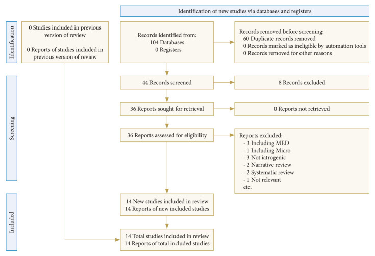
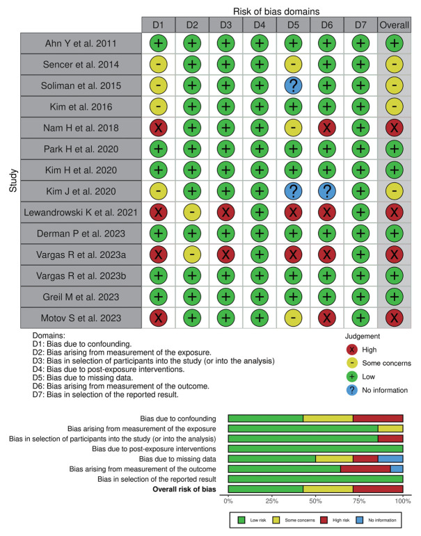
The process of evaluating studies according to the inclusion and exclusion criteria is demonstrated in Fig. 1, using the PRISMA 2020 flow diagram for updated systematic reviews [8]. From the initial results of 104 studies from the databases, after screening the abstracts and reviewing the complete texts, 14 studies with a total of 68,546 patients were ultimately included in this review [4,9-21]. A total of 8 retrospective cohorts, 2 prospective cohorts, 2 case series, 1 case report, and 1 retrospective surgeon survey were included. Most of the indications for surgery were lumbar disc herniation, followed by lumbar spinal canal stenosis, or both. Furthermore, most of the ESS systems reported in the included studies were uniportal or full-endoscopy (12 studies, 85.71%). All the details of the included studies are shown in Table 1. A total of 4 studies were considered to have a moderate level RoB, which is commonly due to the possible contributing confounding factors observed in their respective studies. Another 4 studies were considered to have high-level RoB and were also commonly observed from the possible contributing confounding factors and concerns from the measurement of the outcomes. The overall RoB was considered moderate or possessed some concerns when considering all of the 7 domains altogether, which could be expected from the inclusion of nonrandomized studies. The details of the RoB assessment visualized using the Robvis tool are demonstrated in Fig. 2.
Fig. 1.
Flowchart diagram demonstrating the articles included in this systematic review using the PRISMA (Preferred Reporting Items for Systematic Reviews and Meta-analyses) 2020 guideline.
Table 1.
Studies included in this systematic review and its characteristics
| Study | Study design | No. | Diagnosis | ESS system | No. of tear | Convert to open? (n) | Technique of dural closure? (n) | Bed rest? (n) | Drain? | Symptoms | Revision surgery? (n, cause) |
|---|---|---|---|---|---|---|---|---|---|---|---|
| Ahn et al. [9] (2010) | Retrospective | 816 | LDH | Uni | 9 | No | Suture (2); cadaveric dura; fibrin glue; and TachoComb | NA | NA | Headache, sensory deficit, leg pain, back pain | Yes (7, delayed diagnosis) |
| Sencer et al. [10] (2014) | Prospective | 163 | LDH | Uni | 6 | Yes (1); No (5) | NA (1); No (5) | 5 Days (1); 2 days (5) | Lumbar drainage (1); No (5) | NA | Yes (1) |
| Soliman [11] (2015) | Prospective | 104 | LSS | Uni | 6 | No | No | 3 Days | NA | Headache | No |
| Kim et al. [12] (2016) | Retrospective | 18 | LDH | Uni | 1 | No | NA | NA | Yes | NA | NA |
| Nam et al. [13] (2018) | Case series | 3 | LDH and LSS | Uni | 3 | No | Double-layer TachoSil packing | NA | Yes | Headache, vomiting, leg pain | No |
| Park et al. [14] (2020) | Retrospective | 643 | LSS | Bi | 29 | Yes (1) | No (12); sealants (14); nonpenetrating titanium clip with sealants (2) | 24 Hr (12); 48 hr (2) | NA | No | Yes (1, meningocele) |
| Kim et al. [15] (2020) | Retrospective | 1,551 | LDH and LSS | Bi | 25 | Yes (5) | Patch compression method (21); endoscopic clipping (1); immediate open repair (2); delayed conversion to open repair (2) | 2 Days | No | No | Yes (2, fail blood patch, pseudomeningocele) |
| Kim et al. [16] (2020) | Retrospective | 330 | LSS | Uni | 27 | Yes (2) | Endoscopic patch blocking dural repair (25); open suture repair (2) | NA | NA | NA | No |
| Lewandrowski et al. [4] (2021) | Retrospective survey | 64,470 | NA | Uni and Bi | 689 | NA | No; sealants; suture | 24–48 Hr | NA | Motor weakness, sensory deficit, leg pain | NA |
| Derman et al. [17] (2023) | Retrospective | 295 | LDH and LSS | Uni | 3 | No | Collagen matrix inlay graft (2); collagen matrix inlay graft with PEG hydrogel (1) | 172–1,068 Min (2); overnight (1) | NA | No | No |
| Vargas et al. [18] (2023) | Retrospective | 15 | CDH and LDH | Uni | 3 | No | No | 10 Days (1); 3 days (2) | NA | Seizure, cardiac arrest, diplopia, headache, intracranial air, subarachnoid hemorrhage | No |
| Vargas et al. [19] (2023) | Case series | 3 | CDH | Uni | 3 | NA | NA | NA | NA | Headache, neck pain, seizure, autonomic dysreflexia, hypertension | NA |
| Greil et al. [20] (2023) | Retrospective | 174 | NA | Uni | 13 | No | Dural inlay graft | No (1); 1 day (7); 2 days (4); ≥3 days (1) | NA | NA | No |
| Motov et al. [21] (2023) | Case report | 1 | CFS | Uni | 1 | No | CT-guided epidural fibrin patch | 1 Day | NA | Tingling dysesthesia in both upper extremities, orthostatic headache and neck pain | No |
ESS, endoscopic spine surgery; LDH, lumbar disc herniation; NA, not mentioned or not available; LSS, lumbar spinal canal stenosis; Uni, uniportal; Bi, biportal; CDH, cervical disc herniation; CFS, cervical foraminal stenosis; PEG, polyethylene glycol; CT, computed tomography.
Fig. 2.
Graphic visualization showing the assessment of the risk of bias among the included studies using the ROBINS-E (Risk Of Bias In Non-randomized Studies–of Exposure) tool.
2. Incidence and Clinical Consequences
The incidence and clinical outcomes of ID in spinal surgeries exhibit significant variation across different studies and surgical approaches. A study incorporating the complex full-endoscopic technique of unilateral laminotomy for bilateral lumbar decompression noted an ID rate of 7.5%. It also highlighted that while perioperative durotomy prolongs hospital stays, it does not markedly influence the occurrence of other perioperative complications or the need for revision surgery [20]. Sencer et al. [10] reported a comparable incidence of ID, underscoring that advancements in surgical techniques and management strategies are reducing its adverse effects on patient recovery and functional outcomes. Conversely, Vargas et al. [19] observed that severe irrigation-related complications following the incidental durotomy intraoperatively, such as intracranial air bubble, subarachnoid hemorrhage, meningitis, or pseudomeningocele, can lead to persistent symptoms like headache, neck pain, diplopia, seizure, autonomic dysreflexia, and sensory deficits, or even cardiac arrest if inadequately addressed. However, despite the potential for adverse outcomes, the current literature suggests that with prompt recognition and appropriate management, the long-term effects of incidental durotomy can be significantly decreased, allowing for satisfactory clinical outcomes in the majority of cases [11].
Other included studies offer more insights into the incidence of ID across various endoscopic spinal procedures. Recently, Derman et al. [17] identified an ID incidence of 1.02% in 295 cases of uniportal full ESS. A large number of surveys among endoscopic spine surgeons by Lewandrowski et al. [4] revealed an ID rate of 1.07% in 64,470 lumbar endoscopies, with medium-sized dural tears (1–10 mm) being the most prevalent, whereas another study reported a higher incidence of 4.5% [14]. Moreover, Kim et al. [16] found an even higher rate of 8.18% incidence of ID during endoscopic lumbar decompression procedure in a study of 330 patients. They noted that ID occurrence varied with the surgical level and was more common in the interlaminar decompression compared to the transforaminal approach. Lastly, Kim et al. [15] conducted a study of the recently developed UBE and indicated a similar rate of 1.6% in a total of 1,551 cases.
3. Management
Various management techniques have been described to address ID. Among all the included studies, all variations of management were primarily based on the size of the tear, with different strategies adopted for small, medial, and large-sized tears (roughly as less than 5 mm, 5–10 mm, and more than 10 mm, respectively). For small-sized tears (less than 5 mm), Derman et al. [17] demonstrated that IDs measuring 2 to 2.5 mm could be effectively repaired using a collagen matrix inlay technique. This approach negated the need for prolonged bed rest, and all patients achieved excellent outcomes without further complications, based on their reports. This method is suggested to be suitable for other minimally invasive spine surgery techniques as well. Lewandrowski et al. [4] also observed that most dural tears in their study were small and could be successfully managed with mechanical compression using only gel foam and sealants.
In cases of medium-sized tears (5–10 mm), Kim et al. [16] proposed the Endoscopic Patch Blocking Dura Repair technique, presenting it as an effective method, especially for type 1 to type 3A dural tears based on a classification of dural tears by Blecher et al. [22] This technique was associated with a good prognosis and clinical outcomes. Another study group by Kim et al. [15] reported that 20 cases of IDs measuring less than 10 mm were also successfully treated with just a patch technique. Park et al. [14] have described the medium-sized tear similarly (4–12 mm) and reported a technique of using a fibrin sealant patch (Tachosil, Baxter, Deerfield, IL, USA), complemented by close inpatient observation after the surgery. All of the 14 cases in their study were treated using this fibrin sealant approach with good results.
When managing large-sized tears (more than 10 mm), several different strategies were observed. Park et al. [14] attempted primary closure using the endoscopy for tears exceeding 12 mm. Based on the same classification proposed by Blecher et al. [22] Kim et al. [16] recommended considering conversion to open repair for type 3B, C, and 4 dural tears, which were usually associated with fair to poor outcomes. Another study by Kim et al. [15] reported that of the 5 cases of IDs measuring 10 mm or more, 3 underwent open repair within a few days. Additionally, 2 cases that did not respond to conservative management required delayed revision surgery due to pseudomeningocele. As a result, the management of ID during ESS thus varies according to the size of the tear, with each tear size category having specific, effective treatment protocols. These strategies range from minimally invasive techniques for smaller tears to more involved surgical interventions for larger tears. This systematic approach to managing IDs is crucial in optimizing patient outcomes and reducing the risk of further complications.
DISCUSSION
The increasing evidence in favor of endoscopic spine procedures (ESS) highlights their growing prominence in the field of spinal surgery, with studies demonstrating outcomes that are either comparable to or surpass those of conventional microscopic surgery [23]. Notably, ESS is associated with numerous postoperative benefits, including diminished pain, decreased reliance on analgesics, smaller incisions, and reduced paraspinal soft tissue damage. These advantages, in turn, contribute to a lower incidence of infection and blood loss [24], facilitating shorter hospital stays and expedited resuming of daily activities. Nonetheless, it is crucial to acknowledge the presence of complications inherent to these procedures. One of the more prevalent perioperative complications encountered in ESS is ID, which, if not properly managed, can lead to serious postoperative issues like pseudo-meningocele, cutaneous fistula, and severe central nervous system infections.
The occurrence of ID in traditional open lumbar spine surgeries is reported to be within the range of 1%–17% [25,26], and that might be considered higher than that observed from ESS in our study. The lower incidence rate of ID observed in ESS can be attributed to several factors. The ESS procedure, which provides a continuous flow of irrigation fluid and significantly enhances magnification power, affords the surgeon superior intraoperative visualization compared to traditional open surgery. However, given that ESS is relatively new in the spine literature, this difference in incidence rates, to be concerned, may also stem from underreporting. Contributing factors to ID in ESS encompass both patient-specific conditions and procedural complexities. These include circumstances such as revision surgeries, multiple cortisone injections [9,14,15], and the inherent learning curve associated with this relatively new technique. The growing adoption of ESS for more intricate cases—such as ossified yellow ligaments, calcified thoracic discectomy, failed back surgery syndrome, or spinal tumors—also plays a significant role. Moreover, the chronic nature of certain pathologies leading to fibrosis post prolonged compression and inflammation, as well as procedures involving bilateral decompression via a unilateral approach, require heightened technical proficiency from surgeons [20].
The management of ID presents a domain that is still in need of definitive guidelines. While some research points out the necessity of converting to open repair in certain instances, the often small size of dural tears renders their endoscopic repair a technically demanding endeavor. A common approach in many studies has been to leave the durotomy untreated or to address it with absorbable hemostatic agents such as gel foam or fibrin sealant patches. One potential explanation for the observed differences compared to traditional surgery is that the ultra-minimally invasive nature of the ESS technique results in smaller soft tissue disruption and dead space relative to more conventional methods, such as open or microsurgery. Consequently, this could contribute to a rapid spontaneous tamponade effect, thereby reducing the likelihood of CSF leakage [16]. Nonetheless, this method must be executed with precision to avoid the risk of introducing intradural foreign bodies.
Recent advancements in spinal endoscopy techniques, including both uniportal and biportal approaches, have significantly enhanced the ability to repair dural defects. These innovations have made it feasible to address not only larger defects, which pose a risk of severe complications, but also smaller ones, particularly those involving an incarcerated nerve root. Our findings indicate that the management strategy for ID largely depends on the size of the tear. For smaller tears, typically less than 5 mm, techniques such as patching techniques, the collagen matrix inlay method, or opting for no intraoperative intervention followed by closed postoperative observation have proven effective in promoting swift recovery without necessitating extended bed rest. For medium-sized tears (usually 5–10 mm), techniques like the Endoscopic Patch Blocking Dura Repair have shown promise. Larger tears (greater than 10 mm) often require more complex approaches, including primary endoscopic closure or, in more challenging cases, conversion to open repair. This distinction in management strategies underscores the necessity for tailored treatment to optimize patient outcomes and minimize the risk of complications.
Several factors should be considered regarding the use of endoscopic suturing in the management of ID. The skill sets and learning curves required for endoscopic dura suture repair vary significantly between uniportal and biportal techniques. Biportal ESS, which permits the use of conventional surgical instruments unlike its uniportal counterpart and demands a skill set similar to that required in conventional surgery, is more accessible and simpler to adopt. Therefore, making direct comparisons between these techniques, especially in contexts requiring suturing during intraoperative dura repair, may not be straightforward. Our systematic review supports the viability of endoscopic repair for medium (5–10 mm) or large (>10 mm) dural tears, although we do not specify a preference for uniportal versus biportal techniques. This is because of the scarcity of reports on dura suturing in uniportal endoscopy and the relative ease of suturing in biportal endoscopy. Moreover, our management recommendations are designed to serve as guidance for clinical decision-making rather than prescriptive mandates. If endoscopic repair is considered unfeasible, we advocate for open repair as a preferable alternative, emphasizing the importance of customized decision-making for each patient. Nevertheless, an in-depth comparison of suturing techniques between uniportal and biportal endoscopy would be beyond the scope and objectives of this study, which aims to establish general recommendations based on the available literature.
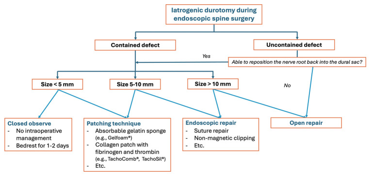
Although not explicitly addressed in the studies we reviewed, the presence of concurrent nerve root herniation is a critical factor influencing the outcomes of iatrogenic dural injury management [27]. This complication often correlates with the size of the dural tear and should be prioritized in treatment. When encountering a “contained defect,” where the nerve root has not herniated outside the dura, management strategies align with those previously outlined. However, in instances of an “uncontained defect” where the nerve root has herniated beyond the dural layer, immediate action is necessary to reposition the nerve root back into the dural sac before any repair or closure attempts. In situations where the herniated nerve root cannot be repositioned within the dural sac, converting to an open surgical procedure is highly advised [28]. This step is crucial to ensure the successful reduction of the herniated nerve root prior to closure, thereby averting further complex complications. Our review introduces a comprehensive flowchart for managing ID, considering both the size of the dural tear and the status of the nerve root (contained or uncontained defect). This flowchart, illustrated in Fig. 3, offers a valuable tool for clinicians, guiding them in selecting the most appropriate management strategy when faced with such intricate complications. This systematic approach aims to optimize treatment outcomes and minimize the risk of further issues arising from the iatrogenic dural injury.
Fig. 3.
A proposed strategic management flowchart for iatrogenic durotomy during endoscopic spine surgery.
Our systematic review offers distinct insights compared to previous analyses, such as the one conducted by Muller et al. [29], which incorporated a broader scope of endoscopic techniques. While their review included both uniportal and tubular-assisted endoscopic techniques, yielding a reported overall dural tear rate of 2.7% (ranging from 0%–8.6%), similar to our findings, it did not distinguish between the nuances of these varying approaches. Likewise, they identified a higher risk of a dural tear in synovial cyst resection and a unilateral approach for bilateral decompression procedures. However, our review focuses explicitly on the more recent advancements in full-endoscopy (uniportal) and UBE (biportal) procedures, excluding the tubular endoscopic-assisted techniques (microendoscopic discectomy/decompression or MED) due to their comparatively limited intraoperative visualization and differing skill requirements. Moreover, as previously mentioned, our systematic review adheres strictly to the Cochrane Handbook for Systematic Reviews and assesses the RoB with greater rigor, thus enhancing its validity. Unlike the previous review, we delve into the specificities of dural tear management based on tear size, offering a nuanced understanding that is critical for clinical application with a proposed structured flowchart for managing dural tears in ESS (Fig. 3). This targeted focus and methodological stringency set our review apart, providing more specific, applicable insights for the management of dural tears in the context of the recent trend of ESS techniques.
Although the incidence of dural tears during ESS is relatively low, future research should aim for more methodologically robust randomized studies. These studies should focus on comparing various techniques for addressing ID intraoperatively and assessing their long-term outcomes to prevent further complications from occurring. It is imperative to interpret the findings from our review with caution due to the moderate level of overall RoB assessed in the included studies. Future research should endeavor to minimize these biases by implementing more stringent controls for confounding factors and selecting more precise methods to evaluate outcomes. Additionally, it is crucial for future authors to comprehensively report all relevant results to reduce the risk of selection bias. This approach will enhance the validity of research in this field, providing clearer insights into the management of ID during ESS.
Our study, while providing valuable insights, is subject to several limitations. Firstly, approximately half of the included studies raised concerns regarding the RoB, predominantly related to potential confounding factors and the selection of clinical measurement tools. Secondly, the relatively small number of studies and participants included in our review could impact its overall validity. This limitation, however, may be partly attributable to the inherently low incidence of dural tears in ESS. Thirdly, although we did not conduct a formal statistical analysis of the included studies, a high degree of heterogeneity is anticipated. This variation primarily results from the inclusion of diverse nonrandomized study designs. Nonetheless, given the low incidence and limited previous literature on iatrogenic dural injury during ESS, our review serves as an essential initial exploration of this complex complication. Finally, considering the increasing global interest in ESS and the retrospective nature of most studies, there is a suspicion that the incidence of ID during ESS may be higher than currently reported, potentially due to information bias. These limitations highlight the need for further research with more rigorous methodologies to provide better quality of the included studies for future studies of systematic review and might contribute to improved patient care and outcomes in this rapidly changing spinal healthcare.
CONCLUSION
The management of incidental durotomies in endoscopic spinal surgery varies widely due to the lack of standardized guidelines. Treatment approaches range from nonintervention for small tears to the use of sealant materials for larger ones. The consideration of contained or uncontained dural defects may also be crucial. Currently, transitioning to open repair is rare, but as endoscopic repair techniques continue to advance, the necessity for open repair may further diminish. These developments hold promise for expanding the capabilities of spinal endoscopy, potentially including more complex procedures such as intradural tumor surgery in the future.
Footnotes
Conflict of Interest
The authors have nothing to disclose.
Funding/Support
This study received no specific grant from any funding agency in the public, commercial, or not-for-profit sectors.
Author Contribution
Conceptualization: WT, JSK, SS; Data curation: WT, AA, KJ, BP, AS; Formal analysis: WT, AA, KJ, BP, AS, YL; Methodology: YL, SS; Project administration: JSK, SS; Visualization: YL, SS; Writing – original draft: WT, AA, KJ, BP, AS; Writing – review & editing: JSK, SS.
REFERENCES
- 1.Jitpakdee K, Liu Y, Heo DH, et al. Minimally invasive endoscopy in spine surgery: where are we now? Eur Spine J. 2023;32:2755–68. doi: 10.1007/s00586-023-07622-7. [DOI] [PubMed] [Google Scholar]
- 2.Liu Y, Kotheeranurak V, Quillo-Olvera J, et al. A 30-year worldwide research productivity of scientific publication in full-endoscopic decompression spine surgery: quantitative and qualitative analysis. Neurospine. 2023;20:374–89. doi: 10.14245/ns.2245042.521. [DOI] [PMC free article] [PubMed] [Google Scholar]
- 3.Ahn Y, Lee S. Uniportal versus biportal endoscopic spine surgery: a comprehensive review. Expert Rev Med Devices. 2023;20:549–56. doi: 10.1080/17434440.2023.2214678. [DOI] [PubMed] [Google Scholar]
- 4.Lewandrowski KU, Hellinger S, De Carvalho PST, et al. Dural tears during lumbar spinal endoscopy: surgeon skill, training, incidence, risk factors, and management. Int J Spine Surg. 2021;15:280–94. doi: 10.14444/8038. [DOI] [PMC free article] [PubMed] [Google Scholar]
- 5.Hutton B, Salanti G, Caldwell DM, et al. The PRISMA extension statement for reporting of systematic reviews incorporating network meta-analyses of health care interventions: checklist and explanations. Ann Intern Med. 2015;162:777–84. doi: 10.7326/M14-2385. [DOI] [PubMed] [Google Scholar]
- 6.Bero L, Chartres N, Diong J, et al. The risk of bias in observational studies of exposures (ROBINS-E) tool: concerns arising from application to observational studies of exposures. Syst Rev. 2018;7:242. doi: 10.1186/s13643-018-0915-2. [DOI] [PMC free article] [PubMed] [Google Scholar]
- 7.Salanti G, Ades AE, Ioannidis JP. Graphical methods and numerical summaries for presenting results from multiple-treatment meta-analysis: an overview and tutorial. J Clin Epidemiol. 2011;64:163–71. doi: 10.1016/j.jclinepi.2010.03.016. [DOI] [PubMed] [Google Scholar]
- 8.Page MJ, McKenzie JE, Bossuyt PM, et al. The PRISMA 2020 statement: an updated guideline for reporting systematic reviews. BMJ. 2021;372:n71. doi: 10.1136/bmj.n71. [DOI] [PMC free article] [PubMed] [Google Scholar]
- 9.Ahn Y, Lee HY, Lee SH, et al. Dural tears in percutaneous endoscopic lumbar discectomy. Eur Spine J. 2011;20:58–64. doi: 10.1007/s00586-010-1493-8. [DOI] [PMC free article] [PubMed] [Google Scholar]
- 10.Sencer A, Yorukoglu AG, Akcakaya MO, et al. Fully endoscopic interlaminar and transforaminal lumbar discectomy: short-term clinical results of 163 surgically treated patients. World Neurosurg. 2014;82:884–90. doi: 10.1016/j.wneu.2014.05.032. [DOI] [PubMed] [Google Scholar]
- 11.Soliman HM. Irrigation endoscopic decompressive laminotomy. A new endoscopic approach for spinal stenosis decompression. Spine J. 2015;15:2282–9. doi: 10.1016/j.spinee.2015.07.009. [DOI] [PubMed] [Google Scholar]
- 12.Kim CH, Chung CK, Woo JW. Surgical outcome of percutaneous endoscopic interlaminar lumbar discectomy for highly migrated disk herniation. Clin Spine Surg. 2016;29:E259–66. doi: 10.1097/BSD.0b013e31827649ea. [DOI] [PubMed] [Google Scholar]
- 13.Nam HGW, Kim HS, Park JS, et al. Double-layer tachosil packing for management of incidental durotomy during percutaneous stenoscopic lumbar decompression. World Neurosurg. 2018;120:448–56. doi: 10.1016/j.wneu.2018.09.040. [DOI] [PubMed] [Google Scholar]
- 14.Park HJ, Kim SK, Lee SC, et al. Dural tears in percutaneous biportal endoscopic spine surgery: anatomical location and management. World Neurosurg. 2020;136:e578–85. doi: 10.1016/j.wneu.2020.01.080. [DOI] [PubMed] [Google Scholar]
- 15.Kim JE, Choi DJ, Park EJ. Risk factors and options of management for an incidental dural tear in biportal endoscopic spine surgery. Asian Spine J. 2020;14:790–800. doi: 10.31616/asj.2019.0297. [DOI] [PMC free article] [PubMed] [Google Scholar]
- 16.Kim HS, Raorane HD, Wu PH, et al. Incidental durotomy during endoscopic stenosis lumbar decompression: incidence, classification, and proposed management strategies. World Neurosurg. 2020;139:e13–22. doi: 10.1016/j.wneu.2020.01.242. [DOI] [PubMed] [Google Scholar]
- 17.Derman PB, Rogers-LaVanne MP, Satin AM. Collagen matrix inlay graft for management of incidental durotomy during full-endoscopic lumbar spine surgery: technique and case series. Int J Spine Surg. 2023;17:399–406. doi: 10.14444/8457. [DOI] [PMC free article] [PubMed] [Google Scholar]
- 18.Vargas RAA, Moscatelli M, Vaz de Lima M, et al. Clinical consequences of incidental durotomy during full-endoscopic lumbar decompression surgery in relation to intraoperative epidural pressure measurements. J Pers Med. 2023;13:381. doi: 10.3390/jpm13030381. [DOI] [PMC free article] [PubMed] [Google Scholar]
- 19.Vargas RAA, Hagel V, Xifeng Z, et al. Durotomy- and irrigation-related serious adverse events during spinal endoscopy: illustrative case series and international surgeon survey. Int J Spine Surg. 2023;17:387–98. doi: 10.14444/8454. [DOI] [PMC free article] [PubMed] [Google Scholar]
- 20.Greil ME, Bergquist J, Kashlan ON, et al. Incidence and management of dural tears in full-endoscopic unilateral laminotomies for bilateral lumbar decompression. Eur Spine J. 2023;32:2889–95. doi: 10.1007/s00586-023-07749-7. [DOI] [PubMed] [Google Scholar]
- 21.Motov S, Stemmer B, Krauss P, et al. Treatment of a symptomatic cervical cerebrospinal fluid fistula after full endoscopic cervical foraminotomy with CT-guided epidural fibrin patch. Eur Spine J. 2024;33:3124–8. doi: 10.1007/s00586-023-07973-1. [DOI] [PubMed] [Google Scholar]
- 22.Blecher R, Anekstein Y, Mirovsky Y. Incidental dural tears during lumbar spine surgery: a retrospective case study of 84 degenerative lumbar spine patients. Asian Spine J. 2014;8:639–45. doi: 10.4184/asj.2014.8.5.639. [DOI] [PMC free article] [PubMed] [Google Scholar]
- 23.Liu Y, Kim Y, Park CW, et al. Interlaminar endoscopic lumbar discectomy versus microscopic lumbar discectomy: a preliminary analysis of L5-S1 lumbar disc herniation outcomes in prospective randomized controlled trials. Neurospine. 2023;20:1457–68. doi: 10.14245/ns.2346674.337. [DOI] [PMC free article] [PubMed] [Google Scholar]
- 24.Mahan MA, Prasse T, Kim RB, et al. Full-endoscopic spine surgery diminishes surgical site infections - a propensity score-matched analysis. Spine J. 2023;23:695–702. doi: 10.1016/j.spinee.2023.01.009. [DOI] [PubMed] [Google Scholar]
- 25.Deyo RA, Cherkin DC, Loeser JD, et al. Morbidity and mortality in association with operations on the lumbar spine. The influence of age, diagnosis, and procedure. J Bone Joint Surg Am. 1992;74:536–43. [PubMed] [Google Scholar]
- 26.Kalevski SK, Peev NA, Haritonov DG. Incidental dural tears in lumbar decompressive surgery: incidence, causes, treatment, results. Asian J Neurosurg. 2010;5:54–9. [PMC free article] [PubMed] [Google Scholar]
- 27.Shanbhag NC, Duyff RF, Groen RJM. Symptomatic thoracic nerve root herniation into an extradural arachnoid cyst: case report and review of the literature. World Neurosurg. 2017;106:1056.e5–1056.e8. doi: 10.1016/j.wneu.2017.07.105. [DOI] [PubMed] [Google Scholar]
- 28.Rahyussalim AJ, Djaja YP, Saleh I, et al. Preservation and tissue handling technique on iatrogenic dural tear with herniated nerve root at cauda equina level. Case Rep Orthop. 2016;2016:4903143. doi: 10.1155/2016/4903143. [DOI] [PMC free article] [PubMed] [Google Scholar]
- 29.Muller SJ, Burkhardt BW, Oertel JM. Management of dural tears in endoscopic lumbar spinal surgery: a review of the literature. World Neurosurg. 2018;119:494–9. doi: 10.1016/j.wneu.2018.05.251. [DOI] [PubMed] [Google Scholar]


