

Abstract
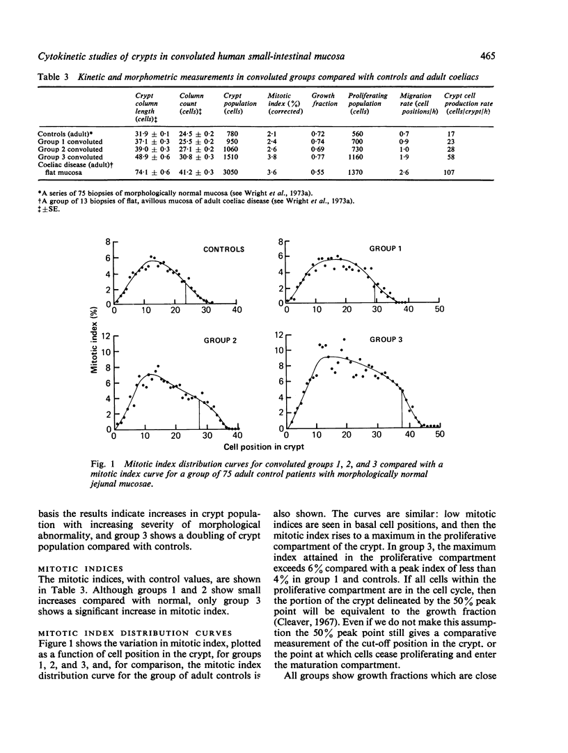
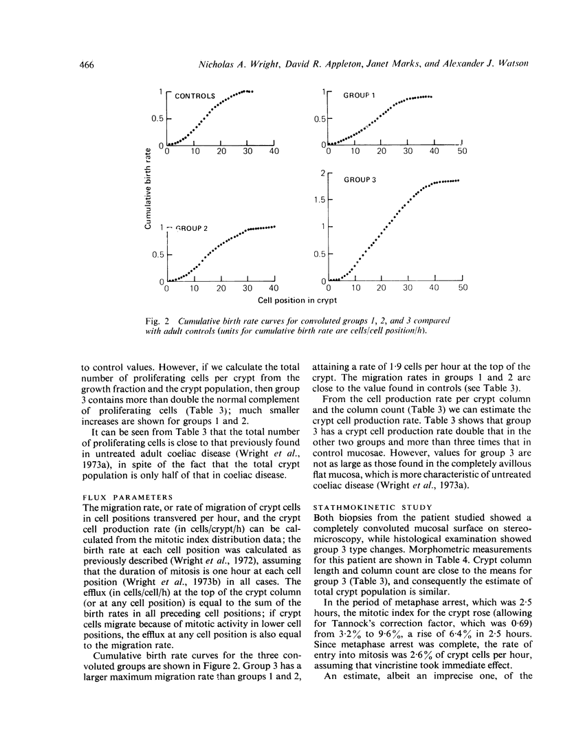
Forty-seven peroral biopsy specimens of duodenojejunal mucosa showing convolutions were obtained from patients with a variety of clinical disorders. These mucosal samples were divided into three groups according to the extent of the convolutions and the severity of the accompanying histopathological changes; the cytokinetic status of the crypts in the three groups was then analysed and compared. Those in which the mucosae were predominantly or totally convoluted (group 3) showed the most notable cytokinetic changes: crypts were hyperplastic and crypt cell production rate was markedly increased compared with the other two groups and with morphologically normal control mucosae.
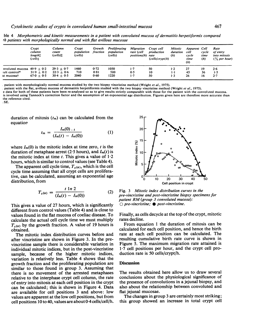
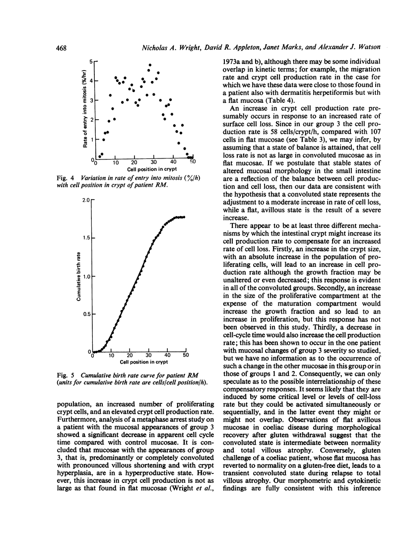
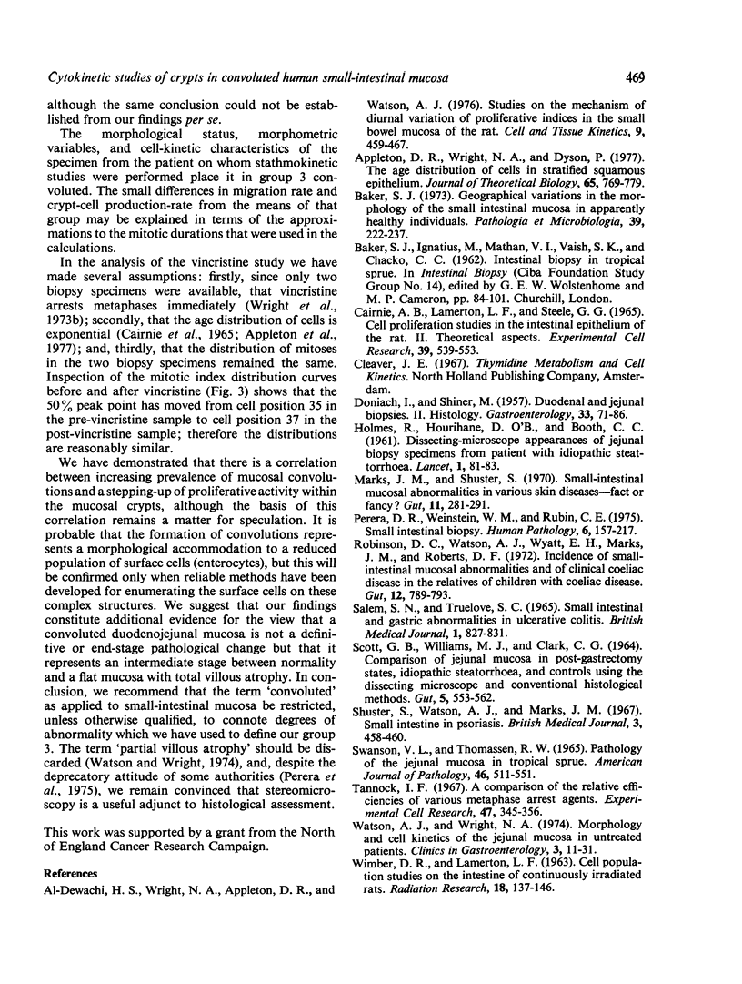
In the case of one patient with mucosal changes of group 3 severity, additional studies were carried out using vincristine to produce metaphase arrest. The cell cycle time of 27 hours was greatly shortened compared with a control value of 45 hours.
We find that the presence of convolutions in small-intestinal mucosal biopsy specimens is accompanied by an increase in the rate of cell production from the crypts, which is presumably related directly or indirectly to the rate of loss of mature enterocytes from the surface of the mucosa. Furthermore, an increase in the proportion of convolutions may reflect an increase in the rates of cell production and cell loss.
In the group 3 convoluted mucosae the cytokinetic profile of the crypts resembled that of some of the flat avillous coeliac mucosae previously studied although the rates of cell production did not reach the levels attained by the most productive of the flat coeliac mucosae.
Full text
PDF
Selected References
These references are in PubMed. This may not be the complete list of references from this article.
- Al-Dewachi H. S., Wright N. A., Appleton D. R., Watson A. J. Studies on the mechanism of diurnal variation of proliferative indices in the small bowel mucosa of the rat. Cell Tissue Kinet. 1976 Sep;9(5):459–467. doi: 10.1111/j.1365-2184.1976.tb01296.x. [DOI] [PubMed] [Google Scholar]
- Appleton D. R., Wright N. A., Dyson P. The age distirubtion of cells in stratified squamous epithelium. J Theor Biol. 1977 Apr 21;65(4):769–779. doi: 10.1016/0022-5193(77)90022-4. [DOI] [PubMed] [Google Scholar]
- Baker S. J. Geographical variations in the morphology of the small intestinal mucosa in apparently healthy individuals. Pathol Microbiol (Basel) 1973;39(3):222–237. doi: 10.1159/000162651. [DOI] [PubMed] [Google Scholar]
- Cairnie A. B., Lamerton L. F., Steel G. G. Cell proliferation studies in the intestinal epithelium of the rat. II. Theoretical aspects. Exp Cell Res. 1965 Sep;39(2):539–553. doi: 10.1016/0014-4827(65)90056-x. [DOI] [PubMed] [Google Scholar]
- DONIACH I., SHINER M. Duodenal and jejunal biopsies. II. Histology. Gastroenterology. 1957 Jul;33(1):71–86. [PubMed] [Google Scholar]
- HOLMES R., HOURIHANE D. O., BOOTH C. C. Dissecting-microscope appearances of jejunal biopsy specimens from patients with "idiopathic steatorrhoea". Lancet. 1961 Jan 14;1(7168):81–83. doi: 10.1016/s0140-6736(61)92123-7. [DOI] [PubMed] [Google Scholar]
- Marks J., Shuster S. Small-intestinal mucosal abnormalities in various skin diseases--fact or fancy? Gut. 1970 Apr;11(4):281–291. doi: 10.1136/gut.11.4.281. [DOI] [PMC free article] [PubMed] [Google Scholar]
- Perera D. R., Weinstein W. M., Rubin C. E. Symposium on pathology of the gastrointestinal tract-Part II. Small intestinal biopsy. Hum Pathol. 1975 Mar;6(2):157–217. doi: 10.1016/s0046-8177(75)80176-6. [DOI] [PubMed] [Google Scholar]
- Robinson D. C., Watson A. J., Wyatt E. H., Marks J. M., Roberts D. F. Incidence of small-intestinal mucosal abnormalities and of clinical coeliac disease in the relatives of children with coeliac disease. Gut. 1971 Oct;12(10):789–793. doi: 10.1136/gut.12.10.789. [DOI] [PMC free article] [PubMed] [Google Scholar]
- SALEM S. N., TRUELOVE S. C. SMALL-INTESTINAL AND GASTRIC ABNORMALITIES IN ULCERATIVE COLITIS. Br Med J. 1965 Mar 27;1(5438):827–831. doi: 10.1136/bmj.1.5438.827. [DOI] [PMC free article] [PubMed] [Google Scholar]
- SCOTT G. B., WILLIAMS M. J., CLARK C. G. COMPARISON OF JEJUNAL MUCOSA IN POST-GASTRECTOMY STATES, IDIOPATHIC STEATORRHOEA, AND CONTROLS USING THE DISSECTING MICROSCOPE AND CONVENTIONAL HISTOLOGICAL METHODS. Gut. 1964 Dec;5:553–562. doi: 10.1136/gut.5.6.553. [DOI] [PMC free article] [PubMed] [Google Scholar]
- SWANSON V. L., THOMASSEN R. W. PATHOLOGY OF THE JEJUNAL MUCOSA IN TROPICAL SPRUE. Am J Pathol. 1965 Apr;46:511–551. [PMC free article] [PubMed] [Google Scholar]
- Shuster S., Watson A. J., Marks J. Small intestine in psoriasis. Br Med J. 1967 Aug 19;3(5563):458–460. doi: 10.1136/bmj.3.5563.458. [DOI] [PMC free article] [PubMed] [Google Scholar]
- Watson A. J., Wright N. A. Coeliac disease. Morphology and cell kinetics of the jejunal mucosa in untreated patients. Clin Gastroenterol. 1974 Jan;3(1):11–31. [PubMed] [Google Scholar]
- Wright N., Morley A., Appleton D. Variation in the duration of mitosis in the crypts of Lieberkuhn of the rat; a cytokinetic study using vincristine. Cell Tissue Kinet. 1972 Jul;5(4):351–364. doi: 10.1111/j.1365-2184.1972.tb00374.x. [DOI] [PubMed] [Google Scholar]
- Wright N., Watson A., Morley A., Appleton D., Marks J. Cell kinetics in flat (avillous) mucosa of the human small intestine. Gut. 1973 Sep;14(9):701–710. doi: 10.1136/gut.14.9.701. [DOI] [PMC free article] [PubMed] [Google Scholar]
- Wright N., Watson A., Morley A., Appleton D., Marks J., Douglas A. The cell cycle time in the flat (avillous) mucosa of the human small intestine. Gut. 1973 Aug;14(8):603–606. doi: 10.1136/gut.14.8.603. [DOI] [PMC free article] [PubMed] [Google Scholar]
- Wright N., Watson A., Morley A., Appleton D., Marks J., Douglas A. The measurement of cell production rates in the crypts of Lieberkuhn. An experimental and clinical study. Virchows Arch A Pathol Anat Histol. 1974;364(4):311–323. doi: 10.1007/BF00432729. [DOI] [PubMed] [Google Scholar]