Abstract
Hashimoto's thyroiditis (HT) is the most frequently diagnosed thyroid disorder worldwide, characterized by hypothyroidy and thyroid autoimmunity. The fibrous variant accounts for a small number of cases. A 48 years old woman, with 20-years history of Hashimoto thyroiditis presented for a large recent goiter with compressive symptoms, in hypothyroidic state and with very high thyroid antibodies antithyroglobulin and antithyroperoxidase. Ultrasound and fine needle aspiration biopsy showed an enlargement of the thyroid gland with nonhomogeneous structure and trachea shifting posteriorly, Bethesda III. CT scan showed similar aspect of the thyroid gland with compression on the trachea and the left common jugular vein. Surgery was performed due to suspicion of malignancy and compression symptoms. Thyroidectomy was uneventful, but the patient developed hypoparathyroid symptoms postoperatively that resolved with high dose calcium, magnesium and vitamin D supplementation. The pathology report was consistent of Hashimoto’s thyroiditis fibrous variant. This case report presents a rare case of the fibrous variant of Hashimoto’s thyroiditis that is rarely taken under consideration in the preoperative setting as diagnosis is hard to establish with the usual algorithm of imaging and FNA biopsy. The multidisciplinary management in pre-and postoperative approach and evaluation are of utmost importance for successful management of such case.
Keywords: Hashimoto’s thyroiditis , fibrous variant , thyroidectomy
Introduction
Hashimoto's thyroiditis (HT) is the most commonly diagnosed thyroid disorder worldwide in the present time.
It was first described by the Japanese physician Hakaru Hashimoto in 1912 as a thyroid disease affecting middle aged women determined by lymphoid infiltration of the thyroid gland that led to goiter with compressive symptoms.
Alternative names are autoimmune thyroid disorder (AITD) or chronic lymphoid thyroiditis and is the main cause of hypothyroidy, affecting mostly women with a ratio of 4:1 or as much as 10:1 depending on the study.
Patients with HT commonly present for mild to moderate hypothyroidy or painless progressive diffuse thyroid swelling.
The main treatment that keeps the disease under control is thyroid hormone supplementation, usually using levothyroxine in doses varying from 25 micrograms to over 100 micrograms daily, for an endpoint of maintaining TSH within normal range.
In rare cases nowadays, HT is progressive under treatment causing a large goiter that causes mass effect in the anterior cervical region, mainly on the trachea and esophagus; the particular fibrous variant is a rare subtype of HT that can mimic thyroid malignancies and impose surgical treatment, this being accounted for up to 10% of the cases of Hashimoto's thyroiditis.
Surgical treatment is reserved for these rare cases or for proved or suspected malignant transformation [1, 2, 3, 4, 5, 6, 7, 8, 9].
This case report has been reported in accordance with the SCARE Criteria [10].
Case presentation
We present the case of a 48 years old woman, smoker, nulliparous, with a 20-years long thyroid disorder diagnosed as Hashimoto thyroiditis, under treatment with substitutive levothyroxine for the entire course of the disease, that was adjusted yearly. A written informed consent was obtained before further considering the case for publication.
The patient described small progressive enlargement of the thyroid gland over the course of years, with recent rapid growth in the last approximately 6 months to a large goiter that caused swallowing difficulties especially for saliva, cervical constriction sensation and dyspnea in supine position.
The clinical exam showed a large goiter evident on inspection that deformed the anterior neck region from the sternum up to the proximity of the mandibula, hard on consistency, with some vertical movement upon swallowing and little to none lateral movement; the mass appeared to protrude behind the sternum as the inferior border could not be properly examined.
Thyroid hormone panel diagnosed hypothyroidy with TSH of 26.2microIU/mL (range 0.34-5.6) and fT4 of 1.07ng/dL (range 0.61-1.12), which led to an increase of dosage for levothyroxine to 100 micrograms daily from 50 micrograms daily.
Dosage of the specific thyroid antibodies-antithyroglobulin and antithyroperoxidase was also performed and the values were very high, over 4000IU/ml and over 2000IU/ml respectively, exceeding the measuring range.
A neck ultrasound was performed that showed enlargement of the thyroid gland with hypoechoic nonhomogeneous structure presenting multiple nodules and mass effect on the trachea shifting it posteriorly; there were no lymphadenopathy identified on the ultrasound exam.
Fine needle aspiration biopsy (FNA) was performed from one of the nodules and the biopsy showed mildly enlarged follicular cells grouped in clusters with slightly enlarged nuclei.
The pathology report was of atypical cellular follicular lesions of undetermined origin-Bethesda III staging.
CT scan of the cervical region was performed that confirmed the enlargement of the thyroid gland (right thyroid lobe of 3,7/5,1cm, left thyroid lobe of 4/6.2cm and isthmus of 2.2/2.3cm) with non-homogenous structure consisting of multiple nodules with different densities; the thyroid gland encompassed cervical and superior thoracic trachea, with posterior shift and a slight stenosis of the cervico-thoracic tracheal junction; the thyroid lobes protruded to the thorax and had a mass effect in the inferior cervical region on the left wall of the esophagus and the left common jugular vein; no lymphadenopathy was identified on CT scan (Figure 1).
Figure 1.
CT scan images of the cervical region
After a month of substitutive treatment that led to a decrease of TSH level to 9.42 microIU/ml the decision was made to perform a total thyroidectomy for suspicion of malignancy due to the uncertain FNA biopsy and for relief of compression symptoms.
The surgery was performed with the patient under general anesthesia, in supine position with hyperextension of the neck region, and underwent accordingly to plan, with some difficulties on dissecting the thyroid gland from the trachea where there were firm adhesions, identifying and preserving the recurrent laryngeal nerves and parathyroid glands.
In the postoperative setting the recovery was smooth for the first three days, on the fourth the patient had a moderate/severe tetany crisis that resolved after calcium and magnesium large dose supplementation, orally and intravenous administered.
The patient was discharged on the fifth postoperative day in stable condition with no residual hypoparathyroid symptoms, under treatment with high dose calcium, magnesium and vitamin D supplementation (2000g calcium, 300 mg magnesium and 4000IU vitamin D daily), apart from substitutive treatment with levothyroxine 100 micrograms daily.
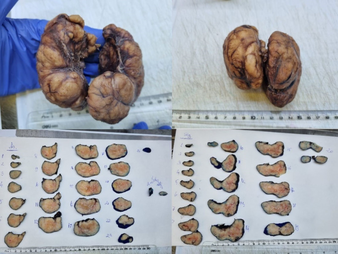
Gross histopathology showed both lobes enlarged (LTL 9/4/6cm, RTL8.5/4.5/5cm, isthmus 0.7/0.6/0.4cm) with intact capsule, solid cut surface with small white-yellowish areas of nodular appearance.
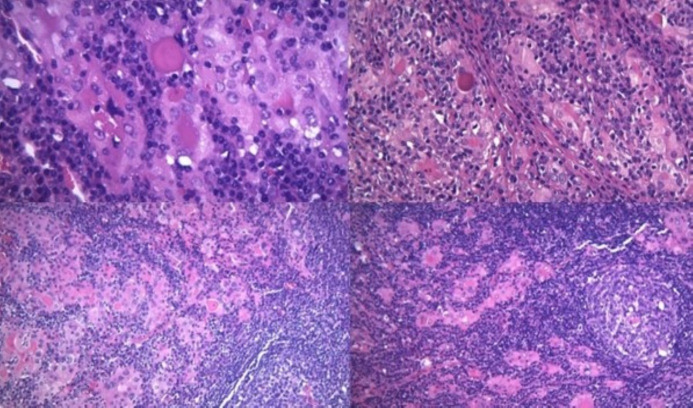
Microscopically, there was a diffuse inflammatory lymphocytic infiltration which formed nodular structures with germinative centers aspect, areas of fibrosis and rare multinuclear giant as well as some atrophic thyroid follicles that appeared bordered by Hurthle cells.
Both thyroid lobes appeared similar in section. One lymph node was found in the histopathology specimen that had normal structure (Figure 2 and Figure 3).
Figure 2.
Gross histopathology specimens
Figure 3.
Histopathology identifies atrophic remnant follicles sometimes bordered by Hurtle cells (A), with interstitial fibrosis (B), surrounded by chronic inflammatory infiltrate (C), with secondary lymph follicle pattern (D). HE staining, A-20×; B-10×; C, D- 4×.
Discussion
Since the publication of Hashimoto’s description of lymphocytic thyroiditis in 1912 and until 1974 when Katz and Vickery et al. reported fifty-six patients with fibrous variants of HT subtype [7], this particular form of thyroiditis was reported mainly as Riedel thyroiditis.
Katz and Vickery described the main characteristics of the fibrous subtype as a rapid enlargement of a previous goiter or the appearance of one that is hard on palpation, causing compression symptoms, with associated hypothyroidy and very high titer of antithyroglobulin antibodies that was correspondent to microscopy findings of fibroid infiltration of the thyroid parenchyma, comprising at least one third of the total, with atrophy of the thyroid follicles that present Hurthle cells specific of HT, and abundant lymphoid infiltration of the gland, sometimes forming germinative centers in inverse proportion to the fibrosis [3, 4, 7, 11, 12, 13].
The differential diagnosis usually comprises of malignant transformation of the thyroid and imaging (ultrasound and CT scan) are performed, as well as fine needle aspiration biopsy that usually is negative or inconclusive in the case of fibrous HT.
Due to compression syndrome or the inability to exclude the malignant transformation, these cases are prone to surgery and the histopathological findings are characteristic of this subtype of HT [2, 3, 7].
Although it was well characterized almost 50 years ago, cases of fibrous HT are rarely reported in literature, as we found only a few since the series forementioned [3, 7, 11, 12, 13].
Our case has all of the clinical characteristics priorly described-rapid enlargement with hard goiter and compression symptoms, hypothyroidy, very high antithyroglobulin antibodies, as well as the main microscopy findings that define the fibrous variant of Hashimoto thyroiditis.
The main treatment for HT, even its fibrous variant, is the replacement hormonal medication, but surgery remains valid for compression symptoms and uncertain FNA biopsy findings.
Surgery in such cases of large goiters and fibrosis is more technically challenging and postoperative complications are more common than in the case of multinodular goiters or other indications of thyroidectomy.
In this case report, postoperative hypocalcemia was present as a mild complication, probably due to compromising the vascular supply to the parathyroid glands, as there were none included in the biopsy specimen [2, 3, 4, 5, 9].
The diagnosis and treatment of the fibrous variant of HT are challenging and need a multidisciplinary approach-endocrinologist, radiologist, pathologist and surgeon for successful management of such cases; post-operatively a close observation is vital in the detection and early intervention of any post-op complication.
Conclusion
In conclusion, this case report presents a rare case of the fibrous variant of Hashimoto’s thyroiditis that is rarely taken under consideration in the preoperative setting.
Patients’ complaints are usually of rapid enlargement of the thyroid gland associated with compressive symptoms that mimic thyroid cancer.
A definitive diagnosis is hard to establish with the usual algorithm of imaging and FNA biopsy and is reserved for thyroidectomy and subsequent histopathology.
The multidisciplinary approach and pre-and postoperative evaluation are of utmost importance for successful management of such case.
Conflict of interests
The authors have no conflict of interest to declare.
References
- 1. Smith PW , Salomone LJ , Hanks JB . In: Sabiston textbook of surgery: the biological basis of modern surgical practice . 19th . Townsend CM , Beauchamp RD , Evers BM , Mattox KL , editors. Elsevier Limited ; 2012 . Thyroid ; pp. 885 – 1011 . [Google Scholar]
- 2. Randolph GW. In: Surgery of the thyroid and parathyroid glands. 1. Randolph GW, editor. Elsevier Inc; 2013 . pp. 104 – 143 . [Google Scholar]
- 3.Almalali MIA, Takrouni AO, Alqattan AA, Alsolami S, Salman JMJ, Aljehani MMM, Albaqami RMJ, Alyahyaei AM, Alrasheed FAA. Autoimmune thyroiditis a fibrous variant of Hashimoto's thyroiditis: A rare case. Ann Med Surg (Lond) 2022;23(79):104019–104019. doi: 10.1016/j.amsu.2022.104019. [DOI] [PMC free article] [PubMed] [Google Scholar]
- 4.Caturegli P. De Remigis A., Rose N.R. Hashimoto thyroiditis: clinical and diagnostic criteria. Autoimmun. 2014;13(4-5):391–397. doi: 10.1016/j.autrev.2014.01.007. [DOI] [PubMed] [Google Scholar]
- 5.Mincer D.L., Jialal I. StatPearls Publishing; Hashimoto Thyroiditis 2022. 2022 Available from: https://www.ncbi.nlm.nih.gov/books/NBK459262/ [PubMed]
- 6.Hiromatsu Y. Satoh H., Amino N. Hashimoto's thyroiditis: history and future outlook. Hormones (Basel) 2013;12(1):12–18. doi: 10.1007/BF03401282. [DOI] [PubMed] [Google Scholar]
- 7.Katz S. M., Vickery A.L., Jr. The fibrous variant of Hashimoto's thyroiditis. Hum. Pathol. 1974;5(2):161–170. doi: 10.1016/s0046-8177(74)80063-8. [DOI] [PubMed] [Google Scholar]
- 8.Bage AM, Kalatharan P. Role of Total Thyroidectomy in Painful (Symptomatic) Hashimoto's Thyroiditis: Descriptive Study. Indian J Otolaryngol Head Neck Surg. 2021 Sep;73(3):296-303. doi: 10.1007/s12070-020-02114-2. Epub 2020 Sep 7. Erratum in: Indian J Otolaryngol Head Neck Surg. 2022;74(2):2608–2608. doi: 10.1007/s12070-020-02114-2. [DOI] [PMC free article] [PubMed] [Google Scholar]
- 9.Guidelines for. Jonklaas, J., Bianco, A.C., Bauer, A.J., Burman, K.D., Cappola, A.R., Celi, F.S., Cooper, D.S., Kim, B.W., Peeters, R.P., Rosenthal, M.S. and Sawka, A.M. [ed.] American Thyroid Association Task Force on Thyroid Hormone Replacement. s.l. American Thyroid Association. 2014;24(12):1670–1751. doi: 10.1089/thy.2014.0028. [DOI] [PMC free article] [PubMed] [Google Scholar]
- 10.Agha R. A., Franchi T., Sohrabi C., Mathew G., Kerwan A., SCARE Group the SCARE 2020 guideline: updating consensus surgical CAse REport (SCARE) guidelines. Int. J. Surg. 2020;84:226–230. doi: 10.1016/j.ijsu.2020.10.034. [DOI] [PubMed] [Google Scholar]
- 11.Baloch Z. W., Saberi M., Livolsi V.A. Simultaneous involvement of Thyroid by Riedel's [correction of Reidel's] disease and fibrosing Hashimoto's thyroiditis: a case report. Thyroid. 1998;8(4):337–341. doi: 10.1089/thy.1998.8.337. [DOI] [PubMed] [Google Scholar]
- 12.Junik R. Juraniec O., Pypkowski J., Krymer A., Marszałek A. A difficult diagnosis: a case report of combined Riedel's disease and fibrosing Hashimoto's thyroiditis. Endokrynol. Pol. 2011;62(4):351–356. [PubMed] [Google Scholar]
- 13.Li Y. Zhou G., Ozaki T., et al. Distinct histopathological features of Hashimoto's thyroiditis with respect to IgG4-related disease. Mod. Pathol. 2012;25:1086–1097. doi: 10.1038/modpathol.2012.68. [DOI] [PubMed] [Google Scholar]


