Abstract
Background
The medial collateral ligament (MCL) is crucial for ensuring implant stability after unicompartmental knee arthroplasty (UKA). Intraoperative MCL lesions can cause valgus instability, affecting function and implant longevity, and thereby negatively impacting the patient’s outcome. Every surgeon who performs UKA may encounter this complication in their daily practice. In this context, this case report presents a rescue technique. The existing literature does not specify a protocol for managing this complication. This article presents the first instance of accidental midsubstance section of the MCL during medial UKA, managed through primary suture and augmentation repair with a fascia lata (FL) autograft. The procedure was subsequently replicated step by step on an anatomical specimen.
Case Description
A 54-year-old woman, previously successfully treated with right medial UKA, was referred to our clinic following an unsuccessful attempt at conservative treatment for osteoarthritis in the left knee. Scheduled for a left medial UKA, an inadvertent midsubstance transection of the deep part of the MCL was encountered during the procedure, resulting in valgus instability. The MCL was promptly repaired and reinforced using an ipsilateral FL augmentation autograft. Subsequent UKA surgery was successfully completed. Follow-up at one year revealed favorable post-operative outcomes, with symmetrical stability on stress radiographs and no indications of early loosening.
Conclusions
To our knowledge, this article represents the first documentation of the direct management for this rare yet severe complication. This case report could therefore inspire any surgeon facing this complication. The technique, grounded in biomechanical principles, ensures direct medial stability whilst allowing uninterrupted continuation of the initial procedure. Characterized by simplicity and reproducibility, the approach demonstrates favorable short-term outcomes. Because the results should be interpreted considering the limited impact of a case report, further prospective studies are essential to substantiate and strengthen these findings.
Keywords: Unicompartmental knee arthroplasty (UKA), medial collateral ligament section (MCL section), autograft augmentation, case report, surgical technique
Highlight box.
Key findings
• Medial collateral ligament (MCL) suture with autograft fascia lata (FL) reinforcement plasty serves as a salvage solution for completing medial unicompartmental knee arthroplasty (UKA) implantation, it provides direct stability, is technically straightforward and reproducible and yields favorable short term clinical and radiological outcomes.
What is known and what is new?
• MCL injury during knee arthroplasty has a negative impact on both postoperative functional results and the rate of adverse outcomes, so its repair is essential.
• This case report is the first to describe the case of accidental sectioning of the MCL during UKA surgery.
• This case is the first to describe the management of this complication by a suture with FL autograft reinforcement plasty.
What is the implication, and what should change now?
• The technique described adds a management option available to any surgeon encountering this complication.
• Further studies are mandatory to validate long-term efficacy.
Introduction
Intraoperative iatrogenic transection of the medial collateral ligament (MCL) during medial unicompartmental knee arthroplasty (UKA) procedure is a rare but very serious complication. The clinical impact of this complication is significant; firstly, the implantation of the UKA can no longer be performed, and secondly, its improper management can affect both the function and longevity of the implant, thereby negatively influencing the outcome of the operated patient, manifesting as instability and chronic pain, possibly requiring early revision. To our knowledge, there are no articles in the current literature dealing with this complication. Its incidence is unknown, but can be extrapolated from cases of total knee arthroplasty (TKA), where it is estimated at 0.8% to 2.7%, with 33.3% to 75.7% involving midsubstance (1). Complete or partial sectioning, which generally occurs during the osseous cuts, induces medial instability. In the case of a UKA, which is an unconstrained implant, this poses several problems. Firstly, the ligament balance, and hence the choice of definitive polyethylene, can no longer be determined. Coronal ligament balance is fundamental to the outcome of the UKA, so it is not possible to increase the thickness of the polyethylene, as is the case with TKAs, at the risk of overstressing the MCL, increasing the risk of implant loosening and osteoarthritis of the opposite compartment (2). Secondly, this significantly increases the contact stress on the polyethylene and on the lateral cartilage, having a negative impact in the future (3). It has been clearly demonstrated that intraoperative injury to the MCL during knee arthroplasty has a negative impact on both postoperative functional results and the rate of adverse outcomes, so its repair is essential (4). Moreover, unlike MCL avulsion, a midsubstance section has less healing potential, making conservative treatment inappropriate (5). In the context of arthroplasty, subsequent reconstructions in the event of instability are ineffective, so deferred management of such an injury is not an option (6). Based on the good results obtained with primary suture in the case of TKA (7), we hypothesized that repair of the MCL during UKA is a valid initial solution for managing this complication, in order to avoid the need to total knee replacement. This article is the first to describe the case of accidental sectioning of the MCL during UKA. It is also the first article to describe the management of this complication by a suture with fascia lata (FL) autograft reinforcement plasty. Given the negative impact of this complication on the patient, the likelihood that any surgeon may encounter it in their daily practice, and the lack of consensus on its management, we decided to report this clinical case, and detail our rescue technique that allowed us to effectively manage this complication. We present this case in accordance with the CARE reporting checklist (available at https://acr.amegroups.com/article/view/10.21037/acr-24-30/rc).
Case presentation
A 54-year-old woman, devoid of relevant comorbidities, presented with severe left medial knee pain accompanied by functional disability. The patient’s medical history includes a successful right medial UKA with favorable progression observed over a span of four years. Clinical examination revealed mild effusion, isolated medial joint space pain, no patellofemoral pain, complete knee stability, and full range of motion (ROM). Preoperative radiographs showed uncomplicated right medial UKA, and severe left medial compartment osteoarthritis. Conservative treatment was attempted for nine months without success. A medial UKA was then planned in our clinic. The patient provided informed consent for both the surgical procedure and research purposes.
Surgical procedure
The procedure started without complications, utilizing the Persona Partial Knee System® (Zimmer Biomet®, Zug, Switzerland) and employing a conventional medial parapatellar approach. Following tibial and femoral cutting, trial implants were selected and positioned. Upon insertion of the trial polyethylene component, a novel medial laxity was noted. Exploration of the medial ligamentous complex, starting from lateral to medial, revealed a sharply transected deep MCL (dMCL) and a partially subtotal section of the superficial MCL (sMCL), both at the tibial cut level, with no loss of substance. These injuries likely occurred during the femoral posterior or chamfer cuts with the saw. We performed two Krackow sutures with FiberWire® (Arthrex, Naples, FL, USA) for each portion, using multiple locking loops inserted on each side of the ligaments. The sutures were then tightened until achieving contact between the two ends. We then applied valgus stress at several degrees to assess our sutures. The superficial portion held, however, the dMCL suture did not resist testing, resulting in residual laxity of the knee during flexion. Consequently, a decision was made to reinforce the dMCL with an autograft augmentation. The initial incision was extended by 1 cm bilaterally, and then a subcutaneous approach was taken laterally. Subsequently, a free distal FL graft measuring 80 mm × 20 mm was harvested, and the donor site was sutured to prevent herniation.
The proximal portion of the graft was tubularized using a FiberLoop® (Arthrex). The femoral insertion point was determined by identifying the Schottle point under fluoroscopy, and the tunnel trajectory was established using a Kirschner wire positioned at a 20 degree proximal orientation in the coronal plane and aligned with the femur in the sagittal plane.
The femoral tunnel was established using a 2 mm diameter drill bit. The medial aspect of the tunnel was enlarged to accommodate the tubular portion of the graft, measuring 5 mm in our case, extending to a depth of 30 mm. Subsequently, the graft was passed through the femoral tunnel and pretensioned using an EndoButton® (Smith & Nephew, Watford, UK). The tibial insertion point was located approximately 6 to 8 mm from the joint line, and the graft was secured to the tibial cortex using two Corkscrew® FT suture anchors (Arthrex), positioned anteriorly and posteriorly with a separation of 10–15 mm. Verification of graft isometric behavior and physiological laxity of 2–3 mm was conducted. Final tensioning was executed with the knee in a neutral rotation position, flexed at 30 degrees with slight varus alignment. The remainder of the procedure proceeded without complications. Postoperative management followed the standard protocol, allowing full weight-bearing and unrestricted mobilization beginning on first postoperative day (Figure 1).
Figure 1.
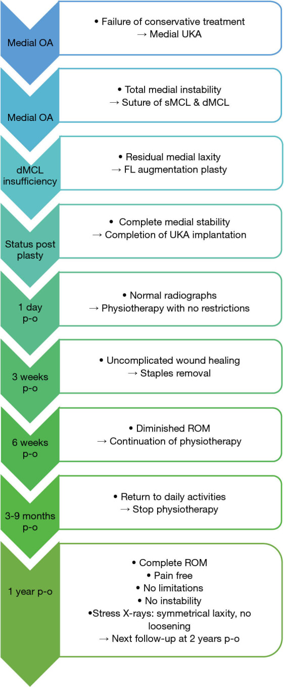
Flowchart illustrating the progression of our case. The blue-green arrows indicate diagnoses and key follow-up and surgical steps, while the boxes contain the progression (point) above and the applied treatment (→) below. OA, osteoarthritis; UKA, unicompartmental knee arthroplasty; MCL, medial collateral ligament; sMCL, superficial MCL; dMCL, deep MCL; FL, fascia lata; p-o, post-operative; ROM, range of motion.
All procedures performed in this study were in accordance with the ethical standards of the institutional and/or national research committee(s) and with the Helsinki Declaration (as revised in 2013). Written informed consent was obtained from the patient for the publication of this case report and accompanying images. A copy of the written consent is available for review by the editorial office of this journal.
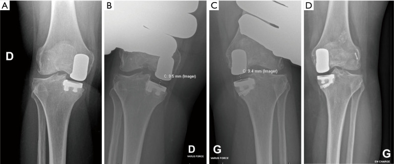
The patient underwent follow-up assessments at tree and six weeks postoperatively, followed by evaluations by the operating surgeon at 3, 6, 9, and 12 months, during which X-rays were taken (Figure 1). There were no immediate postoperative complications observed. Removal of staples at the tree-week mark revealed uncomplicated wound healing. Adherence was complete. Rehabilitation progressed smoothly, albeit achieving full ROM took longer compared to the contralateral side. At the one-year follow-up, the patient reported being pain-free both at the donor site and in the MCL area. There were no functional limitations or sensations of instability noted. Clinical examination showed symmetrical ROM in the knee and stability under valgus stress at 0 and 30 degrees, as well as during rotation. Valgus stress X-rays of the contralateral knee demonstrated symmetrical physiological laxity, without any signs of early implant dislocation (Figures 2,3).
Figure 2.
Lateral and antero-posterior radiographs of the left knee. (A) Pre-operative. (B) Post-operative day 1.
Figure 3.
Bilateral anteroposterior radiographs one year after the described surgery. (A) Standing right knee showing previous unicompartmental knee arthroplasty. (B) Right valgus stress radiograph. (C) Left valgus stress radiographs showing similar physiological laxity. (D) Standing left new unicompartmental knee arthroplasty showing no signs of complication.
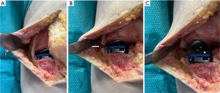
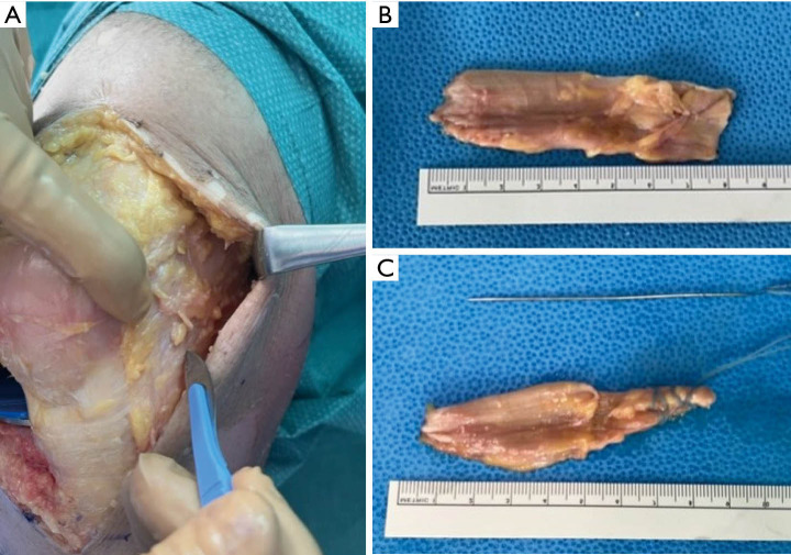
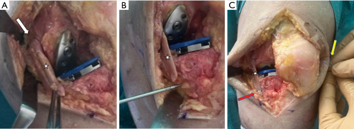
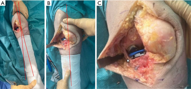
Subsequently, the described FL autograft augmentation procedure was replicated step by step in a cadaveric laboratory setting. The procedure was thoroughly documented, with illustrations provided (Figures 4-7, Tables 1,2).
Figure 4.
Anatomical specimen of the left knee with a parapatellar approach and unicompartmental knee arthroplasty trial implant in place. (A) Intact medial collateral ligament. (B) Midportion section of medial collateral ligament (white arrow). (C) Gap (white star) indicating medial instability in valgus stress.
Figure 5.
Graft harvesting process in an anatomical specimen of the left knee. (A) Lateral extension and fascia lata harvesting. (B) Fascia lata free autograft (8 cm × 2 cm). (C) Proximal part tubularized with FiberLoop® (Arthrex).
Figure 6.
Process of graft placement in an anatomical specimen of the left knee. (A) Fascia lata graft (white star) inserted into the femoral tunnel (white arrow). (B) Fascia lata graft (white star) was fixed on its tibial insertion with 2× Corkscrew FT suture anchor® (Arthrex). (C) Fascia lata graft (white star) attached to the tibia (red arrow) which was tensioned using FiberLoop® (Arthrex) (yellow arrow).
Figure 7.
Clinical assessment of an anatomical specimen of the left knee. (A) Pre-augmentation stage showing valgus instability. (B,C) Post-augmentation with fascia lata graft (white star) showing no medial instability on valgus stress exam.
Table 1. Advantages and disadvantages of MCL suture augmentation with fascia lata autograft.
| Advantages | Disadvantages |
|---|---|
| Acutely feasible | Newly discovered and currently unique technique |
| Technically reproducible and simple | Long-term outcomes not yet acquired |
| No need for special instruments or implants | |
| Simple graft harvesting through the same incision | |
| No need for harvest the medial dynamic stabilizers or allografts | |
| Biological graft, stimulating healing of native MCL | |
| Biomechanical properties close to those of native MCL | |
| Protects the repair, allowing direct ROM to prevent stiffness | |
| No modification of postoperative protocol |
MCL, medial collateral ligament; ROM, range of motion.
Table 2. Surgical pearls and pitfalls of MCL augmentation with fascia lata autograft.
| Pearls | Pitfalls |
|---|---|
| Passing the FiberLoop® wires into the femoral tunnel with aid of an EndoClose® (Medtronic®) | Taking into account the tibial cut thickness to assess the initial height of the joint space |
| Creating the femoral tunnel under fluoroscopy using Schottle’s point | Checking isometry, physiologic laxity and graft tension throughout entire ROM |
| Using native insertions to ensure anatomical graft positioning | Setting final tension with trial implants and the smallest trial polyethylene |
| For sections without loss of substance, using native MCL length to define graft length (+2 cm proximally) | Checking under fluoroscopy that there is no conflict between the screws and the tibial implant |
| Final graft tensioning the knee in neutral rotation, slight varus and flexion (30°) | Ensuring interference-free periosteal positioning of EndoButton® |
| Ensuring absence of other medial complex lesions (e.g., POL) |
ROM, range of motion; MCL, medial collateral ligament; POL, postero-oblique ligament.
Patient perspective
“After a year, I’m generally satisfied with the operation. I’m relieved that the surgeon was able to finish the operation and didn’t have to change the prosthesis. If I compare with the other side, the rehabilitation was more painful and longer, but my knee works now well like the other one.”
Discussion
The significant finding in our case is the successful immediate reinforcement suture for an unintended MCL tear during a medial UKA procedure. To the best of our knowledge, this is the first detailed step-by-step documentation of a procedure addressing such a complication.
While numerous studies have compared various suture techniques, augmentations, and direct MCL reconstructions for TKA, no comparative studies exist for UKA, leaving a gap in the literature regarding this specific scenario. Although no single technique has emerged as superior, certain trends are noteworthy. It has been demonstrated that sutures alone offer less resistance to strain, with a potential for rupture at the tissue-suture interface. Augmentation with internal bracing has shown comparable strength to allograft reconstruction (8).
Studies comparing sutures with augmentation versus reconstructions of the sMCL have found equivalent objective outcomes, yet reconstructions tend to yield superior subjective outcomes (9).
Additionally, the cross-sectional area of the MCL must be considered. Due to the limited healing potential of the MCL midportion, some authors advocate for direct reconstruction with autograft, yielding excellent mid-term clinical results (10,11).
In our case, the MCL tear involved the midportion, which was directly sutured. However, to restore optimal medial stability for both the continuation of the procedure and to ensure long-term stability, we opted for allograft augmentation.
Given the favorable outcomes of the repair, there is no need to transition to a more restrictive implant design, as has been observed in cases of anterior cruciate ligament transection with cruciate retaining (CR) or postero-stabilized (PS) TKA (12). Some studies have reported improved outcomes with a CR design, suggesting that preserving the posterior cruciate ligament, which acts as a medial stabilizer, provides additional stability (1). However, some authors suggest switching to a more constrained implant as a salvage solution only for elderly or less active individuals (13). In the context of a UKA in a young patient, transitioning to a TKA may yield inferior results (14), making it essential to retain the UKA implant in this population.
Various types of autografts and allografts are available for MCL augmentation or reconstruction. While the semitendinosus tendon is commonly used (15), the quadriceps tendon is also utilized (11). Biosynthetic augmentations have been explored as well (16). Additionally, alternative autografts, such as menisci, have been proposed to reinforce the MCL when traditional methods are not feasible (17). Our decision to utilize the FL autograft is based on several factors. Firstly, FL has demonstrated efficacy in numerous reconstructive surgeries, offering favorable outcomes (18-20). Secondly, its ease of harvesting and proximity to the surgical site reduce operative time and eliminate the need for additional incisions. Thirdly, FL harvesting is technically straightforward, requiring no specialized knowledge of ligament surgery. Fourthly, FL harvesting incurs minimal morbidity, as evidenced by its use in upper capsular reconstructions (21,22), where the graft was much longer and larger than in our reinforcement plasty. Fifthly, biomechanically, FL possesses ideal characteristics for ligament autografting, providing immediate strength regardless of the harvesting zone (23). Sixthly, histological studies have shown FL’s excellent remodeling and healing properties, making it closer to the MCL than tendon grafts (24,25).
Numerous strengthening techniques exist, but identifying an optimal approach is challenging due to the limited number of patients and heterogeneity of MCL injuries in studies. To effectively manage this complication, arthroplasty surgeons must possess comprehensive knowledge of medial ligament anatomy. Works by Laprade and Griffith have elucidated the anatomy and stabilizing actions of the medial stabilizing structures, facilitating the development of anatomically-based reconstruction techniques. They also proved the biomechanical effectiveness of their reconstruction based on this anatomical knowledge, helping to develop the concept of “anatomical reconstruction” (26,27). The anatomy of the MCL complex is also quantitatively known, thanks to Liu’s cadaveric study, which defined the length, insertion and relationship to the tibial plateau and other adjacent bony references of the sMCL and dMCL (28). There were, however, some discrepancies between the two studies, notably concerning the proximal insertion of the sMCL. Intraoperatively, in order to achieve the most anatomical reconstruction possible, it is necessary to define which part of the medial complex is affected, as a partial lesion [isolated dMCL with postero-oblique ligament (POL) for example]. In the current case, there was a complete lesion of the dMCL, and a partial lesion of the sMCL, with no obvious damage to the POL requiring targeted reinforcement.
The lateral complex should not be oversimplified to a structure connecting the femur to the tibia. The complexity of anatomical descriptions, particularly regarding the insertions of the sMCL, underscores the challenges in achieving anatomical reconstruction. Given its flat anatomy with multiple insertions, the native MCL exhibits complex tensional behavior throughout flexion. To best reconstruct this complex behavior, intraoperative verification of the most anatomical placement and isometric behavior of the graft is recommended (29). Utilizing flat grafts, such as FL, is biomechanically closer to native structures than tubular grafts. Additionally, replicating the typical “fan shape” of the dMCL (30) with a small proximal insertion and an enlarged distal attachment, as performed in our case, enhances biomechanical fidelity. Furthermore, aligning the graft anatomically is crucial for optimal reconstruction. To achieve this, Athwal et al. provided anatomical references and intraoperative radiological landmarks, such as the use of the Schottle’s point to define the femoral insertion, which is fundamental. Our FL reinforcement plasty technique allows for reconstruction with an anterodistal orientation, resembling the native dMCL (28,31).
Targeted reconstruction of the dMCL can restore anterior rotational stability (32), the significance of which in the context of UKA warrants further investigation. Recent studies (33,34) emphasize the role of hamstrings as dynamic valgus stabilizers, underscoring the importance of preserving medial stability, particularly in cases involving the medial complex. Utilizing FL autografts spares the hamstrings, contributing to improved stability.
Clinical quantification of coronal stability is challenging. In our case, we evaluated medial stability at 0 and 30 degrees of flexion and utilized valgus stress radiographs to assess the suture. Valgus stress and short non-weight knee radiographs are reliable for diagnosing medial instability and assessing coronal alignment (35). Additionally, our patient’s contralateral UKA serves as a point of comparison.
To minimize this rare but debilitating complication, it is crucial to monitor retractor placement during osteotomies and utilize precise oscillating saw blades.
Our article provides detailed documentation of managing this complication during UKA placement, describing the technique of MCL reinforcement plasty using FL. It is the first to describe targeted reconstruction of the deep portion of the MCL complex. The technique is based on current anatomical and biomechanical knowledge and is supported by anatomical specimen reproduction.
However, this study has limitations. A case report precludes the establishment of a consensus decision flowchart. Additionally, intraoperative photographs were unavailable due to an unforeseen circumstance. While the cadaver laboratory session enabled the reproduction and documentation of the entire procedure, it may not perfectly illustrate the procedure performed in our case.
Conclusions
This article is part of the search for effective techniques to resolve and improve the management of rare complications, which can be encountered by any surgeon during professional practice. In view of the encouraging results, the described technique may help surgeons to restore medial stability of the knee during UKA implantation in case of an unintended MCL rupture. Because the results should be interpreted considering the limited impact of a case report, further prospective studies are essential to substantiate and strengthen these findings.
Supplementary
The article’s supplementary files as
Acknowledgments
We thank Dr. Hermes Miozarri (Head of the degenerative knee team, Department of Orthopedic Surgery and Traumatology, Geneva University Hospitals) for his expertise in managing arthroplasty complications, the SFITS Academy (Swiss Foundation For Innovation and Training in Surgery, Geneva) for the supply of anatomical specimen, and Dr. Gilles Dietrich for his review.
Funding: None.
Ethical Statement: The authors are accountable for all aspects of the work in ensuring that questions related to the accuracy or integrity of any part of the work are appropriately investigated and resolved. All procedures performed in this study were in accordance with the ethical standards of the institutional and/or national research committee(s) and with the Helsinki Declaration (as revised in 2013). Written informed consent was obtained from the patient for the publication of this case report and accompanying images. A copy of the written consent is available for review by the editorial office of this journal.
Footnotes
Reporting Checklist: The authors have completed the CARE reporting checklist. Available at https://acr.amegroups.com/article/view/10.21037/acr-24-30/rc
Conflicts of Interest: All authors have completed the ICMJE uniform disclosure form (available at https://acr.amegroups.com/article/view/10.21037/acr-24-30/coif). M.P. serves as an unpaid editorial board member of AME Case Reports from February 2023 to January 2025. The other authors have no conflicts of interest to declare.
References
- 1.Bohl DD, Wetters NG, Del Gaizo DJ, et al. Repair of Intraoperative Injury to the Medial Collateral Ligament During Primary Total Knee Arthroplasty. J Bone Joint Surg Am 2016;98:35-9. 10.2106/JBJS.O.00721 [DOI] [PubMed] [Google Scholar]
- 2.Heyse TJ, El-Zayat BF, De Corte R, et al. Balancing UKA: overstuffing leads to high medial collateral ligament strains. Knee Surg Sports Traumatol Arthrosc 2016;24:3218-28. 10.1007/s00167-015-3848-5 [DOI] [PubMed] [Google Scholar]
- 3.Kwon HM, Kang KT, Kim JH, et al. Medial unicompartmental knee arthroplasty to patients with a ligamentous deficiency can cause biomechanically poor outcomes. Knee Surg Sports Traumatol Arthrosc 2020;28:2846-53. 10.1007/s00167-019-05636-7 [DOI] [PubMed] [Google Scholar]
- 4.Li J, Yan Z, Lv Y, et al. Impact of intraoperative medial collateral ligament injury on outcomes after total knee arthroplasty: a meta-analysis and systematic review. J Orthop Surg Res 2021;16:686. 10.1186/s13018-021-02824-5 [DOI] [PMC free article] [PubMed] [Google Scholar]
- 5.Lee GC, Lotke PA. Management of intraoperative medial collateral ligament injury during TKA. Clin Orthop Relat Res 2011;469:64-8. 10.1007/s11999-010-1502-6 [DOI] [PMC free article] [PubMed] [Google Scholar]
- 6.Pritsch M, Fitzgerald RH, Jr, Bryan RS. Surgical treatment of ligamentous instability after total knee arthroplasty. Arch Orthop Trauma Surg (1978) 1984;102:154-8. 10.1007/BF00575224 [DOI] [PubMed] [Google Scholar]
- 7.Leopold SS, McStay C, Klafeta K, et al. Primary repair of intraoperative disruption of the medical collateral ligament during total knee arthroplasty. J Bone Joint Surg Am 2001;83:86-91. 10.2106/00004623-200101000-00012 [DOI] [PubMed] [Google Scholar]
- 8.Gilmer BB, Crall T, DeLong J, et al. Biomechanical Analysis of Internal Bracing for Treatment of Medial Knee Injuries. Orthopedics 2016;39:e532-7. 10.3928/01477447-20160427-13 [DOI] [PubMed] [Google Scholar]
- 9.LaPrade RF, DePhillipo NN, Dornan GJ, et al. Comparative Outcomes Occur After Superficial Medial Collateral Ligament Augmented Repair vs Reconstruction: A Prospective Multicenter Randomized Controlled Equivalence Trial. Am J Sports Med 2022;50:968-76. 10.1177/03635465211069373 [DOI] [PubMed] [Google Scholar]
- 10.Wang X, Liu H, Cao P, et al. Clinical outcomes of medial collateral ligament injury in total knee arthroplasty. Medicine (Baltimore) 2017;96:e7617. 10.1097/MD.0000000000007617 [DOI] [PMC free article] [PubMed] [Google Scholar]
- 11.Jung KA, Lee SC, Hwang SH, et al. Quadriceps tendon free graft augmentation for a midsubstance tear of the medial collateral ligament during total knee arthroplasty. Knee 2009;16:479-83. 10.1016/j.knee.2009.04.007 [DOI] [PubMed] [Google Scholar]
- 12.Siqueira MB, Haller K, Mulder A, et al. Outcomes of Medial Collateral Ligament Injuries during Total Knee Arthroplasty. J Knee Surg 2016;29:68-73. 10.1055/s-0034-1394166 [DOI] [PubMed] [Google Scholar]
- 13.Dragosloveanu S, Cristea S, Stoica C, et al. Outcome of iatrogenic collateral ligaments injuries during total knee arthroplasty. Eur J Orthop Surg Traumatol 2014;24:1499-503. 10.1007/s00590-013-1330-y [DOI] [PubMed] [Google Scholar]
- 14.Parvizi J, Nunley RM, Berend KR, et al. High level of residual symptoms in young patients after total knee arthroplasty. Clin Orthop Relat Res 2014;472:133-7. 10.1007/s11999-013-3229-7 [DOI] [PMC free article] [PubMed] [Google Scholar]
- 15.Adravanti P, Dini F, Calafiore G, et al. Medial collateral ligament reconstruction during TKA: a new approach and surgical technique. Joints 2015;3:215-7. 10.11138/jts/2015.3.4.215 [DOI] [PMC free article] [PubMed] [Google Scholar]
- 16.LeVasseur MR, Uyeki CL, Garvin P, et al. Knee Medial Collateral Ligament Augmentation With Bioinductive Scaffold: Surgical Technique and Indications. Arthrosc Tech 2022;11:e583-9. 10.1016/j.eats.2021.12.011 [DOI] [PMC free article] [PubMed] [Google Scholar]
- 17.Sun C, Rong W, Du R, et al. Meniscus Graft Augmentation for a Midsubstance Tear of the Medial Collateral Ligament during Total Knee Arthroplasty. J Knee Surg 2022;35:449-55. 10.1055/s-0040-1715115 [DOI] [PubMed] [Google Scholar]
- 18.Ohta S, Ueda Y, Komai O. Postoperative results of arthroscopic superior capsule reconstruction using fascia lata: a retrospective cohort study. J Shoulder Elbow Surg 2024;33:686-97. 10.1016/j.jse.2023.07.021 [DOI] [PubMed] [Google Scholar]
- 19.Park TH. The versatility of tensor fascia lata allografts for soft tissue reconstruction. Int Wound J 2023;20:784-91. 10.1111/iwj.13923 [DOI] [PMC free article] [PubMed] [Google Scholar]
- 20.Espejo-Reina A, Espejo-Reina MJ, Lombardo-Torre M, et al. Anterior Cruciate Ligament Reconstruction Using Combined Graft of Hamstring and Fascia Lata With Extra-articular Tenodesis. A Technique in Case of Insufficient Hamstrings. Arthrosc Tech 2020;9:e1657-63. 10.1016/j.eats.2020.07.007 [DOI] [PMC free article] [PubMed] [Google Scholar]
- 21.Ângelo ACLPG, de Campos Azevedo CI. Minimally invasive fascia lata harvesting in ASCR does not produce significant donor site morbidity. Knee Surg Sports Traumatol Arthrosc 2019;27:245-50. 10.1007/s00167-018-5085-1 [DOI] [PubMed] [Google Scholar]
- 22.Ângelo ACLPG, de Campos Azevedo CI. Donor-Site Morbidity After Autologous Fascia Lata Harvest for Arthroscopic Superior Capsular Reconstruction: A Midterm Follow-up Evaluation. Orthop J Sports Med 2022;10:23259671211073133. 10.1177/23259671211073133 [DOI] [PMC free article] [PubMed] [Google Scholar]
- 23.de Campos Azevedo CI, Leiria Pires Gago Ângelo AC, Quental C, et al. Proximal and mid-thigh fascia lata graft constructs used for arthroscopic superior capsule reconstruction show equivalent biomechanical properties: an in vitro human cadaver study. JSES Int 2021;5:439-46. 10.1016/j.jseint.2021.01.016 [DOI] [PMC free article] [PubMed] [Google Scholar]
- 24.Kuhlmann JN, Luboinski J, Mimoun M, et al. Reconstruction of the medial collateral ligament of the knee in rats using a free autogeneic transplant of fascia lata, ligament or tendon. Acta Orthop Belg 1994;60:10-8. [PubMed] [Google Scholar]
- 25.Kataoka T, Kokubu T, Muto T, et al. Rotator cuff tear healing process with graft augmentation of fascia lata in a rabbit model. J Orthop Surg Res 2018;13:200. 10.1186/s13018-018-0900-4 [DOI] [PMC free article] [PubMed] [Google Scholar]
- 26.Laprade RF, Wijdicks CA. Surgical Technique: Development of an Anatomic Medial Knee Reconstruction. Clin Orthop Relat Res 2012;470:806-14. 10.1007/s11999-011-2061-1 [DOI] [PMC free article] [PubMed] [Google Scholar]
- 27.Griffith CJ, LaPrade RF, Johansen S, et al. Medial knee injury: Part 1, static function of the individual components of the main medial knee structures. Am J Sports Med 2009;37:1762-70. 10.1177/0363546509333852 [DOI] [PubMed] [Google Scholar]
- 28.Liu F, Yue B, Gadikota HR, et al. Morphology of the medial collateral ligament of the knee. J Orthop Surg Res 2010;5:69. 10.1186/1749-799X-5-69 [DOI] [PMC free article] [PubMed] [Google Scholar]
- 29.Kittl C, Robinson J, Raschke MJ, et al. Medial collateral ligament reconstruction graft isometry is effected by femoral position more than tibial position. Knee Surg Sports Traumatol Arthrosc 2021;29:3800-8. 10.1007/s00167-020-06420-8 [DOI] [PMC free article] [PubMed] [Google Scholar]
- 30.Abermann E, Wierer G, Herbort M, et al. MCL Reconstruction Using a Flat Tendon Graft for Anteromedial and Posteromedial Instability. Arthrosc Tech 2022;11:e291-300. 10.1016/j.eats.2021.10.019 [DOI] [PMC free article] [PubMed] [Google Scholar]
- 31.Athwal KK, Willinger L, Shinohara S, et al. The bone attachments of the medial collateral and posterior oblique ligaments are defined anatomically and radiographically. Knee Surg Sports Traumatol Arthrosc 2020;28:3709-19. 10.1007/s00167-020-06139-6 [DOI] [PMC free article] [PubMed] [Google Scholar]
- 32.Cavaignac E, Carpentier K, Pailhé R, et al. The role of the deep medial collateral ligament in controlling rotational stability of the knee. Knee Surg Sports Traumatol Arthrosc 2015;23:3101-7. 10.1007/s00167-014-3095-1 [DOI] [PubMed] [Google Scholar]
- 33.Herbort M, Michel P, Raschke MJ, et al. Should the Ipsilateral Hamstrings Be Used for Anterior Cruciate Ligament Reconstruction in the Case of Medial Collateral Ligament Insufficiency? Biomechanical Investigation Regarding Dynamic Stabilization of the Medial Compartment by the Hamstring Muscles. Am J Sports Med 2017;45:819-25. 10.1177/0363546516677728 [DOI] [PubMed] [Google Scholar]
- 34.Vermorel PH, Testa R, Klasan A, et al. Contribution of the Medial Hamstrings to Valgus Stability of the Knee. Orthop J Sports Med 2023;11:23259671231202767. 10.1177/23259671231202767 [DOI] [PMC free article] [PubMed] [Google Scholar]
- 35.Pan S, Huang C, Zhang X, et al. Non-weight-bearing short knee radiographs to evaluate coronal alignment before total knee arthroplasty. Quant Imaging Med Surg 2022;12:1214-22. 10.21037/qims-21-400 [DOI] [PMC free article] [PubMed] [Google Scholar]


