Abstract
Objectives:
To evaluate the changes in optic nerve sheath diameter (ONSD) caused by this pressure applied to the dura mater and postoperative complications.
Methods:
The study was conducted between 01.01.2022 and 01.06.2022 at Private Medicabil Hospital. The ONSD was measured 3 mm behind the eyeball using US at 5 time points: T1 (in the supine position after anesthesia induction), T2 (after conversion to the prone position), T3 (in the prone position after applying pressure to the dura mater), T4 (in the prone position after the discontinuation of applying pressure to the dura mater), T5 (after conversion to the supine position). Postoperative complications were recorded.
Results:
The ONSD at T3 was higher than those at all time points. For an ONSD value >5.3 mm, the sensitivity, specificity, positive predictive, and negative predictive values were 87.5%, 71.9%, 50.9%, and 94.5%, respectively (Area under the curve 0.830, 95% Confidence Interval: 0.761-0.899, p<0.001)
Conclusion:
We think that the hydrostatic pressure applied to the dura mater in unilateral biportal endoscopic (UBE) surgeries causes changes in the ONSD sheath diameter and that monitoring ONSD with peroperative USG can reduce the possible complications in order to reduce the effects of this pressure on the central nervous system.
Endoscopic spine surgery procedures are becoming even more popular in parallel with the advancements in optical systems and the increasing orientation of spine surgeons to endoscopic anatomy. The tubular or microendoscopic spine surgery technique is an air medium approach, with no use of liquid. However, the liquid medium is used with uniportal full endoscopic and unilateral biportal endoscopic techniques. In these techniques, the liquid medium not only creates a surgical environment by means of hydrostatic pressure but also increases the clarity of view by ensuring that the environment is cleaned of debris and blood.
In uniportal endoscopic techniques, the water is continuously drained off from the working portal, which is always open. Therefore, these techniques may cause excessive levels of fluid pressure in the epidural space. However, in the unilateral biportal endoscopic (UBE) technique, the working portal is created percutaneously and independently of the endoscopic portal. Fluid drainage takes place through the percutaneous working portal. Therefore, excessive pressure is likely to occur in the epidural surgical environment due to possible blockage or defects of the working portal. Intracranial pressure (ICP) increases especially in prolonged operations and in patients who are operated under high water pressure, resulting in postoperative symptoms such as dizziness, nausea, and headache. 1 Although there is not enough evidence in the literature, it is recommended that the water pressure be kept between 30 and 50 mmHg during the surgery. 2
The gold standard for ICP measurement is invasive ICP monitoring through an intraventricular catheter. This technique has a risk of serious complications such as infection and bleeding. 3,4 Recently, ocular ultrasound (US) evaluation of ONSD have been shown to be a fast, reliable, reproducible, and effective noninvasive technique to demonstrate changes in ICP. 5,6
The aim of this study was to evaluate the effect of hydrostatic pressure applied to the dura mater on ICP during endoscopic spine surgery with liquid medium by measuring ONSD, to follow up potential complications by intraoperative monitoring of ICP increase, and to prevent potential postoperative complications.
Methods
This retrospective study was conducted After obtaining the approval for the study from the Non-Interventional Research Ethics Committee of Uludag University Faculty of Medicine (decision number:2011-KAEK-26/419), data of patients who underwent UBE surgery with preoperative and intraoperative optic nerve sheath diameter measurements in between 01.01.2022 and 01.06.2022 at Private Medicabil Hospital in accordance with the Declaration of Helsinki and Good Clinical Practice Guidelines. The study included ASA I-II patients over 18 years of age. Patients with intraocular pathology, idiopathic intracranial hypertension, glaucoma treatment, and ocular pathology associated with systemic diseases, Body Mass İndex(BMI)>30 were not included in the study.
Anesthesia management
After establishing vascular access in the operating room, 0.01 mg/kg intravenous midazolam was administered for premedication, as the standard protocol. Standard monitoring, including electrocardiogram (ECG), noninvasive blood pressure, peripheral oxygen saturation, end-tidal CO2 pressure (ETCO2) and bispectral index (BIS, Aspect 1000TM, Aspect Medical Systems Inc., Natick, MA, USA) was performed in all patients. Propofol 2 mg/kg, fentanyl 2 mcg/kg, and rocuronium 0.6 mg/kg were intravenously administered for anesthesia induction. The airway was secured by endotracheal intubation two minutes after the induction. In the maintenance of anesthesia, TIVA was administered with an infusion of propofol 10 mg/kg/h for the first 10 minutes, 8 mg/kg/h for the next 10 minutes, and 6 mg/kg/h thereafter until the end of the surgery, in a controlled mode of ventilation with 50% oxygen and 50% air, a tidal volume of 8 mL/kg, PEEP of 5 cm H2O, and a respiratory rate to maintain ETCO2 pressure between 32-35 mmHg. Mean arterial pressures were adjusted during the case to be greater than 65 mmHg and less than a 20% decrease in mean arterial pressure relative to the entry measurement value.
The patients were placed in the prone position for surgery. During the prone position, the arms were positioned with the shoulder in 90 degrees of abduction and the elbow in 90 degrees of flexion. The heads of the patients were placed on a face down pillow in a neutral position without pressure on the eye, nose, and neck. Head position and pressure were checked at frequent intervals during the surgical procedure. To prevent intraabdominal pressure increase, abdominal compression was prevented by supporting the chest and pelvis with a soft paddle.
For measurement of optic nerve sheath diameter, measurement of ONSD in the prone position was performed by an operator (E.C), while an assistant rotated the head 30 degrees to the right (to measure right ONSD) and then 30 degrees to the left (to measure left ONSD) so as not to obstruct venous return.
Surgical procedure
The patients were fixed on the Jackson table in the prone position, with the hip joint in semiflexion. After the sterilization of the dorsal and lumbar areas, portals were created for UBE surgery in accordance with the relevant level rules. Surgery was performed with a stable infusion of isotonic solution by means of an arthropump at a pressure of 35-45 mmHg. In patients undergoing stenosis surgery, unilateral laminotomy, contralateral sublaminar bone decompression, total flavectomy, and bilateral foraminotomy were performed. Discectomy was performed in patients with disc pathology, while an interbody cage was placed following endplate preparation after discectomy in unstable patients. In stabilization patients, percutaneous posterior instrumentation was performed after endoscopic procedures, and stabilization was completed. In patients with disc pathology, the disc fragment or fragments were removed through the window opened on the ligamentum flavum after limited laminotomy.
Measurement of optic nerve Sheath diameter
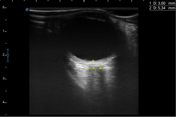
Optic nerve imaging was performed with a high-frequency linear transducer (7.5 MHz Linearprop, Sonoscape E1 Exp, Shenzhen, China) on the transverse axis by applying a thick layer of US gel over the closed eye. Anechoic vitreous fluid in the eyeball and echogenic papilla on the posterior wall were visualized. Hypoechoic optic nerve complex surrounded by echogenic retrobulbar fat was identified. The optic nerve sheath was visualized as a thin echolucent structure extending from both sides of the hypoechoic optic nerve. The ONSD was measured 3 mm behind the papilla, perpendicularly to the cursors (Figure 1).
Figure 1.

- Measurement of optic nerve sheath diameter by ultrasound technique. 1, 3 mm behind globe; 2, optic nerve sheath diameter.
Measurements were performed starting before the surgery until the patient was fully awakened on the recovery table after the surgery was completed. For this purpose, ONSD was measured at following time points: T1, in the supine position after induction of anesthesia; T2, after conversion to the prone position; T3, in the prone position after applying pressure to the dura mater; T4, in the prone position after the discontinuation of applying pressure to the dura mater; T5, after conversion to the supine position.
Intraoperative and postoperative follow-up
Operative time, arterial blood pressure, the rate of change in blood pressure and required fluid pressure, abrupt changes in fluid pressure performed during surgery when necessary were intraoperatively recorded, and the effect of abrupt pressure changes on ONSD and whether there is a change in ONSD depending on time were followed up by US.
After the operation, the patient was transferred to the post-anesthesia care unit in the early postoperative period. Visual Analogue Scale (VAS) scores of 4 and above for pain were evaluated as headache. In addition, low back pain, leg pain, nausea, and vomiting were questioned and noted.
Statistical analysis
Although the sample size calculation was not performed for our study in the pre-study period, post hoc power analyses were carried out to examine whether the sample size was sufficient after the study. The effect sizes for ONSD, eyeball transverse diameter (ETD), and ONSD/ETD ratio were 3.70, 0.67, and 2.49, respectively. A sample size of 64 patients was estimated to provide a power of 99% for all three variables, with an alpha error of 5%. Power analyses were carried out using the G*Power version 3.1 software. The measurement data of the study are shown with median (minimum-maximum) values. The Kolmogorov-Smirnov test was used to check the assumption of normality for the measurements. Since the data were non-normally distributed, the Mann-Whitney U test was used to compare the measurements between the groups, and the Friedman test was used for the temporal comparison of the measurements within the group. The ROC curve analysis was used to determine the threshold value for headache. A p-value <0.05 was considered statistically significant. In post hoc analyses, Bonferroni correction was performed for p-values. All analyses were carried out using the SPSS version 20 software.
Results
The study was conducted with data from 64 patients with a median age of 42.5 (range, 29.0-68.0) years, median BMI 24,9 (range,23.8-26.0), median heights 170(range, 165-176)cm, median weights 71(range, 68-74)kg. Headache complaints were experienced by 16 (25.0%) of the patients. The evaluation of the chronic diseases revealed that 6 (9.4%) patients had hypertension and 5 (7.8%) patients had diabetes. The comparison of the right and left eye data showed similar values for ETD, ONSD and the ratio of these 2 measurements (p>0.05). No visual problems and seizures were observed in any of the patients.
The analysis of the change in right ONSD measurements by time unveiled significant correlations between ONSD and ETD data. According to the results of post hoc analysis, there were statistically significant differences in median ONSD values in all paired comparisons of time points, except for paired comparisons of T1 (in the supine position after induction of anesthesia) and T5 (after conversion to the supine position) time points and T2 (after conversion to the prone position) and T4 (in the prone position after the discontinuation of applying pressure to the dura mater) time points (p<0.001). The median ONSD at T3 (in the prone position after applying pressure to the dura mater) was higher than those at all other time points, while the median ONSD at T2 was lower than those at all other time points, except for T4 (Table 1).
Table 1.
- Change in right ONSD measurements by time.
| Time | Median (Min-Max) | P-value* |
|---|---|---|
| T1 ONSD | 4.4 (3.9-5.7) | <0.001 a |
| T2 ONSD | 4.1 (3.5-5.2) | |
| T3 ONSD | 5.3 (4.2-6.0) | |
| T4 ONSD | 4.2 (3.5-5.2) | |
| T5 ONSD | 4.5 (3.9-5.8) | |
| a Post hoc comparisons: All significant, except for paired comparisons of T1-T5 and T2-T4 | ||
| T1 ETD | 22.0 (20.4-25.0) | <0.001b |
| T2 ETD | 22.0 (19.0-24.8) | |
| T3 ETD | 22.0 (20.0-24.0) | |
| T4 ETD | 22.0 (18.0-24.5) | |
| T5 ETD | 21.9 (19.5-25.3) | |
| bPost hoc comparisons: Paired comparisons of T1-T2 and T1-T4 are significant | ||
| T1 ONSD/ETD ratio | 0.20 (0.18-0.25) | <0.001 a |
| T2 ONSD/ETD ratio | 0.18 (0.16-0.24) | |
| T3 ONSD/ETD ratio | 0.24 (0.19-0.27) | |
| T4 ONSD/ETD ratio | 0.19 (0.17-0.24) | |
| T5 ONSD/ETD ratio | 0.21 (0.18-0.27) | |
Post hoc comparisons: All significant, except for paired comparisons of T1-T5 and T2-T4,
Freidman test.
ONSD, optic nerve sheath diameter; ETD, eyeball transverse diameter.
According to the results of post hoc analysis, the median ETD at T1 was the same, but the distribution range was found to be narrower and higher than those at T2 and T4 time points (p<0.001) (Table 1).
The results of post hoc analysis revealed that the median ratio of ONSD to ETD (will only be indicated as the ratio in the continued part) was statistically significant in all paired comparisons of time points, except for paired comparisons of T1 and T5 time points and T2 and T4 time points (p<0.001). The median ratio at T3 was higher than those measured at all other time points, while the median ratio at T2 was lower than those at all other time points, except for T4 (Table 1).
The analysis of the change in left ONSD measurements by time showed significant correlations between ONSD and ETD data. According to the results of post hoc analysis, the differences in the median ONSD were statistically significant in all paired comparisons of time points, except for paired comparison of T1 and T5 (p<0.001). The median ONSD at T3 was higher than those at all other time points, while the median ONSD at T2 was lower than those at all other time points (Table 2).
Table 2.
- Change in left ONSD measurements by time.
| Time | Median (Min-Max) | P-value* |
|---|---|---|
| T1 ONSD | 4.4 (3.9-5.7) | <0.001 a |
| T2 ONSD | 4.1 (3.6-5.2) | |
| T3 ONSD | 5.3 (4.2-6.0) | |
| T4 ONSD | 4.3 (3.5-5.2) | |
| T5 ONSD | 4.5 (3.9-5.8) | |
| T1 ETD | 22.0 (20.0-25.3) 5.3 | <0.001 b |
| T2 ETD | 22.0 (19.2-24.8) 5.6 | |
| T3 ETD | 22.0 (19.5-24.0) | |
| T4 ETD | 22.0 (20.0-24.5) 4.5 | |
| T5 ETD | 22.0 (19.5-25.3) 5.8 | |
| T1 ONSD/ETD ratio | 0.20 (0.17-0.25) | <0.001 c |
| T2 ONSD/ETD ratio | 0.19 (0.16-0.24) | |
| T3 ONSD/ETD ratio | 0.24 (0.19-0.27) | |
| T4 ONSD/ETD ratio | 0.19 (0.17-0.24) | |
| T5 ONSD/ETD ratio | 0.21 (0.18-0.27) |
Freidman test.
ONSD, optic nerve sheath diameter; ETD, eyeball transverse diameter,
Post hoc comparisons: All significant, except for paired comparison of T1-T5,
Post hoc comparisons: Paired comparisons of T1-T2, T1-T4, and T1-T5 are significant,
Post hoc comparisons: All significant. except for paired comparisons of T1-T4 and T2-T4
The results of post hoc analysis showed that the median ETD at T1 was the same, but the distribution range was narrower and higher than those at T2 and T5 time points and wider than that at T4 (p<0.001) (Table 2).
According to the results of post hoc analysis, the differences in the median ratio were statistically significant in all paired comparisons of time points (p<0.001), except for paired comparisons of T1 and T4 time points and T2 and T4 time points. The median ratio at T3 was higher than those at all other time points, while the median ratio at T2 was lower than those measured at all other time points, except for T4 (Table 2).
The analysis of the ONSD measurements after applying pressure to the dura mater in the prone position by the development of headache showed that the median ONSD and the ratio were significantly higher on the side of headache than on the opposite side (p<0.001 for all). ETD was similar between patients with and without headache on both sides (p>0.05) (Table 3).
Table 3.
- The ONSD measurements by headache development.
| Time | Headache | P-value* | |
|---|---|---|---|
| No | Yes | ||
| Median (Min-Max) | Median (Min-Max) | ||
| Right | |||
| T3 ONSD | 5.1 (4.2-6.0) | 5.6 (5.2-6.0) | <0.001 |
| T3 ETD | 22.0 (20.0-24.0) | 22.0 (21.0-23.0) | 0.513 |
| T3 ONSD/ETD ratio | 0.23 (0.19-0.27) | 0.25 (0.23-0.27) | <0.001 |
| Left | |||
| T3 ONSD | 5.1 (4.2-5.9) | 5.6 (5.2-6.0) | <0.001 |
| T3 ETD | 22.0 (19.5-24.0) | 22.0 (21.0-23.0) | 0.627 |
| T3 ONSD/ETD ratio | 0.23 (0.19-0.27) | 0.25 (0.23-0.27) | <0.001 |
Mann-Whitney U test.
ONSD, optic nerve sheath diameter; ETD, eyeball transverse diameter.
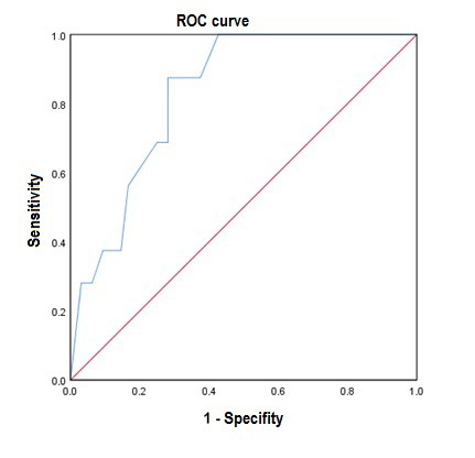
To examine whether a threshold value can be determined for ONSD for the development of headache, ROC curve analysis was performed and the optimal threshold value was found to be 5.3 (Area under the curve 0.830, 95% Confidence Interval: 0.761-0.899, p<0.001). For the determined value of 5.3, the sensitivity, specificity, positive predictive, and negative predictive values were 87.5%, 71.9%, 50.9%, and 94.5%, respectively (Table 4, Figure 2).
Table 4.
- Threshold value for ONSD values for the development of headache (ROC curve analysis).
| THV | AUC | 95% CI | p-value | Sen | Spe | PPV | NPV | |
|---|---|---|---|---|---|---|---|---|
| Lower limit | Upper limit | |||||||
| 5.3 | 0.830 | 0.761 | 0.899 | <0.001 | 87.5 | 71.9 | 50.9 | 94.5 |
THV, threshold value; AUC, area under the curve; CI, confidence interval; Sen, sensitivity, Spe, Specificity; PPV, positive predictive value; NPV, negative predictive value.
Figure 2.

- ROC curve analysis
Discussion
In our study, it has been shown that when pressure is applied to the dura mater, it causes changes in the diameter of the optic nerve sheath and may be associated with complications due to increased intracranial pressure in the postoperative period.
The optic nerve sheath is the part of the dura mater extending to the back of the eye. Thus, the subarachnoid space can extend to the back of the eyeball. It has been demonstrated that normal ONSD is 4.0 mm in children younger than 1 year, 4.5 mm in children aged 1-18 years, and 5.0 mm in adults. 7,8 An ONSD value greater than 5.7 mm has been associated with elevated ICP. 9 In our study, ONSD measured at all time points was below the threshold value for increased ICP.
It has been shown that ONSD measurement with ultrasound correlates with invasive ICP measurements in the follow-up of increased intracranial pressure in patients with subarachnoid hemorrhage. 10 In meta-analyses comparing ultrasound ONSD measurements with the gold standard invasive ICP measurement devices; It has been shown that there is a correlation with the invasive method. 11,12 It was thought that it could be used due to its potential benefit in cases where invasive ICP cannot be measured. 12 In the study evaluating the effect of prone position on optic nerve diameter in acute respiratory stress patients in intensive care; It was observed that prone position did not make any statistical difference in optic nerve sheath diameter measurements. In the same study, ONSD measurements were observed to be lower than the initial value. 13
It has been found that ICP increases in patients who are turned to the prone position under anesthesia, and 4.3 mm is the cut-off value for the ICP increase in the prone position.14 In our study, the ONSD diameter measured in the prone position was 4.1 mm. We think that the reason for this is the application of TIVA to the patients. It was thought that TIVA would be protective against the increase in ICP in preventing the intracranial increase in full endoscopic spine surgery on animals. 15 It has been shown that propofol-based TIVA application has lower ONSD measurements compared to sevoflurane inhalation anesthesia in robotic laparoscopic surgeries that cause increase in ICP. 16,17 In spine surgery, the prone position does not increase ICP in short and moderately long operations; however, changes have been shown in ONSD in very long operations. 18 The results of our study showed no difference between T1-T5 measurements, similar to the literature.
A study evaluating the effect of body position on intraocular pressure (IOP) and ICP reported decreased ICP in the prone position but no change in IOP. 19 In animal studies, it was measured that there was no statistically significant difference in ICP, IOP and translaminar pressure in the eye in the prone position, but ICP and IOP were lower in the supine position. 20 In our study, the decrease in T2 measurements of patients compared to T1 could be explained by the decrease in ICP.
It has been shown that hydrostatic pressure applied to the dura mater in endoscopic spine surgeries increases the pressure in the epidural space. 21 In a series of 27 patients, it was shown that the hydrostatic pressure applied to the dura mater returned to baseline after surgery. 22 Our study showed that the ONSD values were higher at T3 measurements when pressure was applied to the dura mater, while they returned to baseline when the effect of hydrostatic pressure on the dura mater was ruled out.
Headache, neck pain, and seizures can develop after endoscopic spine surgeries. A study of 28 patients showed that 8 patients developed headaches after surgery. 23 Another study of 33 patients reported head and neck pain in 8 patients and seizures in 4 patients. 24 In our study, none of the patients developed seizures, and headaches were observed in 16 patients in the early postoperative period; however, the patients did not have a headache during discharge.
It is believed that intraoperative increase in epidural pressure may be responsible for the development of headaches. 21 In our study, an ONSD value >5.3 had a sensitivity of 87.5% and a specificity of 71.9% to demonstrate the development of headaches.
In conclusion, the effects of hydrostatic pressure applied to the dura mater in endoscopic spine surgeries on the central nervous system are unknown. The general opinion is that the hydrostatic pressure should be kept below 30 mmHg, although its effects cannot be followed instantly. We are of the opinion that ONSD, which is evaluated by ocular US to monitor the pressure applied to the dura mater in endoscopic spine surgeries, is an important technique that provides an effective, noninvasive, and continuous monitoring opportunity to detect an intraoperative increase in ICP early and to reduce the development of postoperative complications. The major limitation of our study is the inability to perform simultaneous control with invasive measurements. Larger patients studies to be conducted in the future will help determine different threshold values by investigating the effects of different intraoperative hydrostatic pressure values on ONSD.
Footnotes
References
- 1. Rehder D. Idiopathic Intracranial Hypertension: Review of Clinical Syndrome, Imaging Findings, and Treatment. Curr Probl Diagn Radiol 2020; 49: 205-214. [DOI] [PubMed] [Google Scholar]
- 2. Kim JE, Choi DJ. Unilateral biportal endoscopic decompression by 30° endoscopy in lumbar spinal stenosis: Technical note and preliminary report. J Orthop 2018; 15: 366-371. [DOI] [PMC free article] [PubMed] [Google Scholar]
- 3. Le Roux P, Menon DK, Citerio G, Vespa P, Bader MK, Brophy G, et al. The International Multidisciplinary Consensus Conference on Multimodality Monitoring in Neurocritical Care: a list of recommendations and additional conclusions: a statement for healthcare professionals from the Neurocritical Care Society and the European Society of Intensive Care Medicine. Neurocrit Care 2014; 21 Suppl 2: 282-296. [DOI] [PMC free article] [PubMed] [Google Scholar]
- 4. Yuen J, Selbi W, Muquit S, Berei T. Complication rates of external ventricular drain insertion by surgeons of different experience. Ann R Coll Surg Engl 2018;100:221-225. [DOI] [PMC free article] [PubMed] [Google Scholar]
- 5. McLaughlin D, Anderson L, Guo J, McNett M. Serial Optic Nerve Sheath Diameter via Radiographic Imaging: Correlation With ICP and Outcomes. Neurol Clin Pract 2021; 11: e620-e626. [DOI] [PMC free article] [PubMed] [Google Scholar]
- 6. Toscano M, Spadetta G, Pulitano P, Rocco M, Di Piero V, Mecarelli O, et al. Optic Nerve Sheath Diameter Ultrasound Evaluation in Intensive Care Unit: Possible Role and Clinical Aspects in Neurological Critical Patients’ Daily Monitoring. Biomed Res Int 2017; 2017: 1621428. [DOI] [PMC free article] [PubMed] [Google Scholar]
- 7. Le A, Hoehn ME, Smith ME, Spentzas T, Schlappy D, Pershad J. Bedside sonographic measurement of optic nerve sheath diameter as a predictor of increased intracranial pressure in children. Ann Emerg Med 2009; 53: 785e91. [DOI] [PubMed] [Google Scholar]
- 8. Blaivas M, Theodoro D, Sierzenski PR. Elevated intracranial pressure detected by bedside emergency ultrasonography of the optic nerve sheath. Acad Emerg Med 2003; 10: 376e81. [DOI] [PubMed] [Google Scholar]
- 9. Geeraerts T, Launey Y, Martin L, Pottecher J, Vigué B, Duranteau J, et al. Ultrasonography of the optic nerve sheath may be useful for detecting raised intracranial pressure after severe brain injury. Intensive Care Med 2007; 33: 1704-1711. [DOI] [PubMed] [Google Scholar]
- 10. Kim KH, Kang HK, Koo HW. Prediction of Intracranial Pressure in Patients with an Aneurysmal Subarachnoid Hemorrhage Using Optic Nerve Sheath Diameter via Explainable Predictive Modeling. J Clin Med 2024; 13: 2107. [DOI] [PMC free article] [PubMed] [Google Scholar]
- 11. Xu J, Song Y, Shah Nayaz BM, Shi W, Zhao Y, Liu Y, et al. Optic Nerve Sheath Diameter Sonography for the Diagnosis of Intracranial Hypertension in Traumatic Brain Injury: A Systematic Review and Meta-Analysis. World Neurosurg 2024; 182: 136-143. [DOI] [PubMed] [Google Scholar]
- 12. Robba C, Santori G, Czosnyka M, Corradi F, Bragazzi N, Padayachy L, et al. Optic nerve sheath diameter measured sonographically as non-invasive estimator of intracranial pressure: a systematic review and meta-analysis. Intensive Care Med 2018; 44: 1284-1294. [DOI] [PubMed] [Google Scholar]
- 13. A Demir U, Taşkın Ö, Yılmaz A, Soylu VG, Doğanay Z. Does prolonged prone position affect intracranial pressure? prospective observational study employing Optic nerve sheath diameter measurements. BMC Anesthesiol 2023; 14; 23: 79. [DOI] [PMC free article] [PubMed] [Google Scholar]
- 14. Xu N, Zhu Q. Optic nerve sheath diameter measured by ultrasonography versus Magnetic Resonance Imaging for diagnosing increased intracranial pressure: a systematic review and meta-analysis. Med Ultrason 2023; 29; 25: 270-278. [DOI] [PubMed] [Google Scholar]
- 15. Amato MCM, Carneiro VM, Fernandes DS, de Oliveira RS. Intracranial Pressure Evaluation in Swine During Full-Endoscopic Lumbar Spine Surgery. World Neurosurg 2023; 179: e557-e567. [DOI] [PubMed] [Google Scholar]
- 16. Sujata N, Tobin R, Tamhankar A, Gautam G, Yatoo AH. A randomised trial to compare the increase in intracranial pressure as correlated with the optic nerve sheath diameter during propofol versus sevoflurane-maintained anesthesia in robot-assisted laparoscopic pelvic surgery. J Robot Surg 2019; 13: 267-273. [DOI] [PubMed] [Google Scholar]
- 17. Kim JE, Koh SY, Jun IJ. Comparison of the Effects of Propofol and Sevoflurane Anesthesia on Optic Nerve Sheath Diameter in Robot-Assisted Laparoscopic Gynecology Surgery: A Randomized Controlled Trial. J Clin Med 2022; 12; 11: 2161. [DOI] [PMC free article] [PubMed] [Google Scholar]
- 18. Gültop F, Tekin EA. The impact of prone position on optic nerve sheath diameter. Arch Clin Exp Med 2021; 6: 96-99. [Google Scholar]
- 19. Jasien JV, Samuels BC, Johnston JM, Downs JC. Effect of Body Position on Intraocular Pressure (IOP), Intracranial Pressure (ICP), and Translaminar Pressure (TLP) Via Continuous Wireless Telemetry in Nonhuman Primates (NHPs). Invest Ophthalmol Vis Sci 2020; 61: 18. [DOI] [PMC free article] [PubMed] [Google Scholar]
- 20. Jasien JV, Samuels BC, Johnston JM, Downs JC. Effect of Body Position on Intraocular Pressure (IOP), Intracranial Pressure (ICP), and Translaminar Pressure (TLP) Via Continuous Wireless Telemetry in Nonhuman Primates (NHPs). Invest Ophthalmol Vis Sci 2020; 1; 61: 18. [DOI] [PMC free article] [PubMed] [Google Scholar]
- 21. Kang T, Park SY, Lee SH, Park JH, Suh SW. Assessing changes in cervical epidural pressure during biportal endoscopic lumbar discectomy [published online ahead of print, 2020 Oct 30]. J Neurosurg Spine 2020; 1-7. [DOI] [PubMed] [Google Scholar]
- 22. Kang MS, Park HJ, Hwang JH, Kim JE, Choi DJ, Chung HJ. Safety Evaluation of Biportal Endoscopic Lumbar Discectomy: Assessment of Cervical Epidural Pressure During Surgery. Spine (Phila Pa 1976) 2020; 45: E1349-E1356. [DOI] [PubMed] [Google Scholar]
- 23. Joh JY, Choi G, Kong BJ, Park HS, Lee SH, Chang SH. Comparative study of neck pain in relation to increase of cervical epidural pressure during percutaneous endoscopic lumbar discectomy. Spine (Phila Pa 1976) 2009; 34: 2033-2038. [DOI] [PubMed] [Google Scholar]
- 24. Choi G, Kang HY, Modi HN, Prada N, Nicolau RJ, Joh JY, et al. Risk of developing seizure after percutaneous endoscopic lumbar discectomy. J Spinal Disord Tech 2011; 24: 83-92. [DOI] [PubMed] [Google Scholar]


