

Abstract
Introduction
The toes are the distal extension of the foot increasing its weight-bearing area and the security of the stance. The nails are the acral part of the toes and are so intimately linked with the distal interphalangeal joint, its ligaments, and tendons that they were also called musculoskeletal appendages. The big toe is of particular importance for gait and stance. Anatomic alterations of the foot and toe lead to pathological changes of the nails, both directly as well as indirectly.
Methods
The author evaluated the clinical photographs of 1,663 patients examined for toenail conditions.
Results
It was found that a normal straight axis of the first ray of metatarsal and phalanx bones occurred in less than 10% of the patients with hallux valgus and hallux valgus interphalangeus being extremely frequent. The commonest nail changes observed were compression nail (n = 247), ingrown nail (196), onychomycosis (192), disappeared nail bed (191), congenital malalignment (118), pincer nails (118), and nail overcurvature (114). The most frequent foot-toe abnormalities were hallux valgus (775) and hallux valgus interphalangeus (1,277).
Conclusion
Although a direct causal relationship is hard to prove it was realized that most of the nail changes were associated with foot and toe abnormalities. The study reveals that assessing toenail changes requires examination of the entire foot, best in relaxed, standing, and walking conditions.
Keywords: Toenails, Toes, Foot alterations, Congenital malalignment, Compression nail, Retronychia, Ingrown nail, Nail tumors
Plain Language Summary
The foot of man is a strong organ consisting of 26 bones, 33 joints, of which 20 are actively movable, and more than 100 muscles, tendons and connective tissue bands keeping this complex body part together. The foot is usually divided into three parts, the hind, mid, and forefoot. The latter contains the toes as their most forward-pointing parts. They are covered by the nails, which are plates of hard horn substance. The toes increase the weight-bearing surface of the foot and are thus important for balance while standing and walking, for running and jumping. If the foot is not healthy this will seriously influence its function and also change the toenails. An examination of more than 1,600 persons’ toenail photographs was performed to find out, whether foot anomalies can be found in patients with toenail changes. This was in fact the case in a very high number of the patients.
Introduction
The human foot is a very complex and strong structure, a real bio-technical marvel. It consists of 26 bones plus a variable number of sesamoids, 33 joints, of which 20 – the ankle, subtalar joint, and interphalangeal joints – are actively articulated, and more than a hundred muscles, tendons, ligaments, and aponeuroses. The talus and calcaneus form the hindfoot. The midfoot is made up by the cuboid, navicular, and 3 cuneiform bones. These bones are tightly connected by ligaments and form arches that act as a shock absorber. Distal to these bones is the forefoot made up of the five metatarsals and the phalanges that form the toes. The big toe has two phalanges, all other four toes have three. Beneath the bones is a specialized fibrofatty tissue acting as a cushion. The foot has several functions of which locomotion is the most obvious; however, we need them to stand safely and their unique innervation makes the sole of the foot a sensory organ allowing to perceive where we are, what our present position is on the earth, and to keep the balance. The toes are the distal extension of the foot increasing the weight-bearing area and stabilizing balance. The nails are the toes’ most acral parts. They are so intimately connected to the distal phalanx, the tendons, entheses, ligaments, and aponeuroses that the nails were also – rightfully – called musculoskeletal appendages [1, 2].
The big toe is an extremely important part of the foot, both for forward movement as well as for secure stance. It is estimated that up to 90% of the stability of the foot derives from the flexibility of the hallux. Any disturbance of the big toe, particularly hallux valgus (HV) and hallux rigidus, has a serious impact on the stance and the whole kinematic of gait, from the foot to the ankle, knee to the hip and probably the spine, which is actually the basis for the concept of the asymmetric gait – nail unit syndrome (AGNUS) [3, 4]. On the other hand, a big toe that is out of the correct axis cannot correctly fulfill its physiological function. It is clear that anomalies of the toes and feet may have a profound impact on the nails [5, 6].
The gait is usually divided into phases beginning with the heel strike, leading to forefoot loading, the full foot, heel lift, and finally toe off. At this last part, most of the body weight is on the tip of the big toe multiplied by a factor of about 2.5 due to the kinetic energy of the propulsion/forward thrust; this is even higher during running and jumping reaching several times the entire body weight. Some developmental biologists claim that the stubby strong big toe was the prerequisite for the human bipedal gait [7]. The hallux nail is strong enough to give sufficient counterpressure to the toe tip’s soft tissue to prevent it from being dislodged dorsally and forming a false distal nail fold or distal bulge [8].
Foot and toe deformation thus may cause nail abnormalities. This has hitherto rarely been investigated [3, 5, 6, 9]. We have therefore searched the personal photo-archive of the specialized nail clinics held at the Departments of Dermatology, Inselspital, Univ Berne, Switzerland, University Hospital Ghent, Belgium, Dermatology Center Epidermis, Instituto CUF, Porto, Portugal, and Klinika Manola, Zagreb, Croatia, between October 2018 and December 2023.
Methods
The author’s archive containing photographs of all patients personally seen by the author between October 2018 and December 2023 in 4 different specialized nail clinics in four countries was searched for toenail changes (n = 1,663). Photographs were taken in a manner displaying at least both the mid- and forefoot as well as the toes thus allowing it to be evaluated for skeletal anomalies. Particular attention was paid to the toenail alterations. Patients with insufficient photos were excluded (n = 11) because their photographs did not allow a clinical evaluation of the forefoot skeleton. Photographs of patients seen twice or several times were counted as 1 patient if they did not have different diagnoses. Radiographs were taken in 48 cases and magnetic resonance in 1 patient, in most cases to confirm the diagnosis of a subungual exostosis or of osteophytes in patients with pincer nails and disappeared nail bed. The photos were then analyzed for forefoot and toe deformations.
Site of nail clinic, gender, age at and date of consultation, clinical nail diagnosis, presence or absence of foot and toe abnormality, potential co-involvement of finger nails, onychological co-morbidities, other relevant systemic diseases, X-ray results, and treatments were listed. The many different nail conditions and foot anomalies were grouped to allow a trend to be calculated.
First, age and gender of the patients were listed, then the toenail diagnosis was noted, finally the foot was clinically analyzed for the absence or presence of the following abnormalities: HV, HV interphalangeus (HVI), hallux erectus (HE); inward rotation of the big toe and outward rotation of the little toe were interpreted as signs of a splayfoot.
The following definitions were used.
-
1.
Malalignment of the toenail: visible deviation of the nail unit’s long axis from that of the distal phalanx.
-
2.
HV: deviation of the proximal hallux phalanx of more than 3° from the longitudinal axis of the first metatarsal bone.
-
3.
HVI, also called lateral displacement of the distal phalanx: deviation of the longitudinal axis of the distal phalanx of >3° from the axis of the proximal phalanx.
-
4.
HE, also called hyperextension: defined as a lack of contact of the big toe pulp with the ground and visibly strained extensor tendons of the toes under relaxed condition.
-
5.
Inward rotation (tilting) of the big toe: the transverse axis of the toe is rotated >3° medially.
-
6.
Outward rotation of the little toe: the transverse axis of the little toe is tilted >15°.
-
7.
The concomitant rotation of both the big and the little toes was interpreted as a sign of a splayfoot.
Secondary nail and relevant additional systemic diagnoses were listed. The deviation of the long axis of the big toe’s proximal phalanx from that of the first metatarsal bone as well as that of the distal phalanx from the proximal phalanx of the big toe was measured on radiographs by drawing a line along the metatarsal bone and the phalanges and using the turn function in the layout of Microsoft Word, clinically by palpating the dorsal aspect of the metatarsal and proximal phalanx bones and estimating the bone direction of the distal phalanx (Fig. 1).
Fig. 1.

Clinical (blue lines) and radiographic (yellow lines) photographs with lines drawn to show how the measurements for the deviation of the bones’ long axis were made. a Female patient with overcurvature of the left hallux and short left big toenail. The proximal phalanx has a medial deviation of 2° from the metatarsal bone; the distal phalanx exhibits a deviation of 13°. b Radiograph of a 38-year-old woman with overcurvature of the hallux nails, a medial deviation of the first metatarsal bone in relation to the os cuneiforme I and a HVI with a lateral deviation of the distal phalanx of 18° in relation to the long axis of the proximal phalanx.
Results
Of the 1,663 toenail patients in the archive, eleven were excluded as their photographs did not show enough of the forefoot to evaluate potential foot anomalies. Thus, 1,652 photo-documented cases were available for the study.
There were 1,031 female (62.4%) and 621 male (36%) patients (p < 0.02) (Table 1). The age of the patients spanned from several weeks to over 90 years with a peak between 6 and 30 years (Table 2).
Table 1.
Summary of study data: demographic data
| Patients with toenail changes, N | 1,663 |
| Patients excluded because of insufficient data, N | 11 |
| Patients evaluable, N | 1,652 |
| Gender | Female 1,031 (64%) | Male 621 (37.6%) | p < 0.01 |
| Current gender distribution in the countries of most patients | |||
| Germany | Population 84,360,000 | Female:male = 50.7:49.3 | |
| Switzerland | Population 8,820,000 | Female:male = 50.4:49.6 | |
| Portugal | Population 10,421,117 | Female:male = 52.3:47.7 | |
| Croatia | Population 3,899,000 | Female:male = 51.9:48.1 | |
Table 2.
Patients’ age at presentation (age distribution of the study group)*
| Age group | n (%) | Relation female:male |
|---|---|---|
| 0–6 | 77 (4.6) | (26 f: 51 m) |
| 6–16 | 320 (19.4) | (196 f: 124 m) |
| 17–30 | 371 (22.5) | (269 f: 102 m) |
| 31–40 | 221 (13.4) | (138 f: 83 m) |
| 41–50 | 239 (14.5) | (147 f: 92 m) |
| 51–60 | 183 (11.0) | (126 f: 57 m) |
| 61–70 | 156 (9.4) | (96 f: 60 m) |
| 70–80 | 60 (3.6) | (25 f: 35 m) |
| 81–90 | 11 (0.6) | (6 f: 5 m) |
| >90 | 1 (0.1) | (1 m) |
f, female; m, male.
*The exact age could not be retrieved in some patients.
There were 51 boys and 26 girls up to the age of 6, and 124 boys and 196 girls between 6 and <17 years. Of the 371 patients between 17 and 30 years (22.5%), 269 were female and 102 male patients.
The main diagnoses (Table 3) were congenital nail abnormalities (118) that often appeared during childhood and were seen in the younger age group with the exception of pincer nails (118), onychomycoses (192), and traumatic nail changes. Almost all of the alterations due to repeated trauma, such as compression nails (247), disappeared nail bed (191), and AGNUS (89), were associated with marked deviations of the forefoot skeleton, mostly HV and HVI.
Table 3.
Most common nail diagnoses
| Compression nail | 247 |
| Ingrown nail | 196 |
| Onychomycosis | 192 |
| Disappeared nail bed | 191 |
| Congenital malalignment | 118 |
| Pincer nails | 118 |
| Nail overcurvature | 114 |
| AGNUS | 89 |
| Melanonychia | 83 |
| plus 2 Laugier-Hunziker-Baran syndrome with toenail involvement | |
| Psoriasis | 50 |
| Double nail of the little toe | 47 |
| Subungual exostosis | 36 |
| Ungual fibrokeratoma | 22 |
| Trapezoid nail | 22 |
| Melanoma of the toenail | 14 |
| Myxoid pseudocyst | 14 |
| Onychogryposis | 12 |
| Acrodermatitis continua suppurativa | 7 |
| Eczema, different clinical forms | 7 |
| Yellow nail syndrome | 6 |
| Onychoschizia | 4 |
| Onychopapilloma | 4 |
| Pachydermoperiostosis | 4 |
| COVID toe | 3 |
| Subungual glomus tumor | 2 |
| Onychomatricoma | 2 |
| Onychoteiromania | 2 |
The examination of the photographs for bone anomalies of the metatarsals and phalanges (Table 4) revealed that a “normal straight first ray” was rather an exception. Adding normal (46) and mild HVI (104) yielded 150 near-normal feet out of 1,652 corresponding to only 9.1%. HV was seen in 775 cases, HVI 1277, HVE 237, the combination of HV and HVI 222, and that of HV, HVI and HVE 207 times. The combinations of HV plus HE (9) as well as HVI plus HE (2) were very rare (Table 4).
Table 4.
Frequency of foot and toe anomalies, all patients n = 1,652
| Normal | 46 |
| Mild HVI | 104 |
| Normal plus mild HVI | 150 |
| HV | 775 |
| HVI | 1,277 |
| HE | 237 |
| HV + HVI | 222 |
| HV + HVI + HE | 207 |
| HV + HE | 9 |
| HVI + HE | 2 |
Further single case diagnoses were macro nail due to a subungual metastasis of a mammary carcinoma (F, 53-y-o), subungual osteophyte (F, 76-y-o), nail unit deviation without further nail changes (M, 10-y-o), subungual abscess (F, 26-y-o), hypoplastic little toenail (F, 63-y-o), degenerative eczema (F, 31-yo), postoperative onychodynia (F, 70-y-o), granulating lectitis in a 20-year-old female patient with KID syndrome, pityriasis rubra pilaris of the nail (F, 23-y-o), endovascular papillary proliferation (F, 40-y-o), onychocytic acanthoma (F, 50-y-o), onychotemnomania (M, 50-y-o), congenital broad hallux nail (M, 10-y-o), anonychia in 20-year-old female patient with dystrophic epidermolysis bullosa hereditaria and one after phenolization (F, 18-y-o), post-traumatic pyogenic granuloma of the fifth toe (M, 50-y-o), Bowen disease plus bullous pemphigoid (F, 72-y-o), bacterial chromonychia (M, 67-y-o), hallux tip eczema after bone fracture (M, 20-y-o), koilonychia in iron and zinc deficiency (F, 36-y-o), total diffuse leukonychia (F, 54-y-o), watch-glass nails (M, 61-y-o), subungual corn (F, 20-y-o), and onychoptosis (F, 52-y-o).
Discussion
This study has shown that the prevalence of toenail diseases in a specialized nail clinic may considerably differ from the average in common dermatologic practice. It has also demonstrated that the toenails should not be observed isolated but together with the toe position and probably the entire foot [6]. In contrast to frequently repeated claims, onychomycoses were by far not the commonest diagnoses.
In the young age groups, congenital malalignment was very frequent (Fig. 2) [10, 11]. This is in accordance with another study noting that this condition is a frequent reason for parents to bring their child for a nail consultation [12]. Malalignment of the big toenail is an idiopathic lateral deviation of the nail apparatus that is often inborn and has a strong genetic background [13]. A nail unit out of the orthograde axis, virtually always exacerbated by a lateral deviation of the distal phalanx and often the entire big toe, is subject to chronic repeated microtrauma rendering the nail prone to onycholysis, onychomadesis, retronychia, paronychia, and nail growth disturbance (Fig. 3) [9, 14]. Any frontal compression produces torque to the nail damaging the matrix and nail bed adherence. However, in children between 3 and 10 years of age, there was often a “mixed diagnosis” of malalignment and considerable nail dystrophy, or near-total onycholysis without obvious nail malalignment. This leads to lack of counterpressure of the nail for the soft tissue of the toe tip during the toe-off phase of gait allowing it to be dislodged dorsally and thus causing the development of a distal bulge and disappeared nail bed (Fig. 4) [15, 16]. In our experience, the disappeared nail bed is not primarily due epidermal metaplasia of the nail bed epithelium, but a result of the shrinkage of the nail bed due to lack of support by a functioning adherent nail [6]. An onycholytic big toenail may be seen as “functionally dead.” The border between the ridged skin of the toe tip and the nail bed always remains visible although the thick onycholytic nail has usually to be cut away to reveal this. Often, the lateral margins of the nail plate can then be seen as “ungual impressions” (Fig. 5).
Fig. 3.

a, b Unilateral congenital malalignment of the left big toenail. There is almost complete onycholysis, a considerable shrimp-nail dystrophy, the nail bed has disappeared and a distal bulge developed. The toe shapes vary between the normal and the involved toes. This example shows that onycholysis is the most important factor for the development of nail dystrophy in this condition.
Fig. 4.

Right big toenail of a 7-year-old boy with mild HV and HVI. The nail is completely onycholytic, very thick, yellow, with a proximal deep transverse furrow, and a disappeared nail bed and distal bulge.
Fig. 2.

Congenital malalignment of the big toenails in a 3-year-old boy with marked HVI and inward rotation of the toes. The right nail is thick, yellow, transversely ridged, onycholytic, the nail bed is shrunk and there is a distal bulge. The second toe appears shorter and the big toe overrides it partially. The left big toenail is not onycholytic and is therefore not dystrophic.
This study has shown that all these patients had a lateral malalignment of the great toe and its distal phalanx, which is in stark contrast to Buttars et al. [9] who found a straight digit in 22 of 30 photos reviewed for their publication (Fig. 6). It may be speculated that this might, at least in part, be due to the quality of the publication photos reviewed by these authors. As originally stated, big toenails may show other types of congenital nail dystrophy, such as congenital pincer nails and upward growing nails (Fig. 7); however, also other toenails may have an upward growth direction (Fig. 8–10) [17].
Fig. 5.

Left big toenail of a 17-year-old girl with HV and almost complete onycholysis. After clipping the onycholytic nail, the impression of both lateral nail margins can be seen. The nail bed has receded but the border to the ridged skin of the dislocated ridged skin is well recognized.
Fig. 6.
Severe nail changes in a 6-year-old girl with HV and HVI, hallux hyperextension of the right big toe, and early phase of retronychia. The nail is discolored, onycholytic, very thick, there is a distal bulge, and considerable inflammation of the periungual soft tissue.
Fig. 8.

Extremely short upward growing hallux nail in a 9-month-old boy with pes equinus.
Fig. 10.

Upward growing, extremely short big toenails in a 20-year-old woman. Particularly the right hallux shows a distal bulge and a protruding nail bed.
Fig. 7.

Upward growing toenails. a 1-year-old boy. b 2-year-old girl.
Boys were more often seen in the youngest age group, whereas girls between 6 and 16 years were more often afflicted than boys. This continued until the age of 70, where female patients were always in the majority (Table 2). The higher male-to-female ratio under 6 years of age may perhaps be explained by the fact that these patients frequently originated from southern geographic regions (Balkans, near east, north Africa) where girls often do not enjoy the same attention like boys.
Disappeared nail bed as the main diagnosis was observed in 191 patients (Fig. 10, 11). However, this is usually the end result either of chronic onycholysis, the primary cause of which can often no longer be defined, or due to too short a nail or its absence. It was already outlined that the big toe of a normal foot bears 2.5 times the entire body weight at the toe-off phase of the gait. This is increased in many sports activities and dancing [18]. Foot deformities such as HV and HVI shift the weight from the tip of the big toe to the basal joint of the second toe with resultant callus, corns, nail overcurvature, and even onychogryposis [19].
Fig. 9.

Upward growing second toenail. Note the HVI and the distal-medial onycholysis.
Isolated medial deviation of the second toenail only was seen in a patient with a Greek foot. Ingrown nails were seen in 196 patients. They were not specifically subclassified to their type and severity grade (Fig. 12, 13). However, most had both a mild HV and HVI. Abnormal HV and HVI angles were found to be significantly associated with ingrown toenails in one study, but no significant difference to controls in two other studies [20–22].
Fig. 11.

Disappeared nail bed of the left big toenail with distal bulge formation and nail bed hyperkeratosis.
Onychomycosis was proven in 192 cases (Fig. 14). The relatively low percentage may be explained by the fact that onychomycoses are treated by general practitioners and dermatologists and are rarely referred to the special nail clinic.
Fig. 12.

Pincer nails in a 60-year-old woman. The distal nail plate shows a curvature of >270°, there is a marked lateral deviation of the big toenails and a mild medial deviation of the second toenails. a Both feet. b Detail of the left foot.
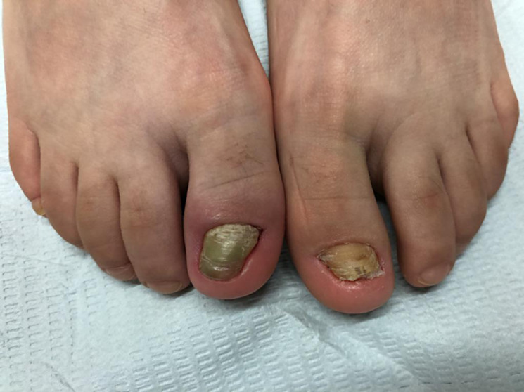
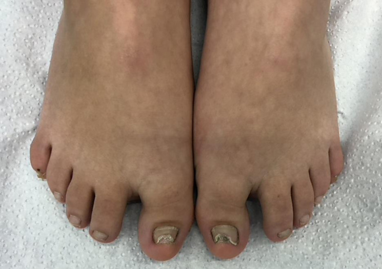
Compression nail is a relatively new concept. It is defined as a thickened, malformed, discolored, often ridged nail, which develops from undue compression in a frontal-to-midfoot direction. Onycholysis is the prerequisite for its development (Fig. 15, 16). The nail is over curved both in longitudinal as well as transverse direction and often bridges a big distal bulge. The onycholytic nail has no support from the nail bed and each compression movement exerts shearing forces on the matrix with the result of “horizontal fractures” and an oyster-shell appearance of the nail. An abnormal big toe with HV and HVI increases these shearing forces by the torque due to the misaligned toe [9]. Thus, it may also be seen as a chronic non-inflammatory variant of repeated retronychia [23]. With 247 out 1,652 cases, it was the most frequent diagnosis. This may sound surprising but may partly be explained by the fact that these patients were seen in a highly specialized nail clinic and had not got any explanation for their distressing nail condition before.
Fig. 15.

Compression nail of the left hallux in an 11-year-old boy. The nail is thickened, discolored, intransparent, more than 75% onycholytic and shows multiple arched transverse furrows; the ridged skin of the toe tip extends under the free end of the nail; there is a distal bulge and the nail bed is considerably decreased in length.
Fig. 16.

Compression nails in a 27-year-old man. The nails are thick, short, intransparent; the nail bed has shrunk, and the nail margins leave impressions in the dorsally dislocated skin of the toe tip.
Fig. 13.

Chronic ingrown toenails in a 27-year-old man. Note the massive fibrosis of the lateral nail folds.
Fig. 14.

Thick discolored nails in a 59-year-old man with onychomycosis.
Lateral deviation of a toenail is virtually always seen in patients with the symmetric type of pincer nails: The big toenail shows a lateral deviation, involved lesser toes display a medial nail deviation, in most cases in a symmetrical fashion (Fig. 12). This is a further hint at the genetic background of pincer nails [24]. Pincer nails were diagnosed 118 times. They are defined as heavily over curved nails with the curvature increasing from proximal to distal and pinching the nail bed. Only symmetrical forms were observed. In all cases, there was HV and marked HVI. Sliding palpation along the sides of the distal interphalangeal joint always revealed a bony excrescence, which is much more marked medially than laterally. This can also be seen in radiographs that usually show the medial one having a hook-like form; it may be the insertion of the interosseous ligament. In cases of involvement of the lesser nails these nails always show a medial deviation. Radiographs of these lesser toes show the distal phalanges to have a pagoda shape [20].
Simple nail overcurvature was defined as a transverse nail curvature >150° but without distal increase of the curvature. This was diagnosed in 114 cases. In contrast to pincer nails, they were asymmetrical in most cases. Patients consulted us because they found this unsightly or had pain. Some tried to help themselves by filing the distal median portion of the nail and padding the toe which often made it worse (Fig. 17).
Fig. 17.
Big toes of a 20-year-old woman with overcurved nails, HVI et erectus. She had tried to take pressure off by filing and cutting the central part of the nail plate.
AGNUS was seen in 89 cases. AGNUS was defined as nail alterations secondary to skeletal abnormalities such as spine deformation, unequal leg length, knee and ankle problems as well as foot problems. They may lead to subconscious abnormal gait and unphysiological distress to the nail unit, mostly in form of localized onycholysis that is mistaken for onychomycosis and treated with antifungal drugs without success [3, 25].
Of the 83 cases of melanonychia of the toenails, approximately half had a racial pigmentation as most of them were immigrated Tamils, persons of north African descent (Maghreb) and a few were East Asians. They were explained the cause of their nail pigmentation and could easily be reassured. The other half of the melanonychia patients were Caucasians (Fig. 18). In most cases, they also had brown streaks in at least one fingernail and dermatoscopy suggested melanocyte activation as the cause in adults and a nevus in children. Two more patients had a Laugier-Hunziker-Baran syndrome, which is characterized by multiple brown spots on the oral mucosa, sometimes also the genital mucosa, and multiple melanonychias [26].
Fig. 18.

Sixty-year-old woman with long-standing longitudinal melanonychia in the right big toenail.
Nail psoriasis is much more frequent on fingers than toes. Thus, 50 patients with toenail psoriasis are still a relatively high proportion but the reason for their consultation was often a misdiagnosed and long and unsuccessfully treated onychomycosis.
Acrodermatitis continua suppurativa of Hallopeau was diagnosed in 7 patients. All had one toenail involved for 8 months to 5 years before the diagnosis was made. Most had been told that they had an onychomycosis and had undergone antifungal therapy in vain.
The little toe may show a rudimentary double nail (Fig. 19). This was seen in 47 patients which is a high number considering the rarity of this changes in the literature. A previous survey had yielded over 70 cases seen in more than 15 years [27]. In this short period, another 47 patients were seen. However, this condition was usually not the primary reason for the nail consultation; instead, it was a chance observation in most patients who had consulted for another nail condition or a first-degree relative accompanying a patient with a double nail was asked to show his or her little toes. Six patients came for a symptomatic double little toenail as they or their doctor had thought of a Durlacher corn. Apparently, this is an autosomal dominant condition. Genetic studies on the SBF 1 gene have not yet been performed. A small lateral bone spike on the apical lateral aspect of the distal phalanx may be seen in good radiographs; this probably induces a small additional nail anlage. If requested, its treatment is by phenolization or complete excision of the additional nail with its matrix [28].
Fig. 19.

Double little toenail in a 19-year-old woman.
Subungual exostosis of a toe was observed 36 times. Most were classically located at the distal medial tip of the distal phalanx (Fig. 20a); however, also lesser toes were involved. The clinical aspect is characteristic with lifting of the distal nail margin by a stone-hard nodule that was covered with a smooth shiny skin surrounded by a very sharp, almost collarette-like border to the toe pulp. A radiograph was taken to confirm the diagnosis and above all to estimate the size and width of bone adhesion as well as to plan the surgery (Fig. 20b, c).
Fig. 20.

Subungual exostosis in a 12-year-old boy. a Clinical aspect. b, c Radiograph. d 8-year-old girl, fourth toe.
Trapezoid nail per se is usually asymptomatic but may cause repeated inflammation of the lateral nail fold similar to a recurrent early ingrown nail. Patient reassurance and taping of the lateral nail folds to keep them away from the margin of the broad nail is usually helpful; if this fails surgical narrowing of the plate by selective lateral matrix horn excision or phenolization is curative.
Melanoma of the toenail unit was seen in 14 cases (Fig. 21). This is a relatively high incidence for the short period of time (there was a 3-month break of nail consultations due to COVID-19 in 2020).
Fig. 21.

Malignant melanoma of the big toenail of a 60-year-old woman. Note the nail dystrophy and the Hutchinson sign.
Myxoid pseudocysts of the toes are relatively rare and were only seen in 14 cases. Most occurred on the second or third toe, one in the deep soft tissue of the big toe. They differ in their clinical appearance from the more common ones on fingers by their blister-like appearance and sharp delimitation, the relatively modest nail change, the high pressure of the content, and their frequent recurrence after surgical removal even after ligation of their potential stalk with the distal interphalangeal joint [29]. They were treated either by steroid injection after expression of the content and a long-term pressure dressing, or with careful excision including the overlying extremely thinned skin, ligation of the connecting stalk, and repair of the primary skin defect with a small transposition flap from the proximal nail fold. The stitches were removed after 2 weeks [30].
Onychogryposis is a ram’s horn-like thickening of the nail in which hundreds of firmly adherent nail lamellae are stacked on each other to form a long, sometimes cork-screw like nail [19]. Onychopapillomas of the toes occurred in 4 patients. They are not really different from those in fingers.
Onychoschizia of the toes was the reason for consultation in 4 little children. No specific therapy was offered.
Pachydermoperiostosis of the big toe was seen in 4 patients. This is clinically characterized by a tender sausage-like swelling of the toe with chronic pain. It is pathogenetically linked to psoriatic arthritis but has often no visible or mild nail changes [31]. This condition has to be differentiated from the genetic hypertrophic pachydermoperiostosis Touraine-Solente-Golé and the cancer-associated Pierre-Marie-Bamberger syndrome [32, 33].
Finally, subungual glomus tumors of the toes were seen twice. As in the much more common fingernail glomus tumors, they stand out by their characteristic pain symptomatology [34]. Surgical removal was uncomplicated and led to immediate disappearance of the pain. Glomus tumors can easily be differentiated from subungual osteoid osteoma seen once in this observational study in the second toe of a 27-year-old woman. It has very characteristic signs and symptoms with enlargement of the distal phalanx, no tenderness on pressure, nagging pain at night usually responding to acetylsalicylic acid and other non-steroidal anti-inflammatory drugs, and radiological features of slight osteolysis with a small denser central core [35]. Complete extirpation led to immediate absence of the pain.
The extremely high percentage of abnormal toe positions was unexpected. Our criteria were relatively strict; however, any deviation from a straight axis leads to the formation of an arc by the first ray of the forefoot. A tendon under tension will increase the angle of the arc meaning that this is a self-aggravating condition. Radiographs of the foot in these patients always show the sesamoid bones out of the axis of the first ray. This has a serious impact on a structure so highly complex like the foot. That is why it is essential to consider the dynamic of the foot and the gait cycle when assessing toenail pathology [36]. The gait is the basis of our daily activity and although appearing as a simple task it is a very complex movement. Its complexity is realized when even one single element of the kinematic chain of gait is impaired. Our locomotor pattern is then adapted to compensate for the dysfunction. The toes together with the first metatarso-phalangeal joint complex are crucial in increasing the weight-bearing area during walking forming an important component in both stance and propulsion during gait. It cannot be overstated that the big toe is a stabilizer for the entire body.
Biomechanically, the foot is a functional unit supporting body weight and serving as a lever to propel the body forward. Its bones form three strong arches: two lengthwise and one across the foot. Both the arch structures and the toes are critical to gait because of the large range of motion displayed by the ankle [37].
Research demonstrated that a partial loss of a big toe both changes the anatomical structure of the foot and modifies the gait pattern [38, 39]. The foot has three pillars, the heel and the metatarsal heads of the pinky toe and of the big toe with the toe itself being an extension of the latter. Lack of the distal half of the big toe shifts the stress concentration area from the tip of the first toe to the head of the third metatarsal bone and the first metatarso-phalangeal joint [40]. The more a HV is out of the straight axis the less it can act in the windlass mechanism of the foot. It can cause many problems when it is not aligned properly or if the muscles connecting with the big toe are strained. Toe pain can arise when pushing off with the hallux, this may even be impossible. Joint swelling, inflammation, hallux rigidus, and big toe arthritis are common consequences. Complete loss of the hallux is an important handicap for secure stance and gait, often accompanied with the transverse arch being lost and by callus formation in the median forefoot [41]. Most nail and soft tissue damage occurs during the propulsive phase of gait.
Another important point is adequate footwear. It should have a low heel, suitable fastening, a wide and deep toe box and a seamless interior. High heels lead to excessive forefoot pressure and trauma to the nail apparatus [42]. Good fastening prevents the foot from sliding forward and thus avoids the development of onycholysis and compression nail. Adequate shoe length minimizes trauma to the toe tips and nails. A wide toe box gives enough space for the toes and avoids rubbing against each other and trauma to the nails. Finally, the shoe interior should not have seams as they might rub against the toes and nails causing subungual hyperkeratosis and even corns [36, 43].
Another problem to be considered is the individual shape of the foot. Feet are so different, often even the left foot is not like the mirror image of the right one. Further, foot shape and function alter with age and disease. Toenail changes should therefore always be examined with the patient sitting or lying, standing, and walking.
The main question arising from this study is whether the observed nail conditions are etiologically or/and pathogenetically linked to the foot and toe deformations seen. It was recognized that although toe anomalies such as HV and HVI are not per se the cause of the nail changes as they might be associated with normal nails many of the ungual abnormalities were never seen without toe and foot anomalies. Thus, it may be speculated that they have one common cause – to use a popular comparison: It is like with a fork, a single prick with a fork will cause two or more wounds. This would mean that foot, toe and toenail changes may have some of their genetic background in common. On the other hand, foot and toe deformities may lead to friction against the nail, toe overriding, tilting of the toes causing subungual hyperkeratosis, which is the immediate cause of onycholysis breaking the barrier of the hyponychium against fungal invasion and being the cause of compression nails.
Limitations of the Study
This is a retrospective single-examiner real-world study from several different European cities with no control group. There is probably a high degree of bias as the patients referred to the highly specialized nail clinic were almost all pre-examined and pretreated and therefore do not reflect the average. No statements can be made about the frequency of certain diseases in the general population.
Strengths of the Study
This is the largest study of its kind up to date. The level of expertise was always the same.
Acknowledgments
I wish to thank the directors of the institutions where the clinics had been held for their never-ending support, cooperation and great help (Prof. Dr. Luca Borradori, Bern, Switzerland, Prof. Dr. Osvaldo Correia, Porto, Portugal, Dr. Ivana Manolo, Zagreb, Croatia, and Prof. Dr. Katia Ongenae, Gent, Belgium) and my various co-workers in their institutions who assisted me during the clinical examinations. Further, I want to thank my patients and their parents for allowing me to use the clinical photos.
Statement of Ethics
All patients were treated according to Best Clinical Practice and no experiments were performed on them. Ethical approval is not required for this retrospective observational study in accordance with local and national guidelines. All patients in this manuscript have given verbal informed consent for participation in the study and the use of their de-identified, anonymized, aggregated data and their case details including photographs for publication; written informed consent to participate was not required in accordance with local guidelines.
Conflict of Interest Statement
The author has no conflict of interest to declare.
Funding Sources
The author received no funding.
Author Contributions
The author saw, examined, and treated all patients himself and solely designed the study.
Funding Statement
The author received no funding.
Data Availability Statement
The data supporting the findings of this study are not publicly available in order to protect the patients’ privacy as the information contained could compromise the informational rights of the research participants but are available from the corresponding author upon reasonable request.
References
- 1. McGonagle D, Tan AL, Benjamin M. The nail as a musculoskeletal appendage: implications for an improved understanding of the link between psoriasis and arthritis. Dermatology. 2009;218(2):97–102. [DOI] [PubMed] [Google Scholar]
- 2. Ash Z, McGonagle D. Joint appendages: the structures which have historically been overlooked in arthritis research and therapy development. Best Pract Res Clin Rheumatol. 2011;25(6):779–84. [DOI] [PubMed] [Google Scholar]
- 3. Zaias N, Rebell G, Casal G, Appel J. The asymmetric gait toenail unit sign. Skinmed. 2012;10(4):213–7. [PubMed] [Google Scholar]
- 4. Kim Y. Effects of foot-toe orthoses on moment and range of motion of knee joint in individuals with hallux valgus. Life. 2023;13(5):1162. [DOI] [PMC free article] [PubMed] [Google Scholar]
- 5. Iorizzo M, Lipner S, Vlahovic TC. Nail dystrophy due to toe malposition in children. Eur J Pediatr. 2017;176(8):1089–91. [DOI] [PubMed] [Google Scholar]
- 6. Haneke E. Toenails: where orthopedics and onychology meet. In: Baran R, editor. Advances in nail disease and management. Switzerland: Springer Nature; 2021. p. 71–86. [Google Scholar]
- 7. Walter C. Thumbs, toes and tears and other traits that make us human. New York: Walker and Co; 2006. [Google Scholar]
- 8. Dawber R, Bristow I, Mooney J. The foot: problems in podiatry and Dermatology. London: M Dunitz; 1996. [Google Scholar]
- 9. Buttars B, Scott SG, Glinka D, Daniel CR, Brodell RT, Braswell MA. Congenital malalignment of the great toenail, the disappearing nail bed, and distal phalanx deviation: a review. Skin Appendage Disord. 2022;8(1):8–12. [DOI] [PMC free article] [PubMed] [Google Scholar]
- 10. Baran R, Bureau H, Sayag J. Congenital malalignment of the big toe nail. Clin Exp Dermatol. 1979;4(3):359–60. [DOI] [PubMed] [Google Scholar]
- 11. Baran R. Congenital malalignment of the big toenail. Arch Dermatol. 1980;116(12):1346. [PubMed] [Google Scholar]
- 12. Tasia M, Lecerf P, Richert B, André J. Paediatric nail consultation in an academic centre in Belgium: a 10-year retrospective study. J Eur Acad Dermatol Venereol. 2019;33(9):1800–5. [DOI] [PubMed] [Google Scholar]
- 13. Baran R, Haneke E. Etiology and treatment of nail malalignment. Dermatol Surg. 1998;24(7):719–21. [DOI] [PubMed] [Google Scholar]
- 14. Kernland Lang K, Haneke E. Congenital malalignment of the big toenail (CMAL) – the result of an orthopedic malformation? 62nd Ann Meet Swiss Soc Dermatol Venereol, Zurich, 2010. [Google Scholar]
- 15. Fowler AW. Excision of the germinal matrix: a unified treatment for embedded toe-nail and onychogryphosis. Br J Surg. 1958;45(192):382–7. [DOI] [PubMed] [Google Scholar]
- 16. Higashi N. Application of artificial nail for the treatment of ingrown nail [Japanese]. Rinhso Derma. 1993;35:417–21. [Google Scholar]
- 17. Samman PD. Great toe nail dystrophy. Clin Exp Dermatol. 1978;3(1):81–2. [DOI] [PubMed] [Google Scholar]
- 18. Eisele SA. Conditions of the toenails. Orthop Clin North Am. 1994;25(1):183–8. [PubMed] [Google Scholar]
- 19. Fabry H. Skin and nail changes in foot deformities. Z Hautkr. 1986;61(1–2):69–74. [PubMed] [Google Scholar]
- 20. Darwish FM, Haddad W, Ammari F, Aoudat Z. Association of abnormal foot angles and onychocryptosis. Foot. 2008;18(4):198–201. [DOI] [PubMed] [Google Scholar]
- 21. Kose O, Celiktas M, Kisin B, Ozyurek S, Yigit S. Is there a relationship between forefoot alignment and ingrown toenail? A case-control study. Foot Ankle Spec. 2011;4(1):14–7. [DOI] [PubMed] [Google Scholar]
- 22. Sarı N, Kurtipek GS, Ünal M, Öztürk M, Sarı İF. Evaluation of foot deformities in patients with ingrown nails. Dermatol Pract Concept. 2024;14(1):e2024049. [DOI] [PMC free article] [PubMed] [Google Scholar]
- 23. Haneke E. Commentary on “Nail unit reconstruction after surgery for refractory retronychia”. Dermatol Surg. 2023;49(11):1035–6. [DOI] [PubMed] [Google Scholar]
- 24. Haneke E. Etiopathogénie et traitement de l'hypercourbure transversale de l'ongle du gros orteil [Etiopathogenesis and treatment of transverse overcurvature of the big toenail]. J Méd Esth Chir Dermatol. 1992;19:123–7. [Google Scholar]
- 25. Zaias N, Escovar SX, Rebell G. Opportunistic toenail onychomycosis. The fungal colonization of an available nail unit space by non-dermatophytes is produced by the trauma of the closed shoe by an asymmetric gait or other trauma. A plausible theory. J Eur Acad Dermatol Venereol. 2014;28(8):1002–6. [DOI] [PubMed] [Google Scholar]
- 26. Haneke E. Laugier-Hunziker-Baran-Syndrom [Laugier-Hunziker-Baran syndrome]. Hautarzt. 1991;42(8):512–5. [PubMed] [Google Scholar]
- 27. Haneke E. Double nail of the little toe. Skin Appendage Disord. 2016;1(4):163–7. [DOI] [PMC free article] [PubMed] [Google Scholar]
- 28. Haneke E. Therapie von Nagelfehlbildungen [Treatment of malformations of the nails]. In: Landthaler M, Hohenleutner U, editors. Fortschritte der operativen Dermatologie. Berlin-Vienna: Blackwell WissVerlag; 1997. Vol. 12. p. 180–7. [Google Scholar]
- 29. Di Chiacchio NG, Fonseca Noriega L, Ocampo-Garza J, Di Chiacchio N. Digital mucous cyst: surgical closure technique based on self-grafting using skin overlying the lesion. Int J Dermatol. 2017;56(4):464–6. [DOI] [PubMed] [Google Scholar]
- 30. Haneke E. Operative Therapie der myxoiden Pseudozyste [Surgical treatment of myxoid pseudocysts]. In: Haneke E, editor. Gegenwärtiger Stand der operativen Dermatologie. Fortschritte der operativen Dermatologie 4. Heidelberg: Springer; 1988. p. 221–7. [Google Scholar]
- 31. Fietta P, Manganelli P. Pachydermoperiostosis and psoriatic onychopathy: an unusual association. J Eur Acad Dermatol Venereol. 2003;17(1):73–6. [DOI] [PubMed] [Google Scholar]
- 32. Joshi A, Nepal G, Shing YK, Panthi HP, Baral S. Pachydermoperiostosis (Touraine-Solente-Gole syndrome): a case report. J Med Case Rep. 2019;13(1):39. [DOI] [PMC free article] [PubMed] [Google Scholar]
- 33. Koliakos E, Chappalley D, Kalogiannis E, Sgardello S, Christodoulou M. Pierre-Marie Bamberger syndrome leading to the diagnosis and surgical treatment of a localized lung cancer. Cureus. 2023;15(11):e48991. [DOI] [PMC free article] [PubMed] [Google Scholar]
- 34. Kimura T, Kubota M, Hattori H, Saito M. Simultaneous glomus tumors of the third and fourth toes: a case report. JBJS Case Connect. 2022;12(2). [DOI] [PubMed] [Google Scholar]
- 35. Mohsen M, Ilaslan H, Davis A, Sundaram M. Subungual osteoid osteoma of the distal phalanx of the great toe. Orthopedics. 2015;38(6):344, 398-9. [DOI] [PubMed] [Google Scholar]
- 36. Murray SC, Dawber RP. Onychomycosis of toenails: orthopaedic and podiatric considerations. Australas J Dermatol. 2002;43(2):105–12. [DOI] [PubMed] [Google Scholar]
- 37. Ridola C, Palma A. Functional anatomy and imaging of the foot. Ital J Anat Embryol. 2001;106(2):85–98. [PubMed] [Google Scholar]
- 38. Hughes J, Clark P, Klenerman L. The importance of the toes in walking. J Bone Joint Surg. 1990;72(2):245–51. [DOI] [PubMed] [Google Scholar]
- 39. Wright WG, Ivanenko YP, Gurfinkel VS. Foot anatomy specialization for postural sensation and control. J Neuphysiol. 2012;107(5):1513–21. [DOI] [PMC free article] [PubMed] [Google Scholar]
- 40. Liu C, Liu L, Liu G, Tian S, Bai J, Yu K, et al. Repair of thumb defect by using the toenail flap: biomechanical analysis of donor foot-a retrospective cohort study. J Orthop Surg Res. 2019;14(1):287. [DOI] [PMC free article] [PubMed] [Google Scholar]
- 41. Quebedeaux TL, Lavery LA, Lavery DC. The development of foot deformities and ulcers after great toe amputation in diabetes. Diabetes Care. 1996;19(2):165–7. [DOI] [PubMed] [Google Scholar]
- 42. Baran R, Dawber RPR, Tosti A, Haneke E. Traumatic disorders of the nail. In: Baran R, Dawber RPR, Tosti A, Haneke E, editors. A text Atlas of nail disorders. Martin dunitz. London; 1997. p. 169–98. [Google Scholar]
- 43. Gibbs RC. Toe nail disease secondary to poorly fitting shoes or abnormal biomechanics. Cutis. 1985;36(5):399–400. [PubMed] [Google Scholar]
Associated Data
This section collects any data citations, data availability statements, or supplementary materials included in this article.
Data Availability Statement
The data supporting the findings of this study are not publicly available in order to protect the patients’ privacy as the information contained could compromise the informational rights of the research participants but are available from the corresponding author upon reasonable request.