Abstract
Background
Primary malignant neoplasms of the spermatic cord are extremely rare, with most reported cases being sarcomas or metastatic carcinomas. However, primary adenocarcinoma of the spermatic cord has not been previously reported.
Case presentation
A 34-year-old male with a solid mass in the right spermatic cord, was eventually diagnosed with primary adenocarcinoma. Histological examination revealed a moderately-to-poorly differentiated adenocarcinoma exhibiting glandular, cribriform, or nested growth patterns, characterized by medium to large-sized cells and focal extracellular mucus. Immunohistochemical analysis demonstrated positive staining for CK (AE1/AE3), CK8/18, CK19, MOC31 (EP-CAM), and Ber-EP4, while negative staining was observed for CK7, D2-40, WT-1, MC, PAX-8, NKX3.1, PSA, CEA, TTF-1, and NapsinA. Furthermore, a complete loss of INI-1 expression and consistent BRG1 expression were noted in all tumor cells. Next-generation sequencing revealed SMARCB1 deletion, low tumor mutation burden (TMB-L), and microsatellite stability (MSS).
Conclusion
We reported the first case of primary adenocarcinoma of the spermatic cord with SMARCB1 (INI-1) deficiency. This case contributes to the expanding understanding of rare neoplasms and underscores the importance of further research into therapeutic strategies targeting SMARCB1-deficient tumors.
Supplementary Information
The online version contains supplementary material available at 10.1186/s13000-024-01558-2.
Keywords: Spermatic cord, Malignant neoplasms, SMARCB1, Immunohistochemistry
Introduction
Tumors located in the spermatic cord are uncommon and mostly are sarcomas and metastatic tumors [1–5]. The primary malignant tumors of the spermatic cord accounting for less than 30% of all spermatic cord tumors [6]. Sarcomas are the most frequently encountered primary malignancy, whereas epithelial tumors, are exceptionally rare. To date, only one case of primary carcinoid tumor on the spermatic cord has been reported in the English literature [7]. Adenocarcinoma of the spermatic cord (ACSC) is typically observed as a metastatic entity, commonly originating from gastrointestinal tract [8–10]. Nevertheless, primary ACSC has not been reported. Here, we present a unique case of primary ACSC, along with a detailed describe of clinical, histopathological, and molecular characteristics.
Case presentation
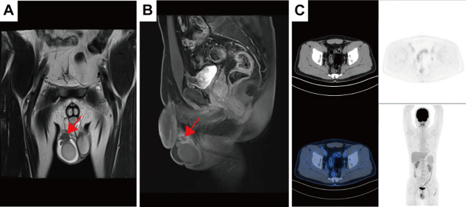
A 34-year-old male present with unilateral nodule of the spermatic cord with enlarged right scrotum and vague pain 1 month ago. Subsequently, enhanced computed tomography (CT) showed a 1.6 cm mass in the right testicular and spermatic cord regions (Fig. 1A and B). Consequently, the patient underwent tumor resection and right inguinal orchiectomy at a local hospital. Following the surgery, the right inguinal canal exhibited typical surgical changes, with no signs of new tumor lesions were detected on whole-body PET-CT scans (Fig. 1C).
Fig. 1.
Preoperative and postoperative imaging results. (A, B) Computed tomography (CT) images of the abdomen/pelvis displaying a 1.6 cm mass in the right testicular and spermatic cord regions (indicated by red arrows). (C) Whole-body PET-CT scan reveals no evidence of new tumor lesions
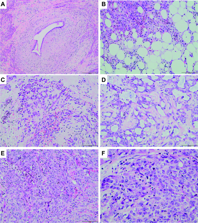
Gross pathological examination revealed a solid tumor, measuring 1.6 cm in greatest diameter. Given the tumor’s proximity to the epididymis, comprehensive sampling and detail histopathological examination were performed on the areas adjacent to the epididymis and the rete testis. No malignant cells were detected in these regions (Supplementary Fig. 1A, 1B). Histological examination confirmed the tumor’s localization within the spermatic cord, as evidenced by the surrounding tumor cells encircling residual vas deferens (Fig. 2A). Invasion into the adjacent fibroadipose tissue was observed (Fig. 2B). The tumor displayed a variety of growth patterns, including cord, cribriform, poorly differentiated solid and nest (Fig. 2C, D and E). Cytologically, epithelioid cells exhibited pale red cytoplasm and round or oval nuclei (Fig. 2F). Interstitial changes included focal necrosis and inflammatory cell infiltration.
Fig. 2.
Predominant histological patterns of adenocarcinoma involving the spermatic cords. (A) Residual vas deferens was surrounded by tumor cells. (B) Invasion of adipose tissue by malignant cells. (C) Epithelioid malignant cells forming interwoven cords. (D) Malignant cells forming tubules and cribriform structures with mucinous content. (E) Areas with poorly differentiated cells in solid or nest patterns. (F) High magnification view of malignant cells
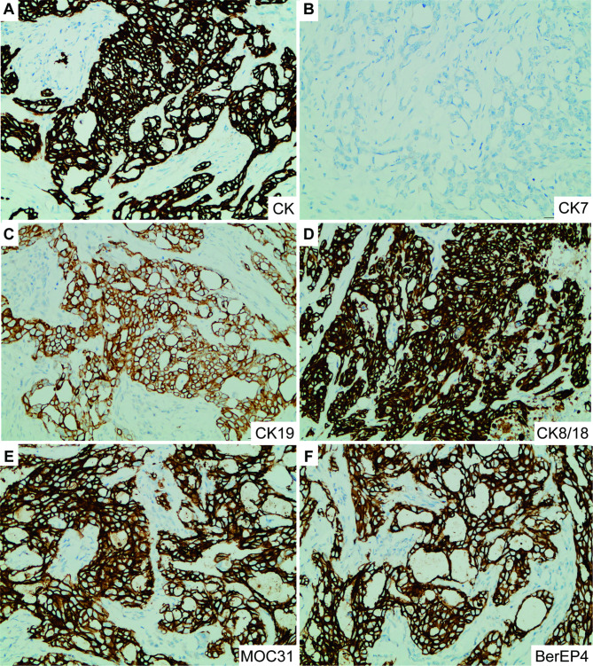
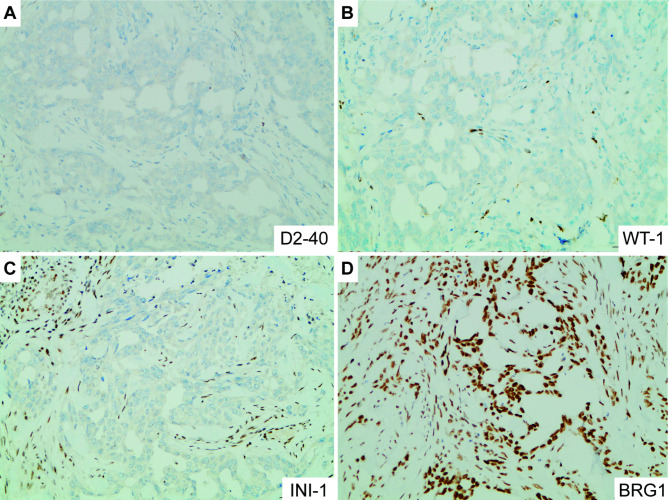
Immunohistochemical staining revealed positive expression for epithelial markers CK (AE1/AE3), CK19, CK8/18, MOC31 and Ber-EP4, while CK7 was negative (Fig. 3). Mesothelioma markers, D2-40, WT1, and Mesothelial Cells (MC) were all negative (Fig. 4A and B). PAX-8 showed positive to the adjacent vas deferens epithelial, but negative to malignant cells (Supplementary Fig. 1C). Additionally, CEA, PSA, NKX3.1, TTF-1, Napsin A, SALL4, Inhibin a, Chromogranin A (CgA), Melan-A, and ALK (D5F3) were all negative. Interestingly, immunohistochemistry revealed complete loss of INI-1 expression and consistent BRG1 expression in all tumor cells (Fig. 4C and D).
Fig. 3.
The expression of epithelial markers. (A) Positive staining for CK in tumor cells. (B) Negative staining for CK7 in tumor cells. (C, D) Positive staining for CK19 and CK8/18 was observed in tumor cells. (E, F) Positive staining for MOC31 and BerEP4 in tumor cells
Fig. 4.
The expression of differential diagnostic and particular molecular markers. (A, B) Negative staining for D2-40 and WT1 in tumor cells. (C) Loss of staining for INI-1 in tumor cells, with endothelial cells and lymphocytes serving as internal positive controls. (D) Consistent expression of BRG1 in tumor cells
For genetic analysis, mutation profiling of DNA obtained from formalin-fixed paraffin-embedded (FFPE) tumor tissue sections was performed using next-generation sequencing (NGS). The NGS tests, which utilized the entire exonic regions of 310 genes and the hotspot mutation regions (exonic, intronic, or promoter regions) of 210 genes associated with PCa and were conducted at a centralized, CLIA-certified, and CAP-accredited clinical testing center (Nanjing Geneseeq Technology Inc., Nanjing, China). The sequencing platform used for the study was Illumina. The final report demonstrated that he had a nonsense mutation (c.842G > A) of the SMARCB1 gene, resulting in an amino acid change at p.W281, with a mutation frequency of 20.36%. Additionally, a missense mutation (c.154G > A) in KLHL6 gene was detected, leading to substitution of threonine for alanine at position 52 (p.A52T), with a mutation frequency of 7.00%. The tumor burden (TMB) was determined to be 1.99 mutations/Mb (TMB-L). Microsatellite stability is indicated by microsatellite stable (MSS), with no embryonic lineage mutations identified.
Currently, the patient remains stable, no evidence of recurrence or distant metastasis observed ten months after radical resection and has not received any postoperative adjuvant therapy.
Discussion
Malignant neoplasms of the spermatic cord are rare, with 352 cases reported in PubMed from 1972 to 2024, including 87 reviews and 232 case reports (Table 1). Sarcomas represent the predominant histological subtype, comprising 79% (278/352) of cases, while epithelial tumors occur less frequently, accounting for 16% (59/352) (Fig. 5A). Among sarcomas, liposarcomas constitute the majority at 52% (145/278), followed by leiomyosarcoma at 18% (50/278), with malignant fibrous histiocytomas ranking third at 12% (34/278) (Fig. 5B). In terms of epithelial neoplasm, metastatic carcinoma is the most prevalent type, primarily originating from the gastrointestinal system, representing 54% (32/59) of cases, followed by the kidney at 21% (12/59) and the pancreas at 12% (7/59), respectively (Fig. 5C). Notably, primary malignant epithelial neoplasm is rare, with only one reported case of a 52-year-old man presenting with a carcinoid tumor [7].
Table 1.
Cases of malignant neoplasms of the spermatic cord in PubMed (1975–2024)
| Histological types | N | Median age (range) | Median tumor size (cm: rang) |
|---|---|---|---|
| Sarcomas (total) | 278 | 62 (4–92) | 6 (0.5–50) |
| Liposarcoma | 145 | 61 (5–92) | 8.5 (1.5–50) |
| Leiomyosarcoma | 50 | 67 (22–83) | 4 (0.5–15) |
| Malignant fibrous histiocytoma | 34 | 69 (32–86) | 5.5 (3-7.5) |
| Rhabdo-myosarcoma | 21 | 17 (5–79) | 4.4 (3–15) |
| Others | 19 | 70 (4–77) | 5 (4–10) |
| Malignant mesenchymoma | 5 | 79 (45–84) | 3.5 (2.5-4) |
| Osteosarcoma | 4 | 55 (50–59) | 5 (4–7) |
| Epithelial neoplasm (total) | 59 | 63 (36–79) | 4 (1.6-8) |
| Metastasis Cancers | 58 | 63 (36–79) | 3.7 (1.6-8) |
| Gastrointestinal Tract | 32 | 63 (36–79) | 3 (1.6-8) |
| Kidney | 12 | 56 (51–60) | 6 (4–8) |
| Pancreas | 7 | 70 (50–71) | 4.9 (3–6) |
| Prostate | 5 | 71 (66–70) | 5.5 (1–9) |
| Others | 2 | 65 (63–68) | 4.5 (2–7) |
| Carcinoid tumor | 1 | 52 | 4 |
| Others | 15 | 63.5 (3–77) | 5.5 (4–10) |
Fig. 5.
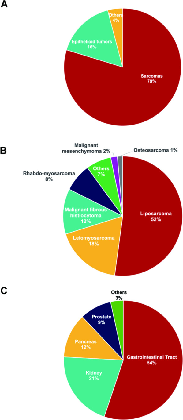
Histologic distribution of malignant spermatic cord tumors reported in PubMed (1975–2024). (A) Distribution of malignant tumors in spermatic cord. (B) Histological classification of spermatic cord sarcoma. (C) Distribution of epithelial neoplasm in spermatic cord
To our knowledge, tumors of the spermatic cord initially presented with painful scrotal and inguinal masses, common symptoms of male genital metastasis [3]. Some patients may exhibit hydrocele and scrotal swelling. While preoperative ultrasonography and CT imaging provide significant diagnostic assistance, the definitive diagnosis necessitates histopathological analysis. The pathological morphology of this ACSC resembles that of moderately to poorly differentiated adenocarcinoma in other organs. Primary adenocarcinoma of the spermatic cord is exceptionally rare and warrants differentiation from malignant mesothelioma, carcinoma with Müllerian differentiation, and metastatic adenocarcinoma. In this case, the absence of D2-40 and WT1 expression, along with positive MOC31 and Ber-EP4 expression, does not support the diagnosis of malignant mesothelioma. The negative expression of PAX-8 in malignant cells does not support a tumor of vas deferens or Müllerian differentiation. Furthermore, negative expression for WT1, NKX3.1, PSA, CEA, TTF-1, and NapsinA helps exclude certain metastatic adenocarcinomas. Additionally, extensive metastatic work-up, including abdomen, pelvis, and thoracic CT scans, as well as whole-body PET-CT imaging, revealed no evidence of new tumor lesions.
Interestingly, the tumor cells exhibited complete loss of INI-1 expression. Molecular analysis further confirmed this as a SMARCB1 (INI-1) deficient adenocarcinoma. SMARCB1 is recognized as a tumor suppression gene [11]. Loss of INI-1 expression as a result of SMARCB1 deletions/mutations has emerged as a defining diagnostic feature in a variety of neoplasms in children and adults, in particular malignant atypical teratoid/rhabdoid tumors of childhood, epithelioid sarcoma, and several epithelial tumor entities in adults and the elderly [12–14]. SMARCB1-deficient tumors often present with a poor prognosis, characterized by widespread metastasis at the time of diagnosis [11]. Considering the rarity of primary SMARCB1-deficient adenocarcinoma of the spermatic cord, there is currently no consensus on adjuvant treatment for this entity. It’s crucial to note the potential for a poor prognosis in such cases, even in the absence of present disease recurrence and progression.
INI-1 (SMARCB1) deficiency is observed in approximately 90% of Epithelioid Sarcoma (ES) [15]. EZH2 plays a role in DNA methylation and transcriptional repression, maintaining the epigenetic silencing of genes [16]. When INI1 loses its regulatory function, EZH2 activity becomes deregulated, allowing EZH2 to play a driving, oncogenic role [17]. With the application of the EZH2 inhibitor Tazemetostat in ES, it has a possibility of promising drug for SMARCB1-deficient tumors [18]. Furthermore, recent data indicate that SMARCB1 loss may augment anti-tumor immunogenicity in specific subtypes of sarcomas and cancers [19]. Hence, epigenetic modulators and immune checkpoint inhibitors could emerge as promising therapeutic modalities for SMARCB1-deficient tumors.
Conclusion
In this study, we present the first case of primary adenocarcinoma of the spermatic cord with SMARCB1 (INI-1) deficiency. Our report encompasses a comprehensive description of its pathological morphology, immunohistology, and molecular characteristics. This case contributes to the expanding understanding of rare neoplasms and underscores the importance of further research into therapeutic strategies targeting SMARCB1-deficient tumors.
Electronic supplementary material
Below is the link to the electronic supplementary material.
Supplementary Material 2: Figure 1. (A) No malignant cells found in the epididymis. (B) No malignant cells found in the rete testis. (C) Normal vas deferens epithelium demonstrated positive expression of PAX-8, while the malignant cells were negative.
Acknowledgements
Not applicable.
Abbreviations
- ACSC
Adenocarcinoma of the Spermatic Cord
- TMB
Tumor Burden
- MSS
Microsatellite Stable
- ES
Epithelioid Sarcoma
Author contributions
QS: writing—review and editing, writing—original draft. YJZ: conceptualization, writing—review and editing. YZY: writing—review and editing. YC: writing—review and editing. XA: writing—review and editing.
Funding
National Natural Science Foundation of China (Grant number 82002668).
Guangdong Basic and Applied Basic Research Foundation (Grant number 2021A1515010579).
Data availability
No datasets were generated or analysed during the current study.
Declarations
Ethics approval and consent to participate
No institutional review board is needed as there is no direct patient intervention.
Consent for publication
Written informed consent was obtained from the patient for publication of this case report and any accompanying images. A copy of the written consent is available for review by the Editor-in-Chief of this journal.
Competing interests
The authors declare no competing interests.
Footnotes
Publisher’s note
Springer Nature remains neutral with regard to jurisdictional claims in published maps and institutional affiliations.
References
- 1.Sogani PC, Grabstald H, Whitmore WF. Jr. Spermatic cord sarcoma in adults. J Urol. 1978;120:301–5. 10.1016/s0022-5347(17)57146-3. [DOI] [PubMed] [Google Scholar]
- 2.Coleman J, Brennan MF, Alektiar K, Russo P. Adult spermatic cord sarcomas: management and results. Ann Surg Oncol. 2003;10:669–75. 10.1245/aso.2003.11.014. [DOI] [PubMed] [Google Scholar]
- 3.Guttilla A, Crestani A, Zattoni F, Secco S, Iafrate M, Vianello F, Valotto C, Prayer-Galetti T, Zattoni F. Spermatic cord sarcoma: our experience and review of the literature. Urol Int. 2013;90:101–5. 10.1159/000343277. [DOI] [PubMed] [Google Scholar]
- 4.Rodriguez D, Barrisford GW, Sanchez A, Preston MA, Kreydin EI, Olumi AF. Primary spermatic cord tumors: disease characteristics, prognostic factors, and treatment outcomes. Urol Oncol. 2014;32:e5219–25. 10.1016/j.urolonc.2013.08.009. [DOI] [PubMed] [Google Scholar]
- 5.Di Franco CA, Rovereto B, Porru D, Zoccarato V, Regina C, Cebrelli T, Fiorello N, Viglio A, Galvagno L, Marchetti C, et al. Metastasis of the epididymis and spermatic cord from pancreatic adenocarcinoma: a rare entity. Description of a case and revision of literature. Arch Ital Urol Androl. 2018;90:72–3. 10.4081/aiua.2018.1.72. [DOI] [PubMed] [Google Scholar]
- 6.Rodriguez D, Olumi AF. Management of spermatic cord tumors: a rare urologic malignancy. Ther Adv Urol. 2012;4:325–34. 10.1177/1756287212447839. [DOI] [PMC free article] [PubMed] [Google Scholar]
- 7.Berdjis CC, Mostofi FK. Carcinoid tumors of the testis. J Urol. 1977;118:777–82. 10.1016/s0022-5347(17)58191-4. [DOI] [PubMed] [Google Scholar]
- 8.Kim JH, Kim DS, Cho HD. MS., L. Late-onset metastatic adenocarcinoma of the spermatic cord from primary gastric cancer. World J Surg Oncol. 12, 128, 10.1186/1477-7819-12-128 [DOI] [PMC free article] [PubMed]
- 9.Fu J, Luo J, Ye H, Chen Y, Xie L. Testicular and Spermatic Cord Metastases from gastric adenocarcinoma: an unusual case. Cancer Manag Res. 2021;13:1897–900. 10.2147/CMAR.S286909. [DOI] [PMC free article] [PubMed] [Google Scholar]
- 10.Dagur G, Gandhi J, Kapadia K, Inam R, Smith NL, Joshi G, Khan SA. Neoplastic diseases of the spermatic cord: an overview of pathological features, evaluation, and management. Transl Androl Urol. 2017;6:101–10. 10.21037/tau.2017.01.04. [DOI] [PMC free article] [PubMed] [Google Scholar]
- 11.Hollmann TJ, Hornick JL. INI1-deficient tumors: diagnostic features and molecular genetics. Am J Surg Pathol. 2011;35:e47–63. 10.1097/PAS.0b013e31822b325b. [DOI] [PubMed] [Google Scholar]
- 12.Woehrer A, Slavc I, Waldhoer T, Heinzl H, Zielonke N, Czech T, Benesch M, Hainfellner JA, Haberler C. Austrian brain tumor, R. Incidence of atypical teratoid/rhabdoid tumors in children: a population-based study by the Austrian brain Tumor Registry, 1996–2006. Cancer. 2010;116:5725–32. 10.1002/cncr.25540. [DOI] [PubMed] [Google Scholar]
- 13.Thway K, Jones RL, Noujaim J, Fisher C. Epithelioid sarcoma: diagnostic features and Genetics. Adv Anat Pathol. 2016;23:41–9. 10.1097/PAP.0000000000000102. [DOI] [PubMed] [Google Scholar]
- 14.Agaimy A, Hartmann A, Antonescu CR, Chiosea SI, El-Mofty SK, Geddert H, Iro H, Lewis JS Jr., Markl B, Mills SE, et al. SMARCB1 (INI-1)-deficient Sinonasal Carcinoma: a Series of 39 cases expanding the morphologic and clinicopathologic spectrum of a recently described Entity. Am J Surg Pathol. 2017;41:458–71. 10.1097/PAS.0000000000000797. [DOI] [PMC free article] [PubMed] [Google Scholar]
- 15.Agaimy A. The expanding family of SMARCB1(INI1)-deficient neoplasia: implications of phenotypic, biological, and molecular heterogeneity. Adv Anat Pathol. 2014;21:394–410. 10.1097/PAP.0000000000000038. [DOI] [PubMed] [Google Scholar]
- 16.Chase A, Cross NC. Aberrations of EZH2 in cancer. Clin Cancer Res. 2011;17:2613–8. 10.1158/1078-0432.Ccr-10-2156. [DOI] [PubMed] [Google Scholar]
- 17.Italiano A. Targeting epigenetics in sarcomas through EZH2 inhibition. J Hematol Oncol. 2020;13. 10.1186/s13045-020-00868-4. [DOI] [PMC free article] [PubMed]
- 18.Rothbart SB, Baylin SB. Epigenetic therapy for Epithelioid Sarcoma. Cell. 2020;181. 10.1016/j.cell.2020.03.042. [DOI] [PubMed]
- 19.Ngo C, Postel-Vinay S. Immunotherapy for SMARCB1-Deficient sarcomas: current evidence and future developments. Biomedicines. 2022;10. 10.3390/biomedicines10030650. [DOI] [PMC free article] [PubMed]
Associated Data
This section collects any data citations, data availability statements, or supplementary materials included in this article.
Supplementary Materials
Supplementary Material 2: Figure 1. (A) No malignant cells found in the epididymis. (B) No malignant cells found in the rete testis. (C) Normal vas deferens epithelium demonstrated positive expression of PAX-8, while the malignant cells were negative.
Data Availability Statement
No datasets were generated or analysed during the current study.


