Abstract
Background & objectives
Cardiovascular diseases (CVDs) are a leading cause of mortality worldwide. The aim of this investigation was to study the role of biological markers in predicting the risk of carotid and coronary artery atherosclerosis.
Methods
A total of 161 males in the age group of 30-65 yr were included in this study. All participants underwent biochemical analyses [cholesterol, low density lipoprotein cholesterol (LDL-C), triglycerides, glucose, (interleukin) IL-8, IL-10, (proprotein convertase inhibitors subtilisin/kexin type 9) PCSK9, sortilin, creatinine]; ECG; echocardiography; coronary angiography; ultrasound doppler of brachiocephalic arteries. Based on PCSK9 levels, participants were divided into four groups: group 1, n=41 individuals with PCSK9 level of 100-250 ng/ml; group 2, n=37 individuals with PCSK9 level of 251-400 ng/ml; group 3, n=51 individuals with PCSK9 level of 401–600 ng/ml and group 4, n=32 individuals with PCSK9 level of 601-900 ng/ml.
Results
Sortilin level was the highest in group 2. Group 3 individuals had the highest level of IL-8. Correlation analysis of the entire data set revealed the relationship of relative left ventricular thickness index with age, cardiovascular risk, body mass index, intima-media thickness and left ventricular mass index; sortilin had a negative relationship of weak strength with age and smoking, a direct relationship between the risk of cardiovascular complications and with IL-10.
Interpretation & conclusions
Sortilin is the innovative marker of CVDs. In the present investigation, we demonstrated the clear increase in the inflammatory markers (IL-8) in individuals with subclinical atherosclerosis. This fact can be explained by the oxygen stress activation. In individuals with coronary artery stenosis (50% and more), the increase in IL-10 levels demonstrates, to our opinion, the activation of antioxidant protection activation.
Keywords: Arterial hypertension, atherosclerosis, cardiovascular risk, PCSK9, sortilin
As per the World Health Organization (WHO), seven out of the 10 leading causes of death in the world are due to non-communicable diseases1. During 2000-2021, four nosologies were in the list of main causes of mortality including cardiovascular diseases (CVDs), cancer, diabetes mellitus and chronic respiratory diseases2. As many years before, CVDs still have the leading position in this context3.CVDs have multiple risk factors (RFs), which have a negative influence on each other3-5. One of the most important modifiable RF for CVDs is arterial hypertension (AH), along with others, such as smoking, obesity and hypercholesterolemia. In numerous investigations, it was estimated that AH is one of the most unfavourable RF4. The basis of the CVDs pathogenesis is an atherosclerotic process – a violation of lipid metabolism. The new approach is aimed at maximising CVR reduction by lowering low density lipoprotein-cholesterol (LDL-C) with proprotein convertase inhibitors subtilisin/kexin type 9 (PCSK9). The regulatory effect of PCSK9 on lipid metabolism was first discovered in 20036. The role of PCSK9 was demonstrated in the atherosclerotic process7. Together with PCSK9, sortilin (a protein regulating intracellular transport through their Vps10p domain) also passes through the Golgi apparatus8. In healthy people, the level of circulating PCSK9 directly correlates with the level of sortilin in plasma9,10. Also, the correlation between sortilin level in serum and AH was revealed11.
Atherogenesis involves the immune system cells, biologically active substances, with a damaging effect on the vascular wall and implementing inflammatory reactions – oxidative stress and phagocytosis with the production of cytokines such as interleukins (IL) 6, 8, 10 and others. Particular attention is paid to the innate and adaptive immune response, which is an attribute of all stages of atherosclerosis development, from initiation to the forming of unstable atherosclerotic plaque12,13.
The present investigation was aimed at studying the role of biological markers in predicting the risk of carotid and coronary artery atherosclerosis.
Material & Methods
The study was carried out by the Department of Cardiology, Russian Railway Medicine, Samara State Medical University from September 2020 – September 2021.
Inclusion and exclusion criteria
The inclusion criteria for the study were as follows: male gender, age 30-65 yr, AH 1-2 grade who provided signed agreement of participation in the study. The exclusion criteria were as follows: patients under 30 and over 65 yr, secondary AH, myocardial infarction or stroke less than six months before the investigation, chronic heart failure NYHA II and more, type 1 diabetes mellitus, toxic diffuse goitre, familial hypercholesterolemia, chronic abdominal ischemia, chronic inflammatory diseases of any localisation, chronic hepatitis, liver cirrhosis and refusal to participate in the study.
A total of 161 males aged 30-65 yr were included in the study. All participants were men because all of them were railway workers – train drivers and assistant drivers. All participants underwent blood biochemical analysis with the determination of total cholesterol, LDL-C, triglycerides, glucose (CLIMA MC-15, IFA-BEST, Russia), interleukin (IL)-8, IL-10, PCSK9 (Quantikine ELISA, USA), sortilin (Aviscera Bioscience, Inc. Santa Clara, CA), creatinine with the calculation of GFR according to the CKD-EPI calculator. The instrumental studies included electrocardiography (ECG) on EK1Т-1/3-07 Aksion, Russia, transthoracic echocardiography (EchoCG) and ultrasound Doppler examination of brachiocephalic vessels with the determination of the thickness of the carotid intima-media complex (CIMT) on Philips EN Visor, USA. In the case of atherosclerotic plaques, the percentage in diameter using the ECST (European Carotid Surgery Trial), NASCET (North American Symptomatic Carotid Endarterectomy Trial) and St. Mary’s ratio criteria. Coronary angiography (CAG) was performed on all the study participants (General Electric Innova 3100, USA).
All participants were divided into four groups in accordance to the level of PCSK9: Group 1 (n=41) –individuals with a level of PCSK9 between 100-250 ng/ml; group 2 (n=37) – PCSK9 between 251-400 ng/ml; group 3 (n=51) – PCSK9 401-600 between ng/ml and group 4 (n=32) – PCSK9 between 601-900 ng/ml. The reason for this dividing was in accordance to the distribution of individuals by quartiles in statistical analysis.
The participants across all the groups were on the standard therapy for AH correction: angiotensin-converting enzyme inhibitors (perindopril), angiotensin II receptor blockers (losartan, valsartan), β-blockers (nebivolol) and statins (simvastatin, atorvastatin).
Statistical analysis
Descriptive statistics was used to evaluate data. The continuous plots were drawn as the mean ± standard deviation (SD), as well as the median and the first and third quartiles. All variables were analysed for normal distribution by the Kolmogorov-Smirnov method. Normally distributed features were compared by one-way ANOVA followed by pairwise comparison with Tukey’s correction and skewed variables were compared by the Kruskal-Wallis test with pairwise comparison by the Dwass-Steele-Critchlow-Fligner method.
Results
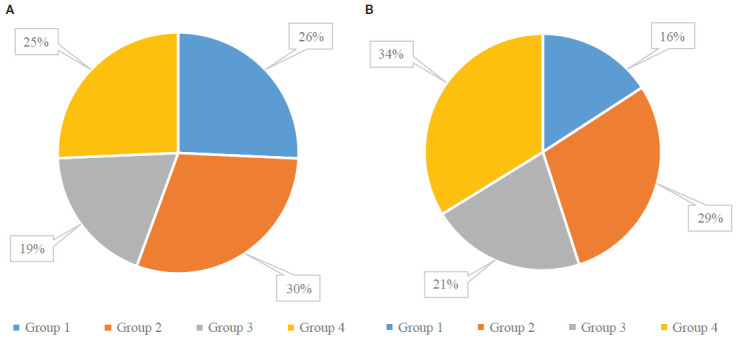
The main clinical characteristics of study groups are seen in Table I. The study groups were homogeneous in gender and comparable in age, glucose level, GFR and lipid spectrum. The smokers were within all studied groups: 51.22 per cent, 59.46 per cent, 37.25 per cent and 53.13 per cent, respectively (Fig. 1). Participants in group 3 had the lowest percentage of smokers; the highest was in group 2 (P=0.03).
Table I.
Clinical characteristics of study participants
| Parameter |
Group 1 (n=41) mean ± SD, median (Q1- Q3) |
Group 2 (n=37) mean ± SD, median (Q1- Q3) |
Group 3 (n=51) mean ± SD, median (Q1-Q3) |
Group 4 (n=32) mean ± SD, median (Q1- Q3) |
P |
|---|---|---|---|---|---|
| Age (yr) |
47.22±12.60 50 (37-56) |
48.62±9.39 50 (46- 54) |
49.33±8.88 52 (45-55) |
51.09±7.29 53 (47.5-56.25) |
χ2=1.408, df=3, P=0.704 |
| Body mass index(kg/m2) |
26.2±3.01 26 (24-28) |
28.292±3.41 29 (26-31) |
27.725±4.21 28 (25-31) |
27.925±4.66 27 (25-29.25) |
χ2=7.482, df=3, P=0.058 |
| Left ventricular mass index, (g/m2) |
118.19±39.82 110 (90-142) |
123.12±39.37 117 (100.5,-136) |
134.98±38.13 136 (106-152.5) |
134.48±32.03 140 (109.75- 152.5) |
χ2=9.261, df=3, P=0.026 |
| Systolic blood pressure (BP), (mm Hg) |
120.48±14.41 119 (110-130) |
125±15.10 122.5 (114-139) |
110.2±8.70 110 (105-113) |
127±1.41 127 (126.5- 127.5) |
χ2=5.358, df=3, P=0.147 |
| Diastolic BP, (mm Hg) |
74.97±7.13 72 (65, 82) |
75.97±7.66 74 (66, 82) |
80.9±7.31 79 (70, 88) |
83.62±7.44 81 (72, 90) |
χ2=4.312, df=3, P=0.218 |
| Heart rate (HR), (beats/min) |
70.02±6.74 69 (63-75) |
69.02±8.32 68 (60-76) |
68.72±8.84 67 (59-75) |
73.03±8.73 72 (63-78) |
χ2=4.165, df=3, P=0.678 |
| Cholesterol, (mmol/l) |
4.96±1.01 4.8 (4.4-5.7) |
4.76±1.06 4.8 (4.2-5.4) |
5.17±0.97 5 (4.5-5.65) |
5.22±0.86 5.15 (4.55- 5.83) |
χ2=4.219, df=3, P=0.239 |
| LDL-Cholesterol, (mmol/l) |
3.29±0.91 3.2 (2.7-3.8) |
3.10±1.09 2.9 (2.3-3.7) |
3.45±0.97 3.4 (2.7-4) |
3.58±0.89 3.6 (3.050-4.2) |
χ2=4.703, df=3, P=0.195 |
| Triglycerides, (mmol/l) |
1.48±0.79 1.2 (0.93-1.89) |
1.67±0.88 1.5 (1.17-1.88) |
1.32±0.55 1.19 (0.94-1.65) |
1.38±0.66 1.2 (0.99-1.76) |
χ2=3.754, df=3, P=0.289 |
| Glucose, (mmol/l) |
5.41±0.81 5.3 (5-5.7) |
5.44±0.91 5.3 (5-5.7) |
5.38±0.63 5.4 (5-5.8) |
5.38±0.66 5.4 (5.075, 5.93) |
χ2=0.606, df=3, P=0.895 |
| eGFR, (ml/min/1,73m2) |
95.07±15.90 99 (80-107) |
93.65±15.58 98 (85-105) |
92±14.62 92 (80-104.5) |
92.66±15.50 93.5 (80-105) |
χ2=1.335, df=3, P=0.721 |
| PCSK9, (ng/ml) |
190.85±43.80 2 (160-24) |
320.62±52.13 3 (280-37) |
493.92±55.61 480 (460-54) |
746.88±88.81 760 (660-83) |
χ2=149.233, df=3, P=0.000 |
| CIMT, (mm) |
1.173±0.34 1.3 (0.7-1.5) |
1.308±0.31 1.5 (1.2-1.5) |
1.19±0.23 1.2 (1.05-1.30) |
1.15±0.21 1.15 (1-1.30) |
χ2=12.625, df=3, P=0.006 |
| Left ventricle posterior wall, (mm) |
9.74±3.63 10 (8-12) |
10.75±2.86 11 (9-12) |
11.39±2.07 12 (10-12.5) |
11.94±2.08 12 (10-13) |
χ2=9.722, df=3, P=0.021 |
LDL, low-density lipoprotein; eGFR, estimated glomerular filtration rate; PCSK9, proprotein convertase subtilisin/kexin type 9; CIMT, carotid intima-media complex; SD, standard deviation
Figure 1.

Pie chart showing the (A) smoking status (B) family history of arterial hypertension among the study participants.
Positive family history positive for AH was observed; in 7.3 per cent in group 1, 13 per cent in the group 2, 9.8 per cent and 15.6 per cent in groups 3 and 4, respectively (Fig. 2). There were no significant differences between the groups (P>0.05).
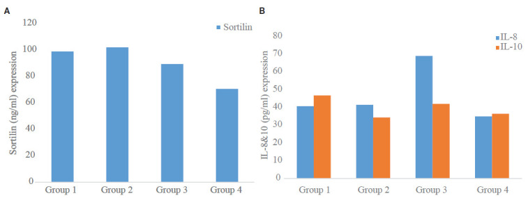
Figure 2.

Expression of (A) sortilin and (B) IL-8,10 levels in group among the study participants.
Among group 4 individuals, systolic BP, diastolic BP and HR were significantly higher in comparison with other groups (P˂0.01). Diastolic BP in the group 4 was significantly higher in comparison with group 1 and 2. Group 3 individuals, had a diastolic BP significantly higher in comparison with group 2 (P˂0.01). Sortilin level was the highest in group 2, however, no statistically significant differences were found. Group 3 individuals had the highest level of IL-8 in comparison with other groups (P=0.01).
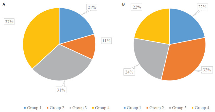
CIMT was the highest in group 2 and was significantly higher in groups 3 and 4 (P=0.04 and P=0.017, respectively). Atherosclerotic plaques of brachiocephalic arteries were found in 70 and 80 per cent of group 1 and 2 participants, respectively, and in 90 per cent of participants in group 3 and 4. The predominance of the brachiocephalic artery stenosis of more than 50 per cent was revealed in 67.57 per cent of the participants in group 2 (Fig. 3).
Figure 3.

Atherosclerotic plaques in carotid arteries among the study participants as shown through Doppler ultrasound of brachiocephalic arteries (A) stenosis <50% (B) stenosis >50%.
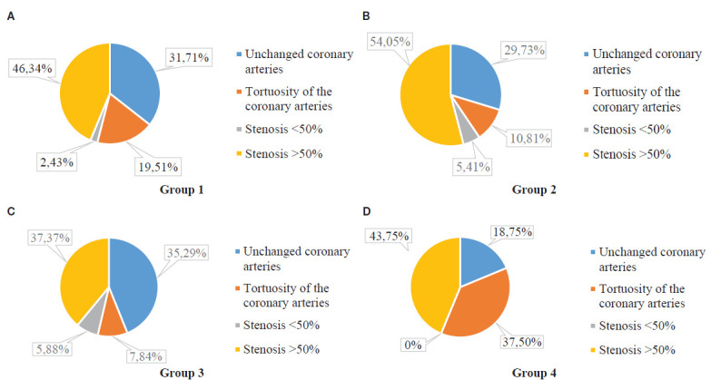
The results of CAG did not demonstrate significant differences between the groups. Coronary artery stenoses were seen in 50 per cent and more in group 1 with 46.4 per cent individuals. In groups 2, 3 and 4 it was 54.05 per cent, 31.37 per cent and 43.75 per cent, respectively (Fig. 4). The highest percentage of stenosis (>50%) at 54.5 per cent was observed in group 2, while the smallest was in group 3 (31.3%). The smallest number of normal coronary arteries was seen in group 2.
Figure 4.

The coronary arteries atherosclerosis and PCSK9 level among study participants as shown through coronary angiography.
Correlation analysis of the entire data set revealed the relationship of relative left ventricular thickness index with age (r=0.43; P=0.005), with CVR (r=0.48; P=0.0005), body mass index (r=0.24; P=0.0024), with the CIMT (r=0.34; P=0.0005), left ventricular mass index (r=0.70; P=0.0004), sortilin had negative relationships of weak strength with age and smoking (r=-0.22; P=0.41; r=-0.26; P=0.14, respectively). Correlation analysis within the groups demonstrated the following tendency: there was a negative relationship between sortilin level and age, a direct relationship between sortilin and the risk of cardiovascular complications (r=-0.37; p=0.24; r=0.352; P=0.24, respectively), sortilin and IL-10 (r=0.43; P=0.04) in group 1.
Intra-group analysis in group 2 revealed an inverse relationship between PCSK9 and left ventricle posterior wall (r=-0.37; P=0.22), sortilin and smoking (r=-0.5; P=0.044). The relationship between PCSK9 and glucose (r=0.30; P=0.02), sortilin with AH family history (r=0.83; P=0.09) and IL-10 (r=0.81; P=0.01), index of relative left ventricular thickness with body mass index (r=0.3; P=0.03), with CVR (r=0.57; P=0.0009), with CIMT (r=0.42; P=0.02). Intra-group analysis of group 3 revealed a strong direct relationship between sortilin and AH family history (r=0.83; P=0.03) and with IL-8 (r=0.82; P=0.01). Participants in group 4 showed an inverse relationship between PCSK9 and HR (r=-0.38; P=0.03).
Discussion
Studying of additional risk factors (RFs) of atherosclerosis is at present a promising branch of cardiological sciences because prevention, diagnostics and early treatment of this pathology is key to decrease the duration and improve the quality of life. The importance of heart arrhythmias in atherosclerosis progression has been reported previously14,15. Over the last few years, there have been several studies dedicated to the clarification of the role of sortilin in some pathologies particularly in AH and subclinical atherosclerosis11,16,17,18, fibrosis19,20, for the protection of neurons in diabetic retina21, and as a potential biomarker and target for glioblastoma22.
A few studies have described sortilin’s immunomodulatory role in atherosclerosis; it is possible that sortilin promotes chronic inflammation and the formation of foam cells in blood vessels, causing atherosclerosis. Suggestively, this chronic inflammation can in turn reduce liver sortilin levels and disruption of the lipoprotein metabolism, further exacerbating atherosclerosis and the progression of CVDs12,18,19. However, given less number of studies on this literature, some controversial results require further studies and clarification.
In this study, we analysed the risk factors for the development and progression of atherosclerotic lesions of the vessels of the carotid and coronary arteries by the levels of PCSK9, sortilin and markers of inflammation. Serum PCSK9 concentration was positively associated with the sortilin levels (r=0.37, P=0.001). In the work of Hu et al23, in 2017 using the multiple regression analysis adjusted for age, sex, LDL-C, smoking and coronary artery disease, they found that the correlation between PCSK9 levels and serum sortilin and PCSK9 levels remained significant in all study participants (P=0.01). In this study, no relationship between sortilin and PCSK9. In the intragroup analysis, at different levels of PCSK9 was observed, but an inverse relationship between age and smoking was found. (in group 1, the minimum and in group 4, the maximum), a negative relationship between the level of sortilin with age and smoking was found, while a direct relationship between sortilin and the risk of cardiovascular complications and family history was observed. Furthermore, Ogawa et al24 2016, in their studies found that the level of sortilin was significantly higher in individuals with CVR, AH and dyslipidaemia. In contrast, Möller et al25 in 2021 showed that none of the risk factors for CHD, such as gender, age and smoking, were associated with plasma sortilin levels.
Previously it has been reported, that the level of PCSK9 is higher in smokers than in non-smokers (P=0.011)7.
In this study, it was found that the lesion of the carotid arteries with stenosis of more than 50 per cent is characteristic precisely for the group of smoking individuals. Coronary artery stenosis (50% and more) was also detected in the same group, which is confirmed by previous studies. Individuals with multiple RFs, such as AH, smoking and obesity, were identified in the two groups, which may explain the greater percentage of carotid and coronary artery lesions in the patients of this particular group.
If during an investigation of the patient we find an increased parameter of CIMT, it is recommended to evaluate the cardiovascular risk for him26. The Gensini scale allows us to determine the value of CIMT>1 mm as a twofold increase in the chance of moderate and severe risk of coronary artery disease26. At the same time, the presence of atherosclerotic lesions in the carotid arteries has a high prognostic value in cardiovascular events. The degree of echogenicity of atherosclerotic lesions in the carotid arteries is an independent predictor of the coronary artery disease severity (P˂0.002)26. Our study demonstrated that in patients in group 2 with the maximum CIMT was revealed the largest number of cases of coronary artery disease (stenosis of more than 50%), in comparison with other groups (Figs. 4 and 5).
In atherosclerosis pathogenetic theories, the risk factors are associated with genetic predisposition, peroxide, lipid infiltration in response to endothelial damage, autoimmune, monoclonal and infectious theories1-3. There is no doubt in the role of the immune-inflammatory mechanism of atherosclerosis12,13,27. Dyslipidaemia and hyperlipidaemia, with the direct participation of LDL-C and VLDL, acquire auto-antigenic properties inducing the formation of an immune response to vascular antigens in case of arterial wall damage, which contributes to the progression of atherosclerosis3.
The main active components of the inflammatory process are immune cells (macrophages, T- and B-cells, etc.), which are attracted to the inflammation focus by pro-inflammatory cytokines. Studies confirm the effect of sortilin on the regulation of cytokine secretion in various immune processes through IL-6, IL-8, IL-10, IL-12, IL-17, tumour necrosis factor alpha (TNF-α) and interferons I, II, III12,13. In experimental mice, the inactivation of sortilin induced a defect in IL-6 secretion (cytokines), at the same time reducing the inflammatory component of atherosclerosis and vascular lesions, regardless of lipid metabolism. Pro-inflammatory cytokines play the main role in plaque progression25. Therefore, sortilin may act as a key regulator of the inflammatory response that enhances atherogenesis.. Our data showed a high level of IL-8 in group 3, where the highest rates of left ventricle hypertrophy, left ventricle mass index and index of relative left ventricular thickness were found.
In this work, we demonstrated a clear increase in inflammatory markers (IL-8) in patients with subclinical atherosclerosis. We can explain with oxygen stress activation. In patients with coronary artery stenosis (50% and more), we revealed an increase of IL-10 that demonstrates, to our opinion, the activation of antioxidant protection. Overall, Sortilin is the innovative marker of CVR in the human population. The role of sortilin in cardiovascular pathology needs furtherinvestigation. Most of the data are contradictory, and they are not enough to make the direct conclusion of its leading role in CVR.
Financial support & sponsorship
None.
Conflicts of Interest
None.
Use of artificial intelligence (AI)-assisted technology for manuscript preparation
The authors confirm that there was no use of AI-assisted technology for assisting in the writing of the manuscript and no images were manipulated using AI.
References
- 1.Timmis A, Vardas P, Townsend N, Torbica A, Katus H, De Smedt D, et al. European Society of Cardiology: cardiovascular disease statistics 2021. Eur Heart J. 2022;43:716–99. doi: 10.1093/eurheartj/ehab892. [DOI] [PubMed] [Google Scholar]
- 2.GBD 2021 Forecasting Collaborators Burden of disease scenarios for 204 countries and territories, 2022-2050: a forecasting analysis for the Global Burden of Disease Study 2021. Lancet. 2024;403:2204–56. doi: 10.1016/S0140-6736(24)00685-8. [DOI] [PMC free article] [PubMed] [Google Scholar]
- 3.Mach F, Baigent C, Catapano AL, Koskinas KC, Casula M, Badimon L, et al. 2019 ESC/EAS guidelines for the management of dyslipidaemias: Lipid modification to reduce cardiovascular risk. Eur Heart J. 2020;41:111–88. doi: 10.1093/eurheartj/ehz455. [DOI] [PubMed] [Google Scholar]
- 4.Mancia G, Kreutz R, Brunström M, Burnier M, Grassi G, Januszewicz A, et al. 2023 ESH Guidelines for the management of arterial hypertension The Task Force for the management of arterial hypertension of the European Society of Hypertension Endorsed by the International Society of Hypertension (ISH) and the European Renal Association (ERA) J Hypertens. 2023;41:1874–2071. doi: 10.1097/HJH.0000000000003480. [DOI] [PubMed] [Google Scholar]
- 5.Germanova O, Shchukin Y, Germanov V, Galati G, Germanov A. Extrasystolic arrhythmia: is it an additional risk factor of atherosclerosis? Minerva Cardiol Angiol. 2022;70:32–9. doi: 10.23736/S2724-5683.20.05490-0. [DOI] [PubMed] [Google Scholar]
- 6.Abifadel M, Varret M, Rabes JP. Mutations in PCSK9 cause autosomal dominant hypercholesterolemia. Nat Genet. 2003;34:154–6. doi: 10.1038/ng1161. [DOI] [PubMed] [Google Scholar]
- 7.Vukolova Y, Gubareva I, Galati G, Germanova O.Proprotein convertase subtilisin kexin type 9 and main artery atherosclerosis in patients with arterial hypertension .Minerva Cardiol Angiol 202371129–34. 10.23736/S2724-5683.22.06008-2 [DOI] [PubMed] [Google Scholar]
- 8.Venkat S, Linstedt AD. Manganese-induced trafficking and turnover of GPP130 is mediated by sortilin. Mol Biol Cell. 2017;28:2569–78. doi: 10.1091/mbc.E17-05-0326. [DOI] [PMC free article] [PubMed] [Google Scholar]
- 9.Siddiq AA, Martin A. Crocetin exerts hypocholesterolemic effect by inducing LDLR and inhibiting PCSK9 and Sortilin in HepG2 cells. Nutr Res. 2022;98:41–9. doi: 10.1016/j.nutres.2021.08.005. [DOI] [PubMed] [Google Scholar]
- 10.Hu D, Yang Y, Peng DQ. Increased sortilin and its independent effect on circulating proprotein convertase subtilisin/kexin type 9 (PCSK9) in statin-naive patients with coronary artery disease. Int J Cardiol. 2017;227:61–5. doi: 10.1016/j.ijcard.2016.11.064. [DOI] [PubMed] [Google Scholar]
- 11.Varzideh F, Jankauskas SS, Kansakar U, Mone P, Gambardella J, Santulli G. Sortilin drives hypertension by modulating sphingolipid/ceramide homeostasis and by triggering oxidative stress. J Clin Invest. 2022;132:e156624. doi: 10.1172/JCI156624. [DOI] [PMC free article] [PubMed] [Google Scholar]
- 12.Rocha VZ, Rached FH, Miname MH. Insights into the role of inflammation in the management of atherosclerosis. J Inflamm Res. 2023;16:2223–39. doi: 10.2147/JIR.S276982. [DOI] [PMC free article] [PubMed] [Google Scholar]
- 13.Liu F, Wang Y, Yu J. Role of inflammation and immune response in atherosclerosis: Mechanisms, modulations and therapeutic targets. Hum Immunol. 2023;84:439–49. doi: 10.1016/j.humimm.2023.06.002. [DOI] [PubMed] [Google Scholar]
- 14.Germanova O, Shchukin Y, Germanov V, Galati G, Germanov A. Extrasystolic arrhythmia: is it an additional risk factor of atherosclerosis? Minerva Cardiol Angiol. 2022;70:32–9. doi: 10.23736/S2724-5683.20.05490-0. [DOI] [PubMed] [Google Scholar]
- 15.Germanova O, Galati G, Germanov A, Stefanidis A. Atrial fibrillation as a new independent risk factor for thromboembolic events: Hemodynamics and vascular consequence of long ventricular pauses. Minerva Cardiol Angiol. 2023;71:175–81. doi: 10.23736/S2724-5683.22.06000-8. [DOI] [PubMed] [Google Scholar]
- 16.Avvisato R, Jankauskas SS, Varzideh F, Kansakar U, Mone P, Santulli G. Sortilin and hypertension. Curr Opin Nephrol Hypertens. 2023;32:134–40. doi: 10.1097/MNH.0000000000000866. [DOI] [PMC free article] [PubMed] [Google Scholar]
- 17.Chu X, Liu R, Li C, Gao T, Dong Y, Jiang Y, et al. The association of plasma sortilin with essential hypertension and subclinical carotid atherosclerosis: A cross-sectional study. Front Cardiovasc Med. 2022;9:966890. doi: 10.3389/fcvm.2022.966890. [DOI] [PMC free article] [PubMed] [Google Scholar]
- 18.Su X, Chen L, Chen X, Dai C, Wang B. Emerging roles of sortilin in affecting the metabolism of glucose and lipid profiles. Bosn J Basic Med Sci. 2022;22:340–52. doi: 10.17305/bjbms.2021.6601. [DOI] [PMC free article] [PubMed] [Google Scholar]
- 19.Ishiyama S, Hasegawa T, Sugeno N, Kobayashi J, Yoshida S, Miki Y, et al. Sortilin acts as an endocytic receptor for α-synuclein fibril. FJ. 2023;37:e23017. doi: 10.1096/fj.202201605RR. [DOI] [PubMed] [Google Scholar]
- 20.Iqbal F, Schlotter F, Becker-Greene D, Lupieri A, Goettsch C, Hutcheson JD, et al. Sortilin enhances fibrosis and calcification in aortic valve disease by inducing interstitial cell heterogeneity. Eur Heart J. 2023;44:885–98. doi: 10.1093/eurheartj/ehac818. [DOI] [PMC free article] [PubMed] [Google Scholar]
- 21.Jakobsen TS, Østergaard JA, Kjolby M, Birch EL, Bek T, Nykjaer A, et al. Sortilin inhibition protects neurons from degeneration in the diabetic retina. Invest Ophthalmol Vis Sci. 2023;64:8. doi: 10.1167/iovs.64.7.8. [DOI] [PMC free article] [PubMed] [Google Scholar]
- 22.Marsland M, Dowdell A, Faulkner S, Gedye C, Lynam J, Griffin CP, et al. The membrane protein sortilin is a potential biomarker and target for glioblastoma. Cancers (Basel) 2023;15:2514. doi: 10.3390/cancers15092514. [DOI] [PMC free article] [PubMed] [Google Scholar]
- 23.Hu D, Yang Y, Peng DQ. Increased sortilin and its independent effect on circulating proprotein convertase subtilisin/kexin type 9 (PCSK9) in statin-naive patients with coronary artery disease. Int J Cardiol. 2017;227:61–5. doi: 10.1016/j.ijcard.2016.11.064. [DOI] [PubMed] [Google Scholar]
- 24.Ogawa K, Ueno T, Iwasaki T, Kujiraoka T, Ishihara M, Kunimoto S, et al. Soluble sortilin is released by activated platelets and its circulating levels are associated with cardiovascular risk factors. Atherosclerosis. 2016;249:110–5. doi: 10.1016/j.atherosclerosis.2016.03.041. [DOI] [PubMed] [Google Scholar]
- 25.Møller PL, Rohde PD, Winther S, Breining P, Nissen L, Nykjaer A, et al. Sortilin as a biomarker for cardiovascular disease revisited. Front Cardiovasc Med. 2021;8:652584. doi: 10.3389/fcvm.2021.652584. [DOI] [PMC free article] [PubMed] [Google Scholar]
- 26.Arnett DK, Blumenthal RS, Albert MA, Buroker AB, Goldberger ZD, Hahn EJ, et al. 2019 ACC/AHA guideline on the primary prevention of cardiovascular disease: A report of the American College of Cardiology/American Heart Association Task Force on clinical practice guidelines. Circulation. 2019;140:e596–e646. doi: 10.1161/CIR.0000000000000678. [DOI] [PMC free article] [PubMed] [Google Scholar]
- 27.Shang D, Liu H, Tu Z. Pro-inflammatory cytokines mediating senescence of vascular endothelial cells in atherosclerosis. Fundam Clin Pharmacol. 2023;37:928–36. doi: 10.1111/fcp.12915. [DOI] [PubMed] [Google Scholar]


