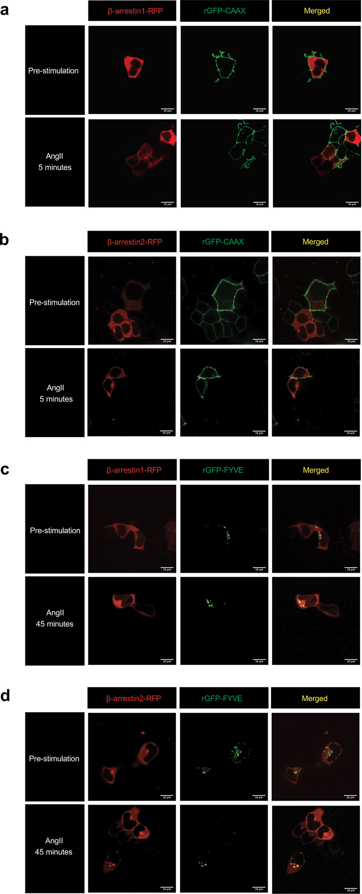
Extended Data Figure 1: Confocal microscopy images of β-arrestin 1 and β-arrestin 2 trafficking to the plasma membrane and early endosomes.

HEK293T cells transfected with FLAG-AT1R, PM marker rGFP-CAAX, and β-arrestin 1-RFP (a) or β-arrestin 2-RFP (b) pre-stimulation and after 5-minute stimulation of 1 μM AngII. HEK293T cells transfected with FLAG-AT1R, early endosomal targeting peptide rGFP-2xFYVE, and β-arrestin 1-RFP (c) or β-arrestin 2-RFP (d) pre-stimulation and after 45-minute stimulation of 1 μM AngII.
