Abstract
Objectives:
To investigate the rate of hospitalized neonates with glucose-6-phosphate dehydrogenase (G6PD) deficiency presented with indirect hyperbilirubinemia at a private tertiary center in Al-Ahsa, Saudi Arabia, over 4 years and to compare the characteristics of G6PD-deficient and normal neonates admitted for indirect hyperbilirubinemia.
Methods:
The retrospective case control study was carried out at Almoosa Specialist Hospital, Al-Ahsa, Saudi Arabia. Data were collected from Yassasi Medical System from 2018-2021 and finalized in 2024. The study included 2 groups: G6PD-normal and G6PD-deficient neonates with indirect hyperbilirubinemia not having recognizable triggers of hemolysis. The analysis focused on serum bilirubin levels, direct bilirubin levels, hematocrit levels, hemoglobin levels, reticulocyte percentage, G6PD levels, duration of phototherapy, and the need for exchange transfusion.
Results:
The study enrolled 3200 neonates with hyperbilirubinemia, of whom 274 met inclusion criteria. A total of 103 (37.6%) neonates were G6PD-deficient, with 77 (74.8%) being male and 26 (25.2%) female. Glucose-6-phosphate dehydrogenase-deficient neonates exhibited significantly higher initial total bilirubin levels and earlier sampling times. There was no significant correlation between G6PD deficiency and hematocrit or hemoglobin levels in hyperbilirubinemic neonates, but 4 neonates required exchange transfusion, demonstrating statistical significance (p=0.009).
Conclusion:
High rate of G6PD deficiency in neonates with indirect hyperbilirubinemia, requiring close monitoring to prevent exchange transfusions, with no significant differences in hematocrit or hemoglobin levels.
Keywords: glucose phosphate dehydrogenase, deficiency, neonatal, hyperbilirubinemia, phototherapy, transfusion
Glucose-6-phosphate dehydrogenase (G6PD) is the primary source of energy from glucose for erythrocytes. 1 This enzyme generates reduced glutathione phosphate and reduced nicotinamide adenine dinucleotide phosphate, crucial for protecting erythrocytes against oxidative damage. Glucose-6-phosphate dehydrogenase deficiency is an enzymopathological disorder induced by mutations in the X-linked gene. Consequently, G6PD deficiency is predominantly reported in males. 2 It ranks among the most prevalent enzymatic disorders across the world, affecting over 400 million people. 3 The G6PD deficiency is highly prevalent in Mediterranean, Asian, and African populations. In the Middle East, its prevalence ranges from 3-29%, while in Asia, it varies between 6.0-15.8%, and in Africa between 3.6-28.0%. 4,5
Neonatal hyperbilirubinemia and kernicterus are frequently associated with G6PD deficiency, often without hematological evidence of hemolysis or identifiable triggers. 6 Research published by the American Academy of Pediatrics reported that G6PD-deficient and G6PD-intermediate infants experienced greater decreases in hematocrit, bilirubin levels, and need for phototherapy than G6PD-normal infants. 7
Additionally, a study involving 100 neonates with moderate to severe indirect hyperbilirubinemia found that 16% were G6PD deficient, a substantial difference from the 6% control group. These neonates had higher serum bilirubin levels, requiring longer phototherapy and hospitalization. However, no significant differences were found in symptoms, reticulocyte counts, or age between G6PD-deficient and non-deficient groups, suggesting that hyperbilirubinemia severity in G6PD-deficient neonates is not solely due to excess hemolysis. 8
A study involving 21,585 participants found that newborns with G6PD deficiency had a higher risk of hyperbilirubinemia with a pooled risk ratio of 3.92 and phototherapy with a pooled risk ratio of 3.01 in contrast to those with normal G6PD levels. 9 A study at Shaikh Zayed Hospital in Lahore, Pakistan, found that 6% of 100 neonates with hyperbilirubinemia were G6PD deficient, with higher bilirubin levels and lower platelet counts. 10 Glucose-6-phosphate dehydrogenase deficiency is prevalent in Al-Hasa, with a high incidence of 36.5% among all regions in Saudi Arabia. 2
The present research study aimed to determine the incidence of hospitalized neonates with G6PD deficiency presenting with indirect hyperbilirubinemia at Almoosa Specialist Hospital, Al-Hasa, Saudi Arabia, over 4 years. Additionally, the study aimed to compare the therapeutic progression of G6PD-deficient and normal neonates admitted for indirect hyperbilirubinemia. The novelty of this research is in evaluating the significance of G6PD-normal and G6PD-deficient levels as quantitative indicators among hyperbilirubinemic males and females at Almoosa Specialist Hospital, Al-Hasa, Saudi Arabia.
Methods
This retrospective case control study was carried out in the Neonatal Intensive Care Unit (NICU) at Almoosa Specialist Hospital in Al-Hasa, Saudi Arabia.
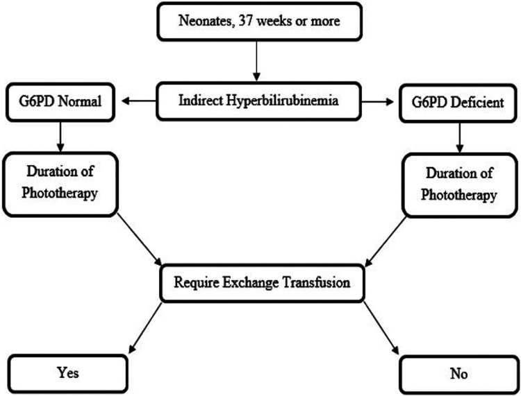
Almoosa Specialist Hospital is a specialized private tertiary hospital in the Eastern Province, equipped with 450 beds, including a Level 3 NICU with 30 beds, operating at an 80% occupancy rate. The hospital delivers approximately 3,500 neonates annually. The study comprised 2 groups with G6PD-deficient and G6PD-normal levels as outlined in Figure 1.
Figure 1.
- Diagram illustrating the study design. G6PD: glucose-6-phosphate dehydrogenase
Data were collected retrospectively from 2018-2021 using the Yassasi Medical System, finalized in 2024.
Dependent variables analyzed included reason for admission, gestational age, birth weight, gender, onset of hyperbilirubinemia, total serum bilirubin (TSB) level, direct bilirubin level, hematocrit level, hemoglobin level, reticulocyte percentage, duration of phototherapy, and requirement for exchange transfusion. Independent variables included G6PD level (deficient or normal).
All neonates with a gestational age of less than 37 weeks and hyperbilirubinemia with additional risk factors other than G6PD deficiency were excluded from the study. These risk factors included ABO incompatibility, Rh incompatibility, polycythemia, sepsis, and any condition known to contribute to hyperbilirubinemia, such as elliptocytosis, spherocytosis, and cephalohematoma. Neonates discharged against medical advice were also excluded to ensure accurate and comprehensive data analysis, particularly concerning the duration of phototherapy.
The study was carried out retrospectively using data collected from electronic databases without direct patient involvement. All collected data were securely stored in electronic systems protected by passwords and accessed only by authorized personnel. Patient identification remained confidential throughout the study. This research received a consent waiver from the institutional review board of Almoosa Specialist Hospital, Al-Hasa, Saudi Arabia, under log number: ARC-22-04-03.
Red blood cell G6PD activity was identified by quantifying the rate of increase in absorbance of nicotinamide adenine dinucleotide phosphate at 340 nm, expressed as units per gram of hemoglobin (U/gHb), following the method described by Beutler et al. 11 Measurements were carried out using a narrow-width (2 nm) spectrophotometer (Beckman model Du 60). A G6PD activity level below 3.36 U/gHb was considered deficient. Total serum bilirubin and conjugated bilirubin levels were determined using a modified diazo method on an automated clinical analyzer (Abbott, Alcyon 300). 12,13
During hospitalization, the type of phototherapy (single, double, triple, or intensive) and the necessity for exchange transfusion were determined based on the TSB levels plotted on the hyperbilirubinemia nomogram used at Almoosa Specialist Hospital in Al-Hasa, Saudi Arabia, adapted from Fanaroff and Martin’s neonatal-perinatal medicine. 14 Total serum bilirubin levels were monitored every 6-8 hours after phototherapy initiation to adjust the treatment intensity as indicated by the nomogram.
Following Kelsey’s methods and considering a 95% confidence interval (CI) and a significance level (α, type 1 error rate) of 5%, a sample size of 268 was determined necessary to achieve 80% power (chance of detecting) for the study, assuming an odds ratio of 2. In this study, 3,200 neonates with hyperbilirubinemia were initially recruited, of which 2,926 did not meet inclusion criteria, primarily owing to incomplete G6PD level data. The final analysis focused on 274 hyperbilirubinemic neonates who fully met the inclusion criteria.
Statistical analysis
The Statistical Package for Social Sciences, version 19.0 (IBM Corp., Armonk, NY, USA) for Windows was used. Data were compared using mean, standard deviation, Student’s t-test, Mann-Whitney-U test, and Chi-square tests. A p-value of <0.05 with a 95% CI was considered significant.
Results
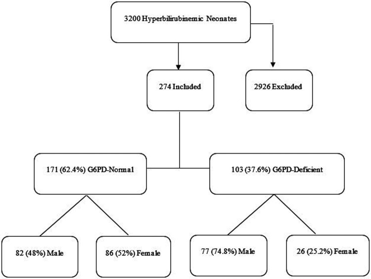
Overall, our study included 274 neonates with indirect hyperbilirubinemia who met the inclusion criteria. Among them, 171 (62.4%) were G6PD-normal, comprising 82 (48%) males and 86 (52%) females. There were a total of 103 (37.6%) G6PD-deficient neonates, including 77 (74.8%) males and 26 (25.2%) females. Figure 2 summarizes the distribution of neonates included in the study.
Figure 2.
- The flowchart illustrating hyperbilirubinemic neonates included in the study categorized by glucose-6-phosphate dehydrogenase status. G6PD: glucose-6-phosphate dehydrogenase
Gender (males and females), gestational age, mode of delivery (normal vaginal delivery [NSVD] or caesarean section [CS]), Appearance, pulse, grimace, activity and respiration (Apgar) scores at 1 and 5 minutes, the onset of hyperbilirubinemia, highest total bilirubin level and its sampling time, direct bilirubin level, initial and lowest hemoglobin level and its sampling time, initial and lowest hematocrit level and its sampling time, percentage of reticulocytes and its sampling time, and duration of phototherapy were not statistically significant when comparing G6PD-normal and G6PD-deficient neonates with indirect hyperbilirubinemia, as presented in Table 1.
Table 1.
- Baseline and laboratory characteristics of hyperbilirubinemic neonates categorized by glucose-6-phosphate dehydrogenase deficiency status.
| Characteristics | G6PD-normal | G6PD-deficient | P-values |
|---|---|---|---|
| Gestational age (weeks) | 38.13±1.3 | 38.53±1.27 | 0.18 |
| Birth weight (kg) | 2.96±0.43 | 3.11±0.37 | 0.004 |
| Weight upon admission (kg) | 2.93±0.42 | 3.07±0.37 | 0.007 |
| Gender, n (%) | |||
| Male | 82 (51.0) | 77 (49.0) | ≤0.0001 |
| Female | 89 (77.4) | 26 (22.6) | |
| Mode of delivery, n (%) | |||
| NSVD | 122 (61.0) | 87 (39.0) | 0.484 |
| CS | 49 (66.2) | 25 (33.8) | |
| Apgar score one minute | 8.58±0.91 | 8.76±0.77 | 0.11 |
| Apgar score 5 minuntes | 9.77±0.7 | 9.88±0.42 | 0.128 |
| Onset of hyperbilirubinemia (hour) | 48±72 | 48±71 | 0.083 |
| Initial total bilirubin level (µmol/L) | 174.3±88.85 | 203.4±121 | 0.023 |
| Initial total bilirubin level sampling time (hour) | 48.15±43.9 | 61.12±52.66 | 0.029 |
| Highest total bilirubin level (µmol/L) | 229.34±88.14 | 253.02±129.8 | 0.74 |
| Highest total bilirubin level sampling time (hour) | 99.2±93.5 | 105.8±65 | 0.52 |
| Direct bilirubin level (µmol/L) | 9.81±10.7 | 9.80±6.3 | 0.995 |
| Initial hemoglobin level (g/dL) | 18.54±11.72 | 18.11±2.09 | 0.71 |
| Initial hemoglobin level sampling time (hour) | 4.87±19.4 | 9.66±33.6 | 0.136 |
| Lowest hemoglobin level (g/dL) | 15.41±3.23 | 14.98±3.63 | 0.451 |
| Lowest hemoglobin level sampling time (hour) | 116.95±140.8 | 126.45±141.2 | 0.7 |
| Initial hematocrit level (%) | 53.6±6.8 | 54.88±6.15 | 0.125 |
| Initial hematocrit level sampling time (hour) | 5.15±19.8 | 9.57±33.8 | 0.175 |
| Lowest hematocrit level (%) | 45.26±10.96 | 46.44±9.35 | 0.501 |
| Lowest hematocrit level sampling time (hour) | 127.54±154.7 | 114.8±117.9 | 0.591 |
| Retics (%) | 4.56±2.31 | 4.19±2.13 | 0.447 |
| Retics sampling time (hour) | 111.65±229.4 | 85.44±72.58 | 0.51 |
| Duration of phototherapy (hour) | 13.39±7.04 | 13.32±10.05 | 0.945 |
| Requiring exchange transfusion, n (%) | 0 (0.0) | 4 (100) | 0.009 |
Values are presented as numbers and percentages (%) or mean ± standard deviation (SD).
G6PD: glucose-6-phosphate dehydrogenase, NSVD: normal spontaneous vaginal delivery, CS: cesarean section
However, birth weight and weight upon admission were significantly higher in G6PD-deficient neonates compared to G6PD-normal hyperbilirubinemic neonates. Initial total bilirubin levels and sampling times were significantly higher in G6PD-deficient hyperbilirubinemic neonates. Out of the 274 neonates included in the study, only 4 required exchange transfusion as they were G6PD deficient, with a mean age of 90 hours, and this need was statistically significant with a p-value of 0.009 (2 [50%] G6PD-deficient males and 2 [50%] G6PD-deficient females).
The intensity of phototherapy, irrespective of gender and G6PD status, did not show statistical significance, as presented in Table 2.
Table 2.
- The intensity of phototherapy in hyperbilirubinemic neonates categorized by glucose-6-phosphate dehydrogenase deficiency status.
| Intensity of phototherapy* | G6PD-normal | G6PD-deficient | Total |
|---|---|---|---|
| Single | 27 (65.5) | 13 (32.5) | 40 (14.6) |
| Double | 62 (64.6) | 34 (35.4) | 96 (35.0) |
| Triple | 56 (62.9) | 33 (37.1) | 89 (32.5) |
| Intensive | 26 (53.1) | 23 (46.9) | 49 (17.9) |
Values are presented as numbers and percentages (%).
Statistically not significant with a p-value of <0.05.
G6PD: glucose-6-phosphate dehydrogenase
When comparing hyperbilirubinemic males and females, there was no significant difference (p=0.793) in the initial total bilirubin level in G6PD-deficient males and G6PD-normal males and female patients (p=0.366). Similarly, there was no statistically significant difference (p=0.29) in the quantitative G6PD in G6PD deficient males compared to that of G6PD deficient females. However, G6PD-normal females had significantly lower G6PD levels compared to G6PD-normal males, as depicted in Table 3.
Table 3.
- Laboratory characteristics and intensity of phototherapy in hyperbilirubinemic neonates with glucose-6-phosphate dehydrogenase deficiency and normal levels, categorized by gender.
| G6PD status | Male | Female | P-values |
|---|---|---|---|
| Initial total bilirubin level (µmol/L), mean±SD | |||
| Deficient | 205.29±124.58 | 198.04±111.95 | 0.793 |
| Normal | 180.7±76.83 | 168.35±98.8 | 0.366 |
| G6PD level (U/gHb), mean±SD | |||
| Deficient | 0.89±1.98 | 1.41±2.66 | 0.29 |
| Normal | 10.57±4.01 | 8.6±4.81 | 0.004 |
| Intensity of phototherapy (G6PD-normal) * | |||
| Single | 16 (19.5) | 11 (12.4) | |
| Double | 27 (35.9) | 35 (39.3) | |
| Triple | 27 (35.9) | 29 (32.6) | |
| Intensive | 12 (14.6) | 14 (15.7) | |
| Intensity of phototherapy (G6PD-deficient) * | |||
| Single | 8 (10.4) | 5 (19.2) | |
| Double | 27 (35.1) | 7 (26.9) | |
| Triple | 23 (29.9) | 10 (38.5) | |
| Intensive | 19 (24.7) | 4 (15.5) | |
Values are presented as numbers and percentages (%) or mean±standard deviation (SD).
Statistically not significant with a p-value of <0.05.
G6PD: glucose-6-phosphate dehydrogenase
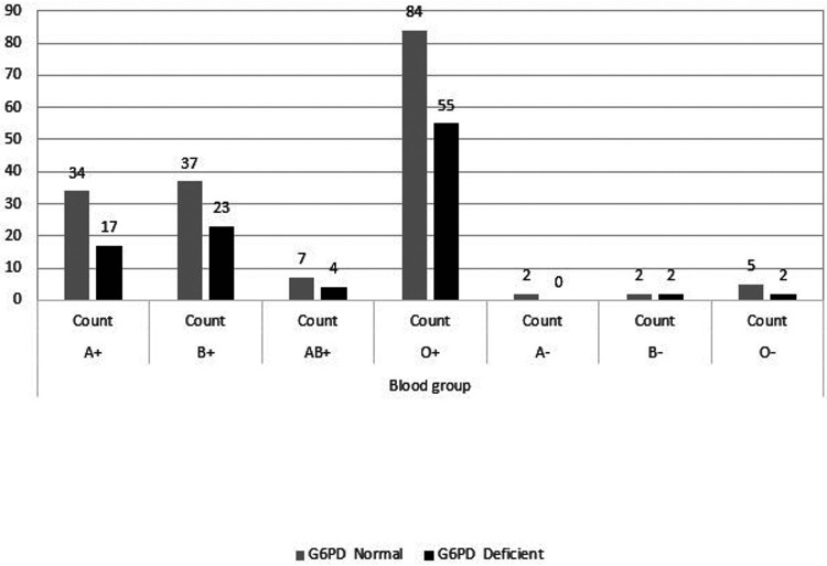
Regarding blood groups and their association with G6PD status, blood group O positive was the most prevalent among both G6PD-normal and G6PD-deficient individuals, as depicted in Figure 3. However, this relationship was not statistically significant (p=0.884).
Figure 3.
- Hyperbilirubinemic neonates with glucose-6-phosphate dehydrogenase deficiency and normal levels, categorized by blood group. G6PD: glucose-6-phosphate dehydrogenase
Discussion
The initial total bilirubin level in G6PD-deficient hyperbilirubinemic neonates was significantly higher than in G6PD-normal hyperbilirubinemic neonates, consistent with previous findings. 10,15,16 Neonatal hyperbilirubinemia is frequently associated with G6PD deficiency and, in many cases, occurs without hematological evidence of hemolysis or an identifiable trigger, as our findings suggest, with an insignificant relation to reticulocyte levels. 17,18 The significance of the sampling time of initial total bilirubin in G6PD-deficient neonates, with a mean age of 61 hours, aligns with the American Academy of Pediatrics’ publication of a study carried out in Nigeria. This study suggests that G6PD-deficient neonates require greater monitoring and early screening in the first week of life, even in the absence of exposure to icterogenic agents. 19
The onset of hyperbilirubinemia, highest bilirubin level, and duration of phototherapy were not significantly related to G6PD status in previous studies or our study. 15,20 Our study also included the intensity of phototherapy categorized as single, double, triple, or intensive which was not statistically significant (p<0.05). This study included males and females and found no significant relationship between G6PD deficiency and hematocrit or hemoglobin levels. This contrasts with a study by Al-Abdi et al 21 in males from the Al-Hasa region, which concluded that G6PD-deficient individuals had higher hematocrit and hemoglobin levels.
Another study concluded that G6PD-deficient individuals had lower hematocrit levels. 22,23 Previous studies have found a high incidence of exchange transfusions among G6PD-deficient neonates, including a study of Sephardic-Jewish infants where only 2 out of 75 G6PD-deficient neonates required such treatment compared to none among 266 with normal G6PD status. 24,25 In our study of 274 neonates, only 4 required exchange transfusion, with a mean age of 90 hours, which was statistically significant (p=0.009) in the G6PD-deficient group: 2 (50%) G6PD-deficient males and 2 (50%) G6PD-deficient females.
While G6PD deficiency is predominantly observed in males, a study carried out in Turkey found that among 46 G6PD-deficient neonates, only 12 (26%) were females, a ratio similar to our findings where 26 (25.2%) out of 103 G6PD-deficient neonates were females, highlighting the importance of including females in G6PD screening. 24 Our study quantitatively assessed G6PD levels, revealing a significant finding: G6PD-normal hyperbilirubinemic females had lower G6PD levels compared to G6PD-normal hyperbilirubinemic males, which was statistically significant ((p=0.004). However, this difference was not significant in the G6PD-deficient group despite lower levels in G6PD-deficient males. Birth weight and weight at admission were significantly higher in G6PD-deficient neonates, suggesting that hyperbilirubinemia in G6PD-normal neonates was often associated with breastfeeding jaundice, especially in early life. Inadequate breastfeeding leads to insufficient caloric intake and dehydration. Insufficient feeding reduces bowel movements, reducing bilirubin elimination through the stool, resulting in an accumulation of unconjugated bilirubin in the bloodstream. Furthermore, these findings indicate that G6PD deficiency alone could account for hyperbilirubinemia without triggering hemolysis. Blood group O positive was the most common blood type associated with G6PD deficiency, consistent with our findings. 21 However, ABO blood type did not show a significant association with hyperbilirubinemia in G6PD-normal and deficient neonates.
Study limitations
It is important to note that our study was retrospective and confined to a single private tertiary centre in Al-Hasa, Saudi Arabia. It did not include neonates with indirect hyperbilirubinemia who might have been readmitted or followed up at other hospitals. Therefore, further research encompassing multiple regions is needed to elucidate the exact relationship and underlying mechanisms of G6PD deficiency in neonates with hyperbilirubinemia.
In conclusion, G6PD deficiency screening should be strongly considered, especially in regions with high prevalence, such as Al-Hasa, Saudi Arabia. As depicted in the present study, G6PD-deficient neonates require more vigilant monitoring for hyperbilirubinemia to enable early detection in the first week of life, ideally by 90 hours of age. This recommendation is supported by specific findings from our study. Firstly, G6PD-deficient neonates exhibited significantly higher initial total bilirubin levels compared to G6PD-normal neonates (203.4±121 µmol/L vs. 174.3±88.85 µmol/L, p=0.023). Additionally, all neonates who required exchange transfusion due to severe hyperbilirubinemia were G6PD-deficient, a finding that was statistically significant (p=0.009). Specifically, out of the 274 neonates included in the study, the 4 who needed exchange transfusion were all G6PD-deficient, with a mean age of 90 hours at the time of the procedure. These results indicate that G6PD-deficient neonates are at a higher risk of developing severe hyperbilirubinemia that may necessitate more intensive treatment, such as exchange transfusion.
Acknowledgment
The authors gratefully acknowledge Falcon Scientific Editing for their English language editing.
Footnotes
References
- 1. Li R, Wang W, Yang Y, Gu C. Exploring the role of glucose–6–phosphate dehydrogenase in cancer (review). Oncol Rep 2020; 44: 2325-2336. [DOI] [PubMed] [Google Scholar]
- 2. Kumar A, Alhiwaishil HM, Alsuliman AA, Alrufayi AH, Hassan HA. Prevalence of glucose-6-phosphate dehydrogenase deficiency in people visiting health care center, KFU, Al-Hasa. Med Forum 2016; 27: 40-43. [Google Scholar]
- 3. Gampio Gueye NS, Peko SM, Nderu D, Koukouikila-Koussounda F, Vouvoungui C, Kobawila SC, et al. An update on glucose-6-phosphate dehydrogenase deficiency in children from Brazzaville, Republic of Congo. Malar J 2019; 18: 57. [DOI] [PMC free article] [PubMed] [Google Scholar]
- 4. Koromina M, Pandi MT, van der Spek PJ, Patrinos GP, Lauschke VM. The ethnogeographic variability of genetic factors underlying G6PD deficiency. Pharmacol Res 2021; 173: 105904. [DOI] [PubMed] [Google Scholar]
- 5. Tantular IS, Kawamoto F. Distribution of G6PD deficiency genotypes among Southeast Asian populations. Trop Med Health 2021; 49: 97. [DOI] [PMC free article] [PubMed] [Google Scholar]
- 6. Lee HY, Ithnin A, Azma RZ, Othman A, Salvador A, Cheah FC. Glucose-6-phosphate dehydrogenase deficiency and neonatal hyperbilirubinemia: insights on pathophysiology, diagnosis, and gene variants in disease heterogeneity. Front Pediatr 2022; 10: 875877. [DOI] [PMC free article] [PubMed] [Google Scholar]
- 7. Badejoko BO, Owa JA, Oseni SB, Badejoko O, Fatusi AO, Adejuyigbe EA. Early neonatal bilirubin, hematocrit, and glucose-6-phosphate dehydrogenase status. Pediatrics 2014; 134: e1082-e1088. [DOI] [PubMed] [Google Scholar]
- 8. Eissa AA, Haji BA, Al-Doski AA. G6PD deficiency prevalence as a cause of neonatal jaundice in a neonatal ward in Dohuk, Iraq. Am J Perinatol 2021; 38: 575-580. [DOI] [PubMed] [Google Scholar]
- 9. Liu H, Liu W, Tang X, Wang T. Association between G6PD deficiency and hyperbilirubinemia in neonates: a meta-analysis. Pediatr Hematol Oncol 2015; 32: 92-98. [DOI] [PubMed] [Google Scholar]
- 10. Akhter N, Habiba U, Mazari N, Fatima S, Asif M, Batool Y. Glucose-6-phosphate dehydrogenase deficiency in neonatal hyperbilirubinemia and its relationship with severity of hyperbilirubinemia. IMJ 2019; 11: 237-241. [Google Scholar]
- 11. Beutler E, Blume KG, Kaplan JC, Löhr GW, Ramot B, Valentine WN. International Committee for Standardization in Haematology: recommended methods for red-cell enzyme analysis. Br J Haematol 1977; 35: 331-340. [DOI] [PubMed] [Google Scholar]
- 12. Klauke R, Kytzia HJ, Weber F, Grote-Koska D, Brand K, Schumann G. Reference measurement procedure for total bilirubin in serum re-evaluated and measurement uncertainty determined. Clin Chim Acta 2018; 481: 115-120. [DOI] [PubMed] [Google Scholar]
- 13. Parviainen MT. A modification of the acid diazo coupling method (Malloy-Evelyn) for the determination of serum total bilirubin. Scand J Clin Lab Invest 1997; 57: 275-279. [DOI] [PubMed] [Google Scholar]
- 14. Martin RJ. Fanaroff and Martin’s neonatal-perinatal medicine. [Updated 2019; 2023 Oct 19]. Available from: https://www.amazon.sa/-/en/Fanaroff-Martins-Neonatal-Perinatal-Medicine-Set/dp/0323567118
- 15. Moiz B, Nasir A, Khan SA, Kherani SA, Qadir M. Neonatal hyperbilirubinemia in infants with G6PD c.563C > T variant. BMC Pediatr 2012; 12: 126. [DOI] [PMC free article] [PubMed] [Google Scholar]
- 16. Isa HM, Mohamed MS, Mohamed AM, Abdulla A, Abdulla F. Neonatal indirect hyperbilirubinemia and glucose-6-phosphate dehydrogenase deficiency. Korean J Pediatr 2017; 60: 106-111. [DOI] [PMC free article] [PubMed] [Google Scholar]
- 17. Javed K. Risk factors associated with alteration of hematological and biochemical parameters in G6PD deficiency. [Updated 2019; 2023 Oct 21]. Available from: https://cust-library.azurewebsites.net
- 18. Yang YK, Lin CF, Lin F, Chen ZK, Liao YW, Huang YC, et al. Etiology analysis and G6PD deficiency for term infants with jaundice in Yangjiang of western Guangdong. Front Pediatr 2023; 11: 1201940. [DOI] [PMC free article] [PubMed] [Google Scholar]
- 19. Wennberg RP, Oguche S, Imam Z, Farouk ZL, Abdulkadir I, Sampson PD, et al. Maternal instruction about jaundice and the incidence of acute bilirubin encephalopathy in Nigeria. J Pediatr 2020; 221: 47-54. [DOI] [PubMed] [Google Scholar]
- 20. Yadav A, Maini B, Gaur BK, Singh RR. Risk factors for serum bilirubin rebound after stopping phototherapy in neonatal hyperbilirubinemia. J Clin Neonatol 2021; 35: 198-202. [Google Scholar]
- 21. Al-Abdi SY, Mousa TA, Al-Aamri MA, Ul-Rahman NG, Abou-Mehrem AI. Hyperbilirubinemia in glucose-6-phosphate dehydrogenase-deficient male newborns in Al-Ahsa, Saudi Arabia. Saudi Med J 2010; 31: 175-179. [PubMed] [Google Scholar]
- 22. Igwilo NH, Salawu L, Adedeji TA. The impact of glucose-6-phosphate dehydrogenase deficiency on the frequency of vasoocclusive crisis in patients with sickle cell anemia. Plasmatology 2021; 15: 26348535211040528. [Google Scholar]
- 23. Francis RO, Jhang JS, Pham HP, Hod EA, Zimring JC, Spitalnik SL. Glucose-6-phosphate dehydrogenase deficiency in transfusion medicine: the unknown risks. Vox Sang 2013; 105: 271-282. [DOI] [PMC free article] [PubMed] [Google Scholar]
- 24. Celik HT, Günbey C, Unal S, Gümrük F, Yurdakök M. Glucose-6-phosphate dehydrogenase deficiency in neonatal hyperbilirubinaemia: Hacettepe experience. J Paediatr Child Health 2013; 49: 399-402. [DOI] [PubMed] [Google Scholar]
- 25. Adissu W, Brito M, Garbin E, Macedo M, Monteiro W, Mukherjee SK, et al. Clinical performance validation of the Standard G6PD test: a multi-country pooled analysis. PLoS Negl Trop Dis 2023; 17: e0011652. [DOI] [PMC free article] [PubMed] [Google Scholar]


