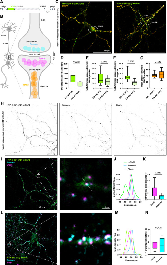
Figure 3: Revealing HTP-mGluR2 localization in hippocampal neurons.
A) Viral DNA expression cassette with HTP-mGluR under hSyn promoter. B) Neural connection via synapses and localization of axonal MAP2, presynaptic Bassoon and postsynaptic Shank proteins (created in biorender.com). C) Confocal imaging of HTP-mGluR2 transduced mouse hippocampal neurons with SiR-d12-SHTL (left) and SiR-d12-HTL (right), co-stained with an antibody against MAP2 for dendrite identification. D-G) Quantification of HTP-mGluR2 labelling in the soma (D), in dendrites (MAP2 positive, E) and in axons (MAP2 negative, F) reveals significantly less signal using SiR-d12-SHTL, while no difference in axonal MAP2 intensity is observed (G). Mean±SD. Student’s t-test. H) Confocal imaging of HTP-mGluR2 transduced mouse hippocampal neurons cells with SiR-d12-SHTL (500 nM), and the pre- and postsynaptic markers Bassoon and Shank, respectively. I) Overlay of images in H. J) Representative line scan of a synapse shows mGluR2 co-localization primarily with the presynaptic marker Bassoon. K) Quantification of mGluR2 localization with respect to Bassoon and Shank. Mean±SD. Student’s t-test. L-M) As for I-K but labelling with SiR-d12-HTL.

