Abstract
Background
Cone-beam computed tomography (CBCT) was quickly adopted in dentistry settings due to its compact size, low cost, and minimal ionizing radiation dose when compared to medical computed tomography. CBCT generates three-dimensional (3D) images that are useful in a variety of oral and maxillofacial circumstances, including diagnosis and assessment of disease severity, treatment planning and delivery, and follow-up. A significant advantage of CBCT lies in its capability to recognize incidental findings. In the realm of dental CBCT imaging, incidental findings often involve the maxillary sinuses, temporomandibular joints, or other craniofacial structures.
Objective
To demonstrate the incidental findings of maxillary sinus disorders in dental patients undergoing CBCT for numerous indications.
Methods
A retrospective cross-sectional study was carried out using data extracted from the Picture Archiving and Communication System (PACS) of Pure Smile Dental Center and Dental College Teaching Hospitals in the Jazan region from September to December 2022. The study sample included a total of 70 patients who underwent orthodontics CBCT, most of whom were female (76%). Statistical analysis was performed using IBM SPSS Statistics for Windows, Version 27 (IBM Corp., Armonk, USA), along with descriptive statistics. Furthermore, chi-square tests and cross-tabulation were done to assess the relation between the study variables. A p-value <0.01 and <0.05 was considered statistically significant.
Results
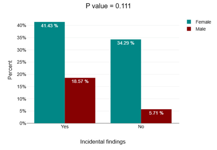
The most common dental CBCT findings were root invasion (18.6%), followed by gingivitis (7.14%), and dental caries (4.29%). Of the 70 patients, 42 (60%) had maxillary sinus findings, including opacification (50%), sinusitis (40%), mucosal thickening (34.3%), and polyps (7.1%). Female patients had more frequency of incidental findings related to males (41.43% and 18.57%, respectively).
Conclusion
A high percentage of dental patients exhibited incidental maxillary sinus findings in CBCT scans, confirming the utility of CBCT as a precise imaging method for detecting paranasal sinus disorders. A holistic documentation of incidental findings is crucial for improving patient outcomes and treatment plans.
Keywords: cbct, incidental findings, maxillary sinuses, opacification, orthodontics
Introduction
Cone-beam computed tomography (CBCT) has emerged as a valuable imaging acquisition in orthodontics, offering detailed three-dimensional (3D) imaging of dental and maxillofacial structures. This technology provides critical information for treatment planning, including tooth position, bone structure, and airway analysis, while minimizing radiation exposure compared to traditional computed tomography (CT) scans [1,2]. The development of specialized CBCT scanners for use in dentistry began in the latter part of the 1990s. Soon after, the usage of CBCT for dental, maxillofacial, and ear-nose-throat applications increased dramatically. CBCT is now widely utilized for a variety of dental applications, including implant design, endodontics, maxillofacial surgery, and orthodontics [2]. Furthermore, CBCT’s expanded field of view (FOV) often reveals incidental findings in adjacent anatomical areas, such as the paranasal sinus, which are beyond the primary scope of orthodontic evaluation [3,4].
The maxillary sinus, due to its proximity to the upper dentition and maxillofacial region, is frequently affected. Incidental findings such as mucosal thickening, sinusitis, and polyps are commonly observed in CBCT scans. Recent literature provides substantial evidence that dental sepsis frequently leads to reactive mucosal thickening in the lower part of the maxillary sinus. It is now recognized that odontogenic factors are a more common cause of maxillary sinusitis, including sinusitis affecting the anterior paranasal sinus, than was previously believed, with incidence rates on the rise. In fact, dental issues account for 75% of opacification in the maxillary sinus and 25-40% of cases involving unilateral anterior sinusitis [5,6]. These findings, while often asymptomatic, may have clinical significance, potentially influencing orthodontic treatment decisions or requiring referral for further medical evaluation. Studies suggest that a substantial proportion of CBCT scans reveal incidental findings in the maxillary sinus, commonly mucosal thickening, highlighting the importance of routine evaluation of these structures during orthodontic assessment [7,8].
Despite the growing use of CBCT in orthodontic practice in Saudi Arabia, particularly in the Jazan region, there is limited data on the prevalence and nature of incidental maxillary sinus findings in this population. Understanding these incidental findings is crucial for promoting interdisciplinary collaboration between orthodontists and other specialists, such as otolaryngologists, for comprehensive patient care. Therefore, the main aim of this study was to determine the prevalence of incidental maxillary sinus findings in CBCT scans among orthodontic patients in the Jazan region.
Materials and methods
Study design and data collection
A cross-sectional retrospective study was conducted at Pure Smile Dental Center and Dental College Teaching Hospitals in the Jazan region between September and December 2022. The study protocol received verbal approval from the ethical committee of the Diagnostic Radiography Technology Department at Jazan University. A total of 70 orthodontic patients, 53 females and 17 males, indicated for CBCT dental scans were included. Patients with known sinus disease and children under 12 years old were excluded. Data were collected using a data collection sheet, including the participant demographic information, dental scan indications, CBCT dental findings, and incidental maxillary sinus disorders.
Equipment and technique
CBCT was performed with a 3D X-ray imaging system (3D Accuitomo 170; J. Morita USA, Inc.) set at 0.4 mm slice thickness, 16x22 FOV, 20 seconds scan time, and 0.49/0.49/0.5 voxel size, 90 kVp, 7 mA, 15.8 seconds exposure time. The CBCT approach performed on patients in sitting position involves a circular or rectangular cone-shaped X-ray beam with a single 360° scan, wherein the X-ray source and a reciprocating array of detectors revolve around the patient's head stabilized by a head holder. Single projection images, known as "basis" images, are obtained at certain degree intervals similar to lateral cephalometric radiography images but slightly offset from one another. The projection data is a collection of such basis projection images used by software programs with sophisticated algorithms to generate a 3D volumetric data set that can be used to provide primary reconstruction images in all three orthogonal planes (axial, sagittal, and coronal). Multiple imaging reconstructions and measurements according to the indications were performed. In CBCT, the smaller FOV is used for periapical evaluation of selected teeth, alveolar bone, and a limited area of maxillary or mandibular bone. On the other hand, larger FOVs include the cervical spine, jaws, paranasal sinuses, skull base, and parts of the cranium. In this study, the 3D CBCT using a larger FOV was diagnosed by an experienced radiologist to assess the incidental findings in maxillary and facial structures.
Data analysis
The data collected in this study was analyzed using IBM SPSS Statistics for Windows, Version 27 (IBM Corp., Armonk, USA) and DATAtab Online Statistics Calculator (DATAtab e.U. Graz, Austria). Descriptive statistics were obtained using frequencies and percentages. Cross-tabulation using chi-square tests was employed to assess the relation between demographic data and incidental CBCT findings. A p-value <0.01 and <0.05 was considered statistically significant.
Results
The results demonstrate that the mean age of orthodontics patients who presented to conduct a CBCT scan was 33.66±13.44 years. Most patients (52.9%) were aged between 15 and 30 years, followed by 28.6% aged between 31 and 46 years. More than two-thirds of the participants in this study were female (75.7%) (Table 1).
Table 1. Frequency distribution of age and gender .
The frequency of age group and gender among 70 orthodontic patients who underwent CBCT.
CBCT: Cone-beam computed tomography
| Demographic characters | Frequency | Percent | |
| Age groups | 15-30 | 37 | 52.9 |
| 31-46 | 20 | 28.6 | |
| 47-62 | 11 | 15.7 | |
| 63-78 | 2 | 2.9 | |
| Gender | Female | 53 | 75.7 |
| Male | 17 | 24.3 | |
| Total | 70 | 100.0 | |
| Mean age ± SD (33.66±13.44 years) | |||
Concerning the dental findings, the study revealed that most of the patients suffered from incidental maxillary sinus disorders (60%) (Table 2).
Table 2. Frequency distribution of detected incidental maxillary sinus findings .
Percentage of incidental findings in maxillary sinuses (60%).
| Incidental maxillary sinus disorders | Frequency | Percent |
| No | 28 | 40.0 |
| Yes | 42 | 60.0 |
| Total | 70 | 100.0 |
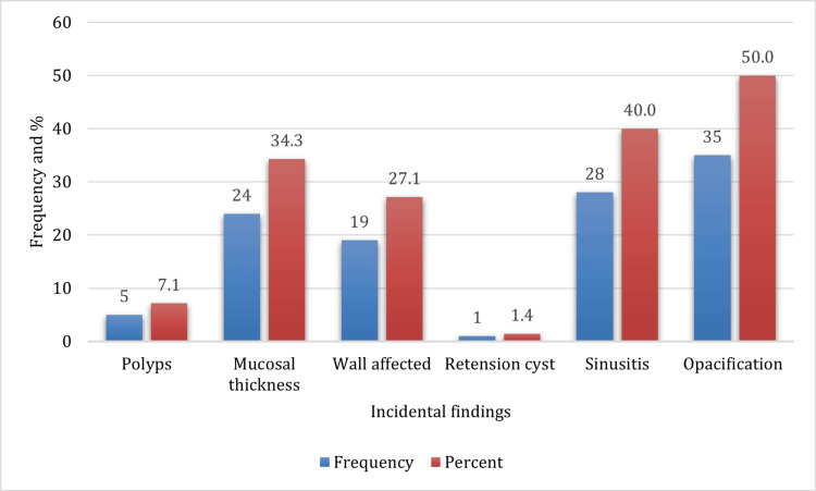
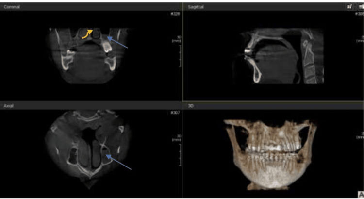
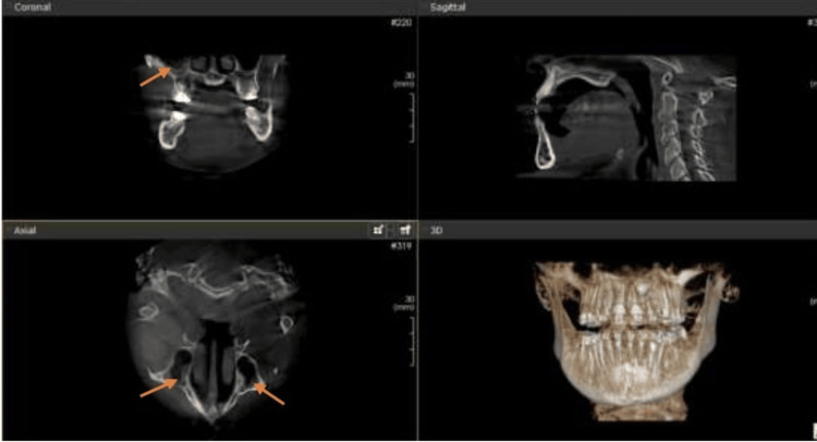
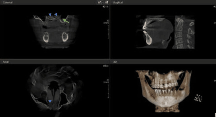
Among the incidental maxillary sinus findings detected, opacification was the most prevalent, occurring in 50% of the cases, followed by sinusitis in 40% and mucosal thickness in 34.3% of the cases. Wall-affected findings were noted in 19% of the cases. The least common associated incidental findings were retention cysts and polyps (Figures 1-4).
Figure 1. Frequency distribution of incidental maxillary sinus findings.
Incidental maxillary sinus disorders in CBCT.
CBCT: Cone-beam computed tomography
Figure 2. Left maxillary sinus opacification (blue arrows in both coronal and axial section) and deviated nasal septum (yellow curved arrow in coronal section) in a patient who presented for nerve treatment.
Coronal, axial, and sagittal CBCT images showing incidental maxillary and nasal findings in an orthodontic patient.
CBCT: Cone-beam computed tomography
Figure 3. Maxillary sinusitis (orange arrows in both coronal and axial sections).
Maxillary sinusitis in coronal and axial CBCT sections of an orthodontic patient.
CBCT: Cone-beam computed tomography
Figure 4. Mucosal thickening (green arrow in coronal section) and hypertrophy of inferior nasal turbinate (blue arrow heads in both coronal and axial sections) .
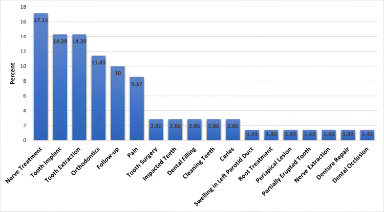
The most common dental indications were nerve treatment (17.14%), tooth extraction (14.29%), tooth implant (14.29%), and follow-up (10%) (Figure 5).
Figure 5. Frequency distribution of dental indications.
Several dental findings were noted, with root invasion being the most common at 18.57%. Other findings included gingivitis (7.14%) and the less frequent ones such as dental caries (4.3%), malocclusion, and irreversible pulpitis. Conditions like impacted teeth, periapical lesions, and periodontitis were noted with lower frequencies (Table 3).
Table 3. Dental findings on CBCT.
CBCT: Cone-beam computed tomography
| Dental findings | Percent |
| Normal | 55.72% |
| Roots invasion | 18.57% |
| Gingivitis | 7.14% |
| Dental caries | 4.29% |
| Impacted tooth | 2.86% |
| Reversible pulpitis | 1.43% |
| Pneumatization | 1.43% |
| Periodontitis | 1.43% |
| Periapical lesion around palatal | 1.43% |
| Palatal impacted | 1.43% |
| Malocclusion | 1.43% |
| Irreversible pulpitis | 1.43% |
| Total | 100% |
In Table 4, when analyzing the types of findings across different age groups, mucosal thickening was more common in the 31-46 and 47-62 age groups, with a total of 24 cases observed. Significant differences were noted among different age groups (p=0.031). Other findings such as polyps, wall defects, retention cysts, and opacifications did not show significant age-related variations. About 35 cases of opacifications were evenly distributed across all age groups, with most cases discovered in age groups 15-30 and 31-46. Sinusitis was more prevalent in age groups 31-46, with no statistically significant differences across different age groups (p=0.327).
Table 4. Cross-tabulation findings and age group.
| Finding | Age group | Total | p-value | ||||
| 15-30 | 31-46 | 47-62 | 63-78 | ||||
| Mucosal thickening | Yes | 8 | 9 | 7 | 0 | 24 | 0.031 |
| No | 29 | 11 | 4 | 2 | 46 | ||
| Polyp | Yes | 4 | 0 | 1 | 0 | 5 | 0.474 |
| No | 33 | 20 | 10 | 2 | 65 | ||
| Wall affected | Yes | 7 | 7 | 5 | 0 | 19 | 0.212 |
| No | 30 | 13 | 6 | 2 | 51 | ||
| Retention cyst | Yes | 1 | 0 | 0 | 0 | 1 | 0.824 |
| No | 36 | 20 | 11 | 2 | 69 | ||
| Opacifications | Yes | 18 | 11 | 5 | 1 | 35 | 0.957 |
| No | 19 | 9 | 6 | 1 | 35 | ||
| Sinusitis | Yes | 1 | 16 | 5 | 6 | 28 | 0.327 |
| No | 1 | 21 | 15 | 5 | 42 | ||
The bar chart demonstrates the distribution of incidental findings in CBCT scans among both genders. It shows that incidental findings were more commonly detected in females, with 41.43% of the total cases (29/70) compared to 18.57% (13/70) in males. Contrariwise, the absence of incidental findings was noted in 34.29% of females (24/70) and only 5.71% of males (4/70). The difference in the incidence of incidental findings between males and females was not statistically significant (p=0.111) (Figure 6).
Figure 6. Cross-tabulation gender and incidental findings .
Bar chart showing females mostly affected by incidental maxillary findings.
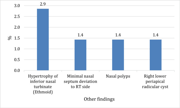
Other incidental findings were noted in the nose and periapical areas such as hypertrophy of the inferior nasal turbinate (ethmoid) with a prevalence of 2.9%, nasal polyps, and right lower periapical radicular cyst (Figure 7).
Figure 7. Other incidental findings .
Bar chart showing other incidental findings.
RT: Right
Discussion
CBCT has been developed in the last few years for applications in dentistry and maxillofacial fields. It is increasingly becoming the preferred imaging technique in particular clinical situations. CBCT offers detailed 3D images of the structures being assessed. CBCT imaging has overcome the limitations associated with two-dimensional (2D) imaging, providing practitioners with high-quality images that possess sub-millimeter resolution, while also ensuring a short scanning duration and reduced radiation exposure. As this technology becomes an integral part of dental practice, reliance on practice-based estimations will diminish, resulting in advantages for both patients and dentists [9]. The maxillary sinuses are the most substantial paranasal sinuses to assess in dentistry because of their closeness to the upper teeth. Consequently, conditions affecting the maxillary antra may mimic odontogenic infections, and issues related to the teeth can also affect the maxillary antra or exhibit symptoms similar to those associated with antral diseases [10]. In this study, the common reasons for CBCT were nerve treatment, tooth implants, and tooth extractions. In contrast, Ritter et al. noted that trauma and dental implants were the primary reasons for CBCT [7]. Meanwhile, Younis et al. reported that the most frequent reason for CBCT was related to tooth implant planning [10].
This study offers a comprehensive analysis of CBCT scans in orthodontic patients, revealing critical insights into incidental findings and dental indications. The mean age of the patients undergoing CBCT scans was 33.66±13.44 years, with the majority (52.9%) aged between 15 and 30 years. Moreover, over two-thirds of the participants were female. Consistent with this study, Lagorsse et al. in a literature review mentioned a higher prevalence of young female patients in orthodontic settings, probably because of their higher aesthetic demands [11].
The incidental maxillary findings detected in a majority of orthodontic patients (60%) underscores the importance of evaluation of CBCT images to assess the associated dental and maxillary abnormalities as early detection can lead to timely diagnosis, treatment, and follow-up. Opacification, sinusitis, and mucosal thickening were the most prevalent in patients undergoing CBCT. In comparison to this study, Biel et al. found that the prevalence of incidental findings in pre-implant treatment was 82%, with the most common incidental finding in CBCT being the thickening of maxillary sinus mucosa detected in 32% of the patients. Kurtuldu et al. mentioned that mucosal thickening was a common maxillary sinus pathology [12,13]. Similarly, the thickening of maxillary sinus mucosa (29.8%) was found as an incidental finding in 300 CBCT scans, and maxillary sinus mucosal thickening was detected as an incidental finding in 691 CBCT scans [14]. Gracco et al., in a study of 513 CBCT scans performed for orthodontic treatment patients, found that the most frequent incidentals include dental abnormalities, maxillary sinus disorders in half of the cases with mucosal thickening (40.1%), and pseudocysts (10.1%) [15]. Pazera et al. mentioned that incidental maxillary sinus disorders were detected in CBCT orthodontic patients with flat mucosal thickening (23.7%), polypoid mucosal thickening (19.4%), and signs of acute sinusitis (3.6%) [16]. Drage et al., agreeing with this study, mentioned that the incidental findings were very common, occurring in 66% of patients undergoing CBCT scans for orthodontic purposes [17]. Inconsistent with the present study in that the incidental findings were dental in nature, including retained deciduous roots, wisdom teeth, dental aplasia, and dislocation, the orthodontist should be aware of this finding, which may require further investigation, treatment, and follow-up [17,18].
When the types of findings across different age groups were analyzed, mucosal thickening was significantly more common in older age groups. This finding is supported by the research of Gracco et al. [15] who also noted increased mucosal thickening in older age groups. Other findings such as polyps, wall defects, retention cysts, and opacifications did not show significant age-related variations, which is in line with previous studies [15,19]. Gender differences in incidental findings showed that females had a higher prevalence (41.43%) compared to males (18.57%), although this difference was not statistically significant. This finding is consistent with the research of Raghav et al. who also found that the maxillary sinus disorders were 59.7%, with no significant gender differences in incidental findings in CBCT scans [19]. Another study mentioned that the prevalence of incidental maxillary sinus disorders in CBCT scans was 73%, with mucosal thickening and polyps as the most common findings, around 41.4% and 16%, respectively, which suggest significant differences in these findings in both genders. Furthermore, an insignificant difference was found between age groups and sinus abnormalities for CBCT scans [20]. Another study by Alotaibi, who conducted a retrospective analysis of 201 CBCT scans, was inconsistent with this study and mentioned that a significant difference exists between both genders, showing a greater existence of sinus pathology in males compared to females, while in this study, the most common incidental finding was detected in females [21]. The inconsistency observed between the findings of Alotaibi's study and this study concerning gender may be attributed to the smaller sample size in this study.
This study confirmed that the most common incidental CBCT findings in the maxillary sinuses were opacification, sinusitis, and mucosal thickening. Because of their closeness to the upper jaw, there may be a link between periapical lesions and maxillary sinusitis. Near the bottom of the maxillary sinus are the roots of the molars and premolars in the upper back. These teeth may have an advanced periapical infection that erodes the bone, separating the root tip from the sinus and opening the door for infection to enter the sinus cavity via the tooth's root; dental symptoms and sinusitis may result from this. Sometimes an infection from a periapical lesion spreads to the maxillary sinus, resulting in symptoms similar to sinusitis. On the other hand, it can occasionally be challenging to identify the precise origin of discomfort directed to the upper back teeth due to maxillary sinusitis. Not all periapical lesions result in maxillary sinusitis and not all maxillary sinusitis is caused by tooth infections. To properly identify and manage these disorders, dental and medical practitioners must provide proper diagnosis and treatment [22-24].
Other incidental findings such as hypertrophy of the inferior nasal turbinate with a prevalence of 2.9%, nasal polyps, and right lower periapical radicular cysts were also noted. These findings are in line with the study by Rheem et al. who reported incidental findings in dentoalveolar, nasal, and airway regions on CBCT [25]. Furthermore, in another study conducted on the Iranian population, it was found that the prevalence of incidental findings in CBCT images of oral and maxillofacial patients was 60%. The principal frequency of incidental findings were periapical lesions, followed by mucous thickening of the maxillary sinus, retained root, and impaction of the third molar [26]. Smith et al. reported that 19.4% of their patients had a deviated septum and 50.0% had mucosal thickness, which is indicative of maxillary sinusitis [27]. Another study mentioned that 52.1% of patients are diagnosed with mucosal thickening and 6.3% with a deviated nasal septum [28]. The results of this study are largely consistent with previous studies, thus reinforcing the importance of comprehensive evaluation of CBCT images to identify incidental findings of maxillary sinuses and maxillofacial region and improve patients' management and treatment outcomes.
This study had several limitations. It was only a two-center, retrospective study with a small sample size compared to similar studies conducted in the literature. Despite these limitations, the study findings demonstrate similar findings that highlight the diagnostic utility of CBCT in detecting incidental findings in maxillary sinuses and maxillofacial structure, which require clinicians to remain cautious concerning these incidental findings. Future research should focus on these findings through more, multi-center studies to improve their significance and pertinency across diverse populations, thereby refining patient management and outcomes in orthodontics and related fields.
Conclusions
This study highlights the prevalence of incidental abnormalities in the maxillary sinus noticed in CBCT scans of orthodontic patients. The most common maxillary sinus abnormalities detected were opacifications, sinusitis, and mucosal thickness. A considerable number of the patients were female, and the incidence of sinus-related abnormalities was predominantly high (60%). Other facial abnormalities were noted rather than maxillary sinus abnormalities, accentuating the need for excluding non-dental findings in dental CBCT. Mucosal thickness varied with age, with a higher prevalence noted among the older population. These findings emphasize the necessity of multidisciplinary collaboration between dentists and otolaryngologists in providing complete treatment to patients. Furthermore, it may be helpful to design guidelines for reporting incidental findings in dental CBCT exams to improve diagnostic precision and patient outcomes.
Acknowledgments
We extend our thanks and appreciation to everyone who contributed and assisted in this research. This research is dedicated to Asma al-Maliki, one of the main researchers of this work, who died in a tragic traffic accident before it was shown. We ask God to have mercy on her, forgive her, and dwell her in his wide paradise.
Disclosures
Human subjects: Consent was obtained or waived by all participants in this study.
Animal subjects: All authors have confirmed that this study did not involve animal subjects or tissue.
Conflicts of interest: In compliance with the ICMJE uniform disclosure form, all authors declare the following:
Payment/services info: All authors have declared that no financial support was received from any organization for the submitted work.
Financial relationships: All authors have declared that they have no financial relationships at present or within the previous three years with any organizations that might have an interest in the submitted work.
Other relationships: All authors have declared that there are no other relationships or activities that could appear to have influenced the submitted work.
Author Contributions
Concept and design: Awadia Gareeballah, Sawsan Saidahmed, Sarra Ali, Meaad Elbashir, Bashair Abu Habiba, Hana Mohammed, Belqees Hakami, Sarra Kamal, Esam S. Alshameri, Asma Alamin, Ali Abdelrazig
Acquisition, analysis, or interpretation of data: Awadia Gareeballah, Sawsan Saidahmed, Sarra Ali, Belqees Hakami
Drafting of the manuscript: Awadia Gareeballah, Sawsan Saidahmed, Sarra Ali, Meaad Elbashir, Hana Mohammed, Sarra Kamal, Asma Alamin, Ali Abdelrazig
Critical review of the manuscript for important intellectual content: Awadia Gareeballah, Sawsan Saidahmed, Meaad Elbashir, Bashair Abu Habiba, Hana Mohammed, Belqees Hakami, Esam S. Alshameri
Supervision: Awadia Gareeballah, Sawsan Saidahmed, Sarra Ali, Bashair Abu Habiba
References
- 1.What is cone-beam CT and how does it work? Scarfe WC, Farman AG. Dent Clin North Am. 2008;52:707–730. doi: 10.1016/j.cden.2008.05.005. [DOI] [PubMed] [Google Scholar]
- 2.Technical aspects of dental CBCT: state of the art. Pauwels R, Araki K, Siewerdsen JH, Thongvigitmanee SS. Dentomaxillofac Radiol. 2015;44 doi: 10.1259/dmfr.20140224. [DOI] [PMC free article] [PubMed] [Google Scholar]
- 3.Clinical significance of pathological and anatomical findings in cone beam CT scans of the maxillary sinus. Kihara E, Chindia M, Ocholla T, Parker M. Open J Stomatol. 2014;4:285–290. [Google Scholar]
- 4.Incidental findings on cone beam computed tomography images. Allareddy V, Vincent SD, Hellstein JW, Qian F, Smoker WR, Ruprecht A. Int J Dent. 2012;2012 doi: 10.1155/2012/871532. [DOI] [PMC free article] [PubMed] [Google Scholar]
- 5.The maxillary sinus: physiology, development and imaging anatomy. Whyte A, Boeddinghaus R. Dentomaxillofac Radiol. 2019;48 doi: 10.1259/dmfr.20190205. [DOI] [PMC free article] [PubMed] [Google Scholar]
- 6.Imaging of odontogenic sinusitis. Whyte A, Boeddinghaus R. Clin Radiol. 2019;74:503–516. doi: 10.1016/j.crad.2019.02.012. [DOI] [PubMed] [Google Scholar]
- 7.Prevalence of pathologic findings in the maxillary sinus in cone-beam computerized tomography. Ritter L, Lutz J, Neugebauer J, et al. Oral Surg Oral Med Oral Pathol Oral Radiol Endod. 2011;111:634–640. doi: 10.1016/j.tripleo.2010.12.007. [DOI] [PubMed] [Google Scholar]
- 8.Association between periapical lesions and maxillary sinus mucosal thickening: a retrospective cone-beam computed tomographic study. Shanbhag S, Karnik P, Shirke P, Shanbhag V. J Endod. 2013;39:853–857. doi: 10.1016/j.joen.2013.04.010. [DOI] [PubMed] [Google Scholar]
- 9.Cone beam computed tomography: basics and applications in dentistry. Venkatesh E, Elluru SV. J Istanb Univ Fac Dent. 2017;51:102–121. doi: 10.17096/jiufd.00289. [DOI] [PMC free article] [PubMed] [Google Scholar]
- 10.Prevalence of incidental maxillary sinus pathology in asymptomatic patient during routine dental examination by cone beam computed tomography (a retrospective study) Younis ST. Int J Med Rev Case Rep. 2022;6:75–78. [Google Scholar]
- 11.Does gender make a difference in orthodontics? A literature review. (Article in French) Lagorsse A, Gebeile-Chauty S. Orthod Fr. 2018;89:157–168. doi: 10.1051/orthodfr/2018011. [DOI] [PubMed] [Google Scholar]
- 12.Incidental findings in cone beam computed tomography (CBCT) scans for implant treatment planning: a retrospective study of 404 CBCT scans. Biel P, Jurt A, Chappuis V, Suter VG. Oral Radiol. 2024;40:207–218. doi: 10.1007/s11282-023-00723-5. [DOI] [PMC free article] [PubMed] [Google Scholar]
- 13.Incidental findings in patients who underwent cone beam computed tomography for implant treatment planning. Kurtuldu E, Alkis HT, Yesiltepe S, Sumbullu MA. Niger J Clin Pract. 2020;23:329–336. doi: 10.4103/njcp.njcp_309_19. [DOI] [PubMed] [Google Scholar]
- 14.Incidental findings on cone-beam computed tomographic images: paranasal sinus findings and nasal septum variations. Avsever H, Gunduz K, Karakoç O, Akyol M, Orhan K. Oral Radiol. 2018;34:40–48. doi: 10.1007/s11282-017-0283-y. [DOI] [PubMed] [Google Scholar]
- 15.Prevalence of incidental maxillary sinus findings in Italian orthodontic patients: a retrospective cone-beam computed tomography study. Gracco A, Incerti Parenti S, Ioele C, Alessandri Bonetti G, Stellini E. Korean J Orthod. 2012;42:329–334. doi: 10.4041/kjod.2012.42.6.329. [DOI] [PMC free article] [PubMed] [Google Scholar]
- 16.Incidental maxillary sinus findings in orthodontic patients: a radiographic analysis using cone-beam computed tomography (CBCT) Pazera P, Bornstein MM, Pazera A, Sendi P, Katsaros C. Orthod Craniofac Res. 2011;14:17–24. doi: 10.1111/j.1601-6343.2010.01502.x. [DOI] [PubMed] [Google Scholar]
- 17.Incidental findings on cone beam computed tomography in orthodontic patients. Drage N, Rogers S, Greenall C, Playle R. J Orthod. 2013;40:29–37. doi: 10.1179/1465313312Y.0000000027. [DOI] [PubMed] [Google Scholar]
- 18.Incidental findings on craniomaxillofacial cone beam computed tomography in orthodontic patients. Kocsis C, Sommerlath Sohns JM, Graf I, et al. https://scholar.google.com/scholar?q=intitle:Incidental%20findings%20on%20craniomaxillofacial%20cone%20beam%20computed%20tomography%20in%20orthodontic%20patients#d=gs_cit&t=1728377488208&u=%2Fscholar%3Fq%3Dinfo%3Adoi2KhfKamQJ%3Ascholar.google.com%2F%26output%3Dcite%26scirp%3D0%26hl%3Den. Int J Comput Dent. 2019;22 [PubMed] [Google Scholar]
- 19.Prevalence of incidental maxillary sinus pathologies in dental patients on cone-beam computed tomographic images. Raghav M, Karjodkar FR, Sontakke S, Sansare K. Contemp Clin Dent. 2014;5:361–365. doi: 10.4103/0976-237X.137949. [DOI] [PMC free article] [PubMed] [Google Scholar]
- 20.Maxillary sinus abnormalities detected by dental cone-beam computed tomography. Elwakeel EE, Ingle E, Elkamali YA, Alfadel H, Alshehri N, Madini KA. Anat Physiol. 2017;7 [Google Scholar]
- 21.Prevalence of incidental maxillary sinus pathologies in dental patients at PAADI using cone‐beam computed tomography (CBCT) Alotaibi F. Clin Oral Implants Res. 2019;30:90. [Google Scholar]
- 22.Evaluation of the effect of periapical lesions and other odontogenic conditions on maxillary sinus mucosal thickness characteristics and mucosal appearance: a CBCT study. Ince Yusufoglu S, Hasanoglu Erbasar GN, Gülen O. J Dent Res Dent Clin Dent Prospects. 2021;15:163–171. doi: 10.34172/joddd.2021.028. [DOI] [PMC free article] [PubMed] [Google Scholar]
- 23.When maxillary sinusitis does not heal: findings on CBCT scans of the sinuses with a particular focus on the occurrence of odontogenic causes of maxillary sinusitis. Vestin Fredriksson M, Öhman A, Flygare L, Tano K. Laryngoscope Investig Otolaryngol. 2017;2:442–446. doi: 10.1002/lio2.130. [DOI] [PMC free article] [PubMed] [Google Scholar]
- 24.Association between cone-beam computed tomography and histological and immunohistochemical features in periapical lesions correlated with thickened maxillary sinus mucosa. Dumitrescu A, Martu MA, Nemtoi A, et al. Medicina (Kaunas) 2021;57 doi: 10.3390/medicina57080840. [DOI] [PMC free article] [PubMed] [Google Scholar]
- 25.Incidental findings in the maxillofacial region identified on cone-beam computed tomography scans. Rheem S, Nielsen I, Oberoi S. https://triggered.edina.clockss.org/ServeContent?url=http%3A%2F%2Fwww.jorthodr.org%2Farticle.asp%3Fissn%3D2321-3825%3Byear%3D2013%3Bvolume%3D1%3Bissue%3D1%3Bspage%3D33%3Bepage%3D39%3Baulast%3DRheem%3Btype%3D2 J Orthod Res. 2013;1:33–39. [Google Scholar]
- 26.Incidental dentomaxillofacial findings on cone beam computed tomography images of Iranian population. Khojastepour L, Haghani J, Mirbeigi S. https://scholar.google.com/scholar?q=intitle:Incidental%20dentomaxillofacial%20findings%20on%20cone%20beam%20computed%20tomography%20images%20of%20Iranian%20population J Oral Health Oral Epidemiol. 2014;3:12–15. [Google Scholar]
- 27.The prevalence of concha bullosa and nasal septal deviation and their relationship to maxillary sinusitis by volumetric tomography. Smith KD, Edwards PC, Saini TS, Norton NS. Int J Dent. 2010;2010 doi: 10.1155/2010/404982. [DOI] [PMC free article] [PubMed] [Google Scholar]
- 28.Prevalence of incidental maxillary sinus anomalies on CBCT scans: a radiographic study. Ahmed J, Gupta A, Shenoy N, Sujir N, Muralidharan A. Diagnostics (Basel) 2023;13 doi: 10.3390/diagnostics13182918. [DOI] [PMC free article] [PubMed] [Google Scholar]


