

Abstract
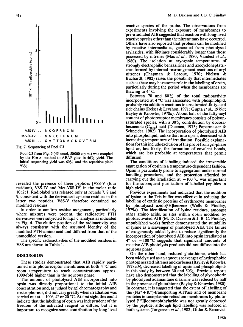
The hydrophobic photosensitive probe 1-azido-4-[125I]iodobenzene (AIB) partitioned preferentially into photoreceptor disc membranes and, upon u.v. irradiation, became covalently bound to opsin and phospholipid. The labelling of both protein and phospholipid was linearly related to AIB concentration. The amount of probe incorporated into protein was not significantly different when membranes were irradiated at -100 degrees, 4 degrees or 25 degrees C, but irreversible aggregation of monomeric opsin was dramatically reduced by performing the photolysis at -100 degrees C. Labelling of opsin after irradiation at -100 degrees or 4 degrees was not significantly reduced by the presence of lysine in the aqueous buffer, indicating that significant amounts of reactive species did not enter the aqueous phase. The incorporation into phospholipid, unlike that into opsin, decreased as the temperature of irradiation increased. Some labelling of opsin occurred on incubation with pre-photoactivated AIB, indicating that reaction may also occur with reactive species of longer lifetimes than the nitrene. Proteolysis of labelled opsin with Staphylococcus aureus V8 proteinase yielded two radiolabelled membrane-bound fragments. The location of the modified sites (cysteine, tryptophan, tyrosine, lysine and histidine residues: all nucleophiles) in the smaller fragment was entirely consistent with putative models for the protein derived from other studies.
Full text
PDF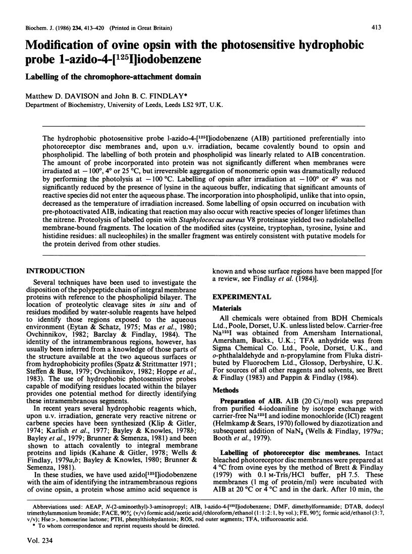


Selected References
These references are in PubMed. This may not be the complete list of references from this article.
- Barclay P. L., Findlay J. B. Labelling of the cytoplasmic domains of ovine rhodopsin with hydrophilic chemical probes. Biochem J. 1984 May 15;220(1):75–84. doi: 10.1042/bj2200075. [DOI] [PMC free article] [PubMed] [Google Scholar]
- Bayley H., Knowles J. R. Photogenerated reagents for membrane labeling. 1. Phenylnitrene formed within the lipid bilayer. Biochemistry. 1978 Jun 13;17(12):2414–2419. doi: 10.1021/bi00605a025. [DOI] [PubMed] [Google Scholar]
- Bayley H., Knowles J. R. Photogenerated reagents for membrane labeling. 2. Phenylcarbene and adamantylidene formed within the lipid bilayer. Biochemistry. 1978 Jun 13;17(12):2420–2423. doi: 10.1021/bi00605a026. [DOI] [PubMed] [Google Scholar]
- Bayley H., Knowles J. R. Photogenerated, hydrophobic reagents for intrinsic membrane proteins. Ann N Y Acad Sci. 1980;346:45–58. doi: 10.1111/j.1749-6632.1980.tb22090.x. [DOI] [PubMed] [Google Scholar]
- Booth A. G., Hubbard L. M., Kenny A. J. Proteins of the kidney microvillar membrane. Immunoelectrophoretic analysis of the membrane hydrolases: identification and resolution of the detergent- and proteinase-solubilized forms. Biochem J. 1979 May 1;179(2):397–405. doi: 10.1042/bj1790397. [DOI] [PMC free article] [PubMed] [Google Scholar]
- Brett M., Findlay J. B. Investigation of the organization of rhodopsin in the sheep photoreceptor membrane by using cross-linking reagents. Biochem J. 1979 Jan 1;177(1):215–223. doi: 10.1042/bj1770215. [DOI] [PMC free article] [PubMed] [Google Scholar]
- Brett M., Findlay J. B. Isolation and characterization of the CNBr peptides from the proteolytically derived N-terminal fragment of ovine opsin. Biochem J. 1983 Jun 1;211(3):661–670. doi: 10.1042/bj2110661. [DOI] [PMC free article] [PubMed] [Google Scholar]
- Brunner J., Semenza G. Selective labeling of the hydrophobic core of membranes with 3-(trifluoromethyl)-3-(m-[125I]iodophenyl)diazirine, a carbene-generating reagent. Biochemistry. 1981 Dec 8;20(25):7174–7182. doi: 10.1021/bi00528a019. [DOI] [PubMed] [Google Scholar]
- Daemen F. J. Vertebrate rod outer segment membranes. Biochim Biophys Acta. 1973 Nov 28;300(3):255–288. doi: 10.1016/0304-4157(73)90006-3. [DOI] [PubMed] [Google Scholar]
- Eytan G. D., Schatz G. Cytochrome c oxidase from bakers' yeast. V. Arrangement of the subunits in the isolated and membrane-bound enzyme. J Biol Chem. 1975 Jan 25;250(2):767–774. [PubMed] [Google Scholar]
- Findlay J. B., Barclay P. L., Brett M., Davison M., Pappin D. J., Thompson P. The structure of mammalian rod opsins. Vision Res. 1984;24(11):1501–1508. doi: 10.1016/0042-6989(84)90312-2. [DOI] [PubMed] [Google Scholar]
- Findlay J. B., Brett M., Pappin D. J. Primary structure of C-terminal functional sites in ovine rhodopsin. Nature. 1981 Sep 24;293(5830):314–317. doi: 10.1038/293314a0. [DOI] [PubMed] [Google Scholar]
- Gitler C., Bercovici T. Use of lipophilic photoactivatable reagents to identify the lipid-embedded domains of membrane proteins. Ann N Y Acad Sci. 1980;346:199–211. doi: 10.1111/j.1749-6632.1980.tb22100.x. [DOI] [PubMed] [Google Scholar]
- Goldman D. W., Pober J. S., White J., Bayley H. Selective labelling of the hydrophobic segments of intrinsic membrane proteins with a lipophilic photogenerated carbene. Nature. 1979 Aug 30;280(5725):841–843. doi: 10.1038/280841a0. [DOI] [PubMed] [Google Scholar]
- Gupta C. M., Radhakrishnan R., Gerber G. E., Olsen W. L., Quay S. C., Khorana H. G. Intermolecular crosslinking of fatty acyl chains in phospholipids: use of photoactivable carbene precursors. Proc Natl Acad Sci U S A. 1979 Jun;76(6):2595–2599. doi: 10.1073/pnas.76.6.2595. [DOI] [PMC free article] [PubMed] [Google Scholar]
- Helmkamp R. W., Sears D. A. A label for the red cell membrane: diazotized diiodosulfanilic acid. Int J Appl Radiat Isot. 1970 Nov;21(11):683–685. doi: 10.1016/0020-708x(70)90127-4. [DOI] [PubMed] [Google Scholar]
- Hoppe J., Friedl P., Schairer H. U., Sebald W., von Meyenburg K., Jørgensen B. B. The topology of the proton translocating F0 component of the ATP synthase from E. coli K12: studies with proteases. EMBO J. 1983;2(1):105–110. doi: 10.1002/j.1460-2075.1983.tb01389.x. [DOI] [PMC free article] [PubMed] [Google Scholar]
- Horn M. J., Laursen R. A. Solid-phase edman degradation: attachment of carboxyl-terminal homoserine peptides to an insoluble resin. FEBS Lett. 1973 Nov 1;36(3):285–288. doi: 10.1016/0014-5793(73)80392-8. [DOI] [PubMed] [Google Scholar]
- Jørgensen P. L., Karlish S. J., Gitler C. Evidence for the organization of the transmembrane segments of (Na,K)-ATPase based on labeling lipid-embedded and surface domains of the alpha-subunit. J Biol Chem. 1982 Jul 10;257(13):7435–7442. [PubMed] [Google Scholar]
- Kahane I., Gitler C. Red cell membrane glycophorin labeling from within the lipid bilayer. Science. 1978 Jul 28;201(4353):351–352. doi: 10.1126/science.663661. [DOI] [PubMed] [Google Scholar]
- Karlish S. J., Jorgensen P. L., Gitler C. Identification of a membrane-embedded segment of the large polypeptide chain of (Na+, K+)ATPase. Nature. 1977 Oct 20;269(5630):715–717. doi: 10.1038/269715a0. [DOI] [PubMed] [Google Scholar]
- Klip A., Gitler C. Photoactive covalent labeling of membrane components from within the lipid core. Biochem Biophys Res Commun. 1974 Oct 8;60(3):1155–1162. doi: 10.1016/0006-291x(74)90433-1. [DOI] [PubMed] [Google Scholar]
- Laemmli U. K., Favre M. Maturation of the head of bacteriophage T4. I. DNA packaging events. J Mol Biol. 1973 Nov 15;80(4):575–599. doi: 10.1016/0022-2836(73)90198-8. [DOI] [PubMed] [Google Scholar]
- Mas M. T., Wang J. K., Hargrave P. A. Topography of rhodopsin in rod outer segment disk membranes. Photochemical labeling with N-(4-azido-2-nitrophenyl)-2-aminoethanesulfonate. Biochemistry. 1980 Feb 19;19(4):684–691. doi: 10.1021/bi00545a012. [DOI] [PubMed] [Google Scholar]
- Ovchinnikov YuA Rhodopsin and bacteriorhodopsin: structure-function relationships. FEBS Lett. 1982 Nov 8;148(2):179–191. doi: 10.1016/0014-5793(82)80805-3. [DOI] [PubMed] [Google Scholar]
- Pappin D. J., Findlay J. B. Sequence variability in the retinal-attachment domain of mammalian rhodopsins. Biochem J. 1984 Feb 1;217(3):605–613. doi: 10.1042/bj2170605. [DOI] [PMC free article] [PubMed] [Google Scholar]
- Pober J. S., Stryer L. Letter to the editor: Light dissociates enzymatically-cleaved rhodopsin into two different fragments. J Mol Biol. 1975 Jul 5;95(3):477–481. doi: 10.1016/0022-2836(75)90204-1. [DOI] [PubMed] [Google Scholar]
- Spatz L., Strittmatter P. A form of cytochrome b5 that contains an additional hydrophobic sequence of 40 amino acid residues. Proc Natl Acad Sci U S A. 1971 May;68(5):1042–1046. doi: 10.1073/pnas.68.5.1042. [DOI] [PMC free article] [PubMed] [Google Scholar]
- Steffens G. J., Buse G. Studies on cytochrome c oxidase, IV[1--3]. Primary structure and function of subunit II. Hoppe Seylers Z Physiol Chem. 1979 Apr;360(4):613–619. [PubMed] [Google Scholar]
- Vandest P., Labbe J. P., Kassab R. Photoaffinity labelling of arginine kinase and creatine kinase with a gamma-P-substituted arylazido analogue of ATP. Eur J Biochem. 1980 Mar;104(2):433–442. doi: 10.1111/j.1432-1033.1980.tb04445.x. [DOI] [PubMed] [Google Scholar]
- Wells E., Findlay J. B. Labelling of the intramembranous region of the major sialoglycoprotein of human erythrocytes with a photosensitive hydrophobic probe. Biochem J. 1979 May 1;179(2):265–272. doi: 10.1042/bj1790265. [DOI] [PMC free article] [PubMed] [Google Scholar]
- Wells E., Findlay J. B. Use of photosensitive hydrophobic probes to label the membrane of the human erythrocyte. Biochem J. 1979 May 1;179(2):257–264. doi: 10.1042/bj1790257. [DOI] [PMC free article] [PubMed] [Google Scholar]
- Zimmerman C. L., Appella E., Pisano J. J. Rapid analysis of amino acid phenylthiohydantoins by high-performance liquid chromatography. Anal Biochem. 1977 Feb;77(2):569–573. doi: 10.1016/0003-2697(77)90276-7. [DOI] [PubMed] [Google Scholar]