Abstract
A neuroadapted strain of yellow fever virus (YFV) 17D derived from a multiply mouse brain-passaged virus (Porterfield YF17D) was additionally passaged in SCID and normal mice. The virulence properties of this virus (SPYF) could be distinguished from nonneuroadapted virus (YF5.2iv, 17D infectious clone) by decreased average survival time in SCID mice after peripheral inoculation, decreased average survival time in normal adult mice after intracerebral inoculation, and occurrence of neuroinvasiveness in normal mice. SPYF exhibited more efficient growth in peripheral tissues of SCID mice than YF5.2iv, resulting in a more rapid accumulation of virus burden, but with low-titer viremia, at the time of fatal encephalitis. In cell culture, SPYF was less efficient in replication than YF5.2iv in all cell lines tested. The complete nucleotide sequence of SPYF revealed 29 nucleotide substitutions relative to YF5.2iv, and these were distributed throughout the genome. There were a total of 13 predicted amino acid substitutions, some of which correspond to known differences among the Asibi, French viscerotropic virus, French neurotropic vaccine, and YF17D vaccine strains. The envelope (E) protein contained five substitutions, within all three functional domains. Substitutions were also present in regions encoding the NS1, NS2A, NS4A, and NS5 proteins and in the 3′ untranslated region (UTR). Construction of YFV harboring all of the identified coding nucleotide substitutions and those in the 3′ UTR yielded a virus whose cell culture and pathogenic properties, particularly neurovirulence and neuroinvasiveness for SCID mice, generally resembled those of the original SPYF isolate. These findings implicate the E protein and possibly other regions of the genome as virulence determinants during pathogenesis of neuroadapted YF17D virus in mice. The determinants affect replication efficiency in both neural and extraneural tissues of the mouse and confer some limited host-range differences in cultured cells of nonmurine origin.
Viruses within the Flavivirus genus of the family Flaviviridae are generally neurotropic, exhibiting various degrees of neurovirulence in rodent and primate hosts (41). Introduction of virus into the murine central nervous system (CNS) causes an acute encephalitis, the outcome of which can be influenced by the dose of virus and the age and strain of the mouse (15). Different strains of yellow fever virus (YFV) can be distinguished by their level of mouse neurovirulence (5, 15, 28, 52–54), and this property can be enhanced by serial passage of the virus in mouse brain (37, 56, 59). This neuroadaptation has also been observed with other flaviviruses (7, 11, 25). Genetic analyses of viruses which differ in their virulence properties have commonly focused on the envelope (E) protein, for which a variety of mutations have been shown to modulate neurovirulence (reviewed in reference 36). Regarding YFV, nucleotide sequence analysis of the partially attenuated French neurotropic virus (FNV) strain of YFV which was derived by mouse brain passage of the wild French viscerotropic virus (FVV) strain, revealed that numerous residues within the E protein, as well as in other regions of the genome, were retained relative to the fully attenuated 17D vaccine (22, 61). FNV caused encephalitis in humans at a rate higher than that which occurs with the YF17D vaccine strain (16), and although the pathophysiologic mechanisms for this phenomenon are not known, mutations in the E protein are presumed to be important. A rare neurovirulent revertant of the YF17D vaccine was shown to contain novel mutations only within the E protein (23). The role of these various mutations in altering those functional properties of the flavivirus E protein which increase neurovirulence is not well understood. It is assumed that receptor binding and subsequent events associated with virus entry, including low-pH-induced conformational changes and fusion with intracellular membranes, are principally involved (6, 32, 34, 35, 45, 46, 52, 54). In contrast to the E protein, neurovirulence determinants in the nonstructural and untranslated regions have been less well studied (8, 14, 25, 31, 38, 43, 47). These are likely to be important in relationship to host factors that affect enzymatic activities associated with viral transcription, translation of the viral polyprotein, or virion assembly or to RNA structures involved in cis-acting regulatory events and/or interactions with host proteins.
In a previous study, the mouse neurovirulence of a neuroadapted strain of YF17D (Porterfield 17D) was initially characterized, and a partial nucleotide sequence including the prM-through-NS2A region was reported (55). This virus causes uniform mortality in young adult mice after intracerebral (i.c.) inoculation of as little as 1 PFU of virus, replicates in mouse brain tissue more rapidly than nonneuroadapted virus, and results in higher peak titers of brain-associated virus and earlier death. Mutations at five positions in the E protein, as well as single substitutions in the NS1 and NS2A proteins, were identified in that study. However, the importance of these substitutions for the mouse-neurovirulent phenotype could not be demonstrated. To further investigate the genetic basis of the enhanced virulence of neuroadapted YF17D virus, we determined the complete nucleotide sequence of a plaque-purified virus (SPYF) after characterization of its virulence properties in mice. We then determined the phenotype of a corresponding virus engineered from cDNA to contain the predicted amino acid substitutions specific to the SPYF strain. In this investigation, adult SCID mice were found to be very sensitive to differences in virulence between neuroadapted and nonneuroadapted virus, developing a fatal encephalitis as early as 8 days after intraperitoneal (i.p.) inoculation of the SPYF strain. This model offers the chance to investigate the virus-host interactions which lead to neuroinvasion in the absence of virus-specific immunity. Use of this model to understand the molecular basis for flavivirus neurovirulence might therefore have implications for engineering live attenuated viral vaccines based on recombinant DNA technology. For instance, a nominal requirement for such vaccines is an acceptable attenuation phenotype based on multiple stable genetic determinants within the viral genome.
MATERIALS AND METHODS
Cells and viruses.
SW-13, BHK-21, Vero, and C6/36 cells were used as previously described (10). The neuroadapted PYF virus, which has undergone more than 20 passages in suckling mouse brain, has been previously described (55). YF5.2iv virus generated from a YF17D molecular clone has been previously described (51). Plaque assays for virus quantitation were routinely done in Vero cells, since the plaquing efficiency of the PYF virus in SW-13 cells is poor. Viruses were diluted in phosphate-buffered saline (PBS) plus 10% fetal bovine serum for injection into mice.
Mouse experiments.
Mice used in these experiments included the ICR strain (Harlan/Sprague-Dawley and Taconic Farms), the SCID/ICR strain (Taconic Farms), and the SCID C.B.-17 strain (Taconic Farms). Mice were used between 3 and 6 weeks of age for studies of neurovirulence. Mice were inoculated by the i.p. route for peripheral inoculation or by the i.c. route, after anesthetization. Mice were observed until the onset of a moribund condition and then sacrificed. Statistical analyses of differences in mortality were done using Fisher's exact test. Differences in survival times were analyzed using Wilcoxon rank-sum tests. For analysis of virus in tissues of infected mice, tissues were recovered by dissection after perfusion with PBS and stored at −70°C until used for virus quantitation. Titration of tissue-associated virus was done by preparing 20% suspensions of tissue homogenates in a Dounce homogenizer in PBS plus 10% fetal bovine serum and then performing plaque assays on Vero cells. The plaque assay was done using serial dilutions of virus plated on cell monolayers under 1.0% ME agarose (SeaKem) in alpha-minimum essential medium plus 5% fetal bovine serum. Plaques were visualized by staining after 5 days with 0.05% neutral red in PBS and then were counted after fixation in 10% formalin and staining with crystal violet.
Derivation of SPYF.
The original Porterfield YF17D strain (55) was used as the starting material. This virus was inoculated at a dose of approximately one million PFU into 5-week-old SCID mice (C.B.-17 strain) by the i.p. route, with onset of encephalitis occurring between 2 and 3 weeks postinoculation. Virus was obtained from the brain of a moribund mouse and a 20% brain suspension was prepared for plaque titration and further passage. After a second round of i.p. inoculation into SCID mice, the brain-associated virus was subjected to three rounds of plaque purification in SW-13 cells, in conjunction with amplification on BHK cells. Media from the third rounds of plaque purifications and amplifications were inoculated into 3-week-old ICR mice by i.c. inoculation, and brains were harvested at the onset of a moribund condition. One of these brain harvests was passaged a final time on Vero cells to provide a working stock of virus, hereafter referred to as SPYF. The nonneuroadapted YF5.2iv virus was derived by transfection of SW-13 cells with SP6 transcripts from a full-length in vitro-ligated cDNA template (51). The progeny virus was plaque purified twice in SW-13 cells, amplified in BHK cells, and passaged once on Vero cells to provide a working virus stock with a passage history which matched that of the SPYF virus, except for the mouse passages.
Molecular cloning and nucleotide sequencing.
RNA was prepared from virus-infected SCID mouse brain at the onset of a moribund condition after i.p. inoculation with the working stock of SPYF virus. RNA was extracted using TRIzol (Life Technologies). The oligonucleotide primers used for reverse transcription-PCR were as follows: minus-strand primers, 10836-10861 (3′-GGAAACCTACTGTTTGTGTTTTGGTGA-5′), 9051-9074 (3′-CGGCACGGTATACCATATACACCG-5′), 6947-6965 (3′-GGTAGATCACGAAGTGGGA-5′), 5228-5250 (3′-GCGAACGCGTGAGAACACAACCG-5′), 2486-2503 (3′-CTCGAGTTCACGCCTCTA-5′), 773-788 (3′-TTGGTACCAAACTTCT-5′); and plus-strand primers, 1-22 (5′-AGTAAATCCTGTGTGCTAATTG-3′), 424-440 (5′-CCATGATGTTCTGACTG-3′), 2482-2503 (5′-GAGAGAGCTCAAGTGCGGAGAT-3′), 4803-4819 (5′-TTGTCGCCTATGGTGGC-3′), 6954-6975 (5′-GTGCTTCACCCTGGAGTTGGCC-3′), 9051-9073 (5′-GCCGTGCCATATGGTATATGTGG-3′).
RNA was reverse transcribed using Superscript-RT (Gibco/BRL) and the YF17D-specific minus-strand primers. The cDNA products were subjected to PCR using Deep Vent DNA polymerase (New England Biolabs) and typical amplification cycles of 2 min of denaturation, 1 min of annealing (55°C), and extension for up to 2.5 min. PCR products were isolated by agarose gel electrophoresis, purified using Wizard PCR preps (Promega), and cloned into Zeroblunt-TOPO plasmids (Invitrogen). At least duplicate reactions were cloned for each sample. Clones containing correctly sized inserts were used for nucleotide sequencing. Nucleotide sequencing was done using Big Dye chain terminator reactions and was analyzed on an Applied Biosystems DNA sequencer. Chromatograms were read manually for detection and verification of all nucleotide substitutions.
Reconstruction of virus.
A molecular clone of the SPYF virus, hereafter referred to as SPYF-MN, was constructed as follows. Construction of YFV plasmids pYF5′3′IV and pYFM5.2 harboring the SPYF substitutions was done using standard cloning techniques to exchange regions from the Zeroblunt-TOPO plasmids into either of these two plasmids. pYF5′3′IV contains YFV nucleotides (nt) 1 to 2271 and 8275 to 10862 (YFV numbering) downstream of an SP6 promoter. pYFM5.2 contains the YFV sequence from nt 1363 to 8704. Construction of pYF5′3′IV(SPYF) was done by incorporation of the SPYF sequence of nt 536 to 1655 and 9058 to 10708 in five steps. An AvaI/NsiI fragment (containing SPYF nt 536 to 1655) was ligated into pYF5′3′(del)-XhoI/SalI, from which an XhoI/SalI fragment (nt 9423 to 10861) had been excised. The deleted XhoI/SalI fragment was then replaced with a NotI/NsiI fragment (nt 6675v to 1655) from pYF5′3′IV (where 6675v identifies the nucleotide position within the vector portion of the plasmid). An NdeI/XbaI fragment containing the SPYF sequence from nt 9058 to 10708 was ligated into pYF5′3′IV(del)-BsmI, from which a BsmI fragment (nt 459 to 1514) had been deleted. The deleted BsmI fragment was then replaced with a NotI/EcoRI fragment (nt 6675v to 2271) from pYF5′3′IV. The EcoRI site in YF5′3′IV lies at the junction of YFV nt 2271 and 8275. The NotI/EcoRI fragment from the plasmid containing the SPYF sequence from nt 536 to 1655 was then ligated to the nt 2271-to-6675v NotI/EcoRI fragment from the plasmid harboring the SPYF sequence from nt 9058 to 10708 to generate the final plasmid pYF5′3′IV(SPYF).
Construction of pYFM5.2(SPYF) required eight steps. The NsiI/SacI fragment containing SPYF nt 1655 to 2486 was ligated into pYFM5.2(del)-SacI, from which two SacI fragments (from nt 2486 to 5108) had been excised. The deleted SacI fragments were then replaced with a partial SacI digestion product containing SPYF nt 2486 to 5108. An NheI/AvaI fragment containing SPYF nt 5459 to 6797 was separately ligated into pYFM5.2(del)-SacI and the SacI deletion was restored with the partial SacI digestion fragment from pYFM5.2. In a third construct pYFM5.2 was digested with SphI and religated to excise the 1684-to-6897 SphI fragment and to produce pYFM5.2(del)-SphI, and the BsgI/EcoRV fragment containing SPYF nt 7444 to 8027 was ligated into this plasmid. A fourth plasmid was produced by partial digestion of pYFM5.2 with BanII to generate pYFM(del)-partial BanII lacking the YFV sequence between nt 1599 and 6330. Into this plasmid an SphI/XbaI fragment containing nt 6897 to 7365v from pYFM5.2(del)-SphI (containing the SPYF sequence from nt 7444 to 8027) was ligated. This resulted in incorporation of the SPYF sequence from 7444 to 8027 into pYFM5.2(del)-partial BanII. Both this plasmid and the pYFM5.2 plasmid containing the SPYF sequence from nt 5459 to 6797 were digested with NgoMIV and XbaI and the 6701-to-7365v fragment from pYFM5.2(del)-partial BanII was ligated into pYFM5.2 containing the SPYF sequence from nt 5459 to 6797, thereby incorporating the SPYF sequences from nt 5459 to 6701 and nt 7444 to 8027 into pYFM5.2. This ligation resulted in the loss of the nonsynonymous SPYF substitution at nt 6758 from this construct. The SPYF sequence from nt 1655 to 5108 was then introduced from pYFM5.2 harboring this SPYF sequence via a common NsiI/NheI fragment (nt 1655 to 5459). The deleted SPYF 6758 substitution was then restored by a three-piece ligation in which both pYFM5.2 containing the SPYF sequences from nt 1655 to 5108, 5459 to 6701, and 7444 to 8027 and the pYFM5.2 plasmid containing the SPYF sequence from nt 5459 to 6797 were digested with NgoMIV and AvaI. The 6701-to-6797 bp fragment from the latter plasmid was ligated into the corresponding site in the first plasmid to produce pYFM5.2 containing the SPYF sequences from nt 1655 to 5108, 5459 to 6797, and 7444 to 8027. This final pYFM5.2 plasmid lacks two synonymous SPYF substitutions at nt 5338 and 8212 which do not alter the predicted amino acid sequence of the SPYF virus. The nt 8212 substitution was omitted since it otherwise restores an intentionally deleted XhoI site in pYFM5.2 which is needed for linearization of full-length templates (51).
The full-length template for synthesis of RNA transcripts was assembled by in vitro ligation of appropriate restriction enzyme fragments as described previously (55). RNA transcription was carried out in the presence of 5′-methyl G-cap analog, SP6 RNA polymerase (New England Biolabs), and 50 to 100 ng of full-length DNA template. RNA was transfected onto Vero cell monolayers using Lipofectamine (Gibco/BRL), and media were harvested between 72 and 120 h postinfection in various experiments. Virus yields were quantitated by plaque assay on Vero cells as described above.
Virus growth curves.
Viruses were inoculated onto monolayers of SW-13, BHK, or Vero cells at 37°C or onto C6/36 cells at 30°C in triplicate wells at low multiplicities of infection (see legends to Fig. 4 and 6), and media were harvested at 24-h intervals, followed by replacement with fresh media. Virus yields were determined by plaque assay on Vero cells.
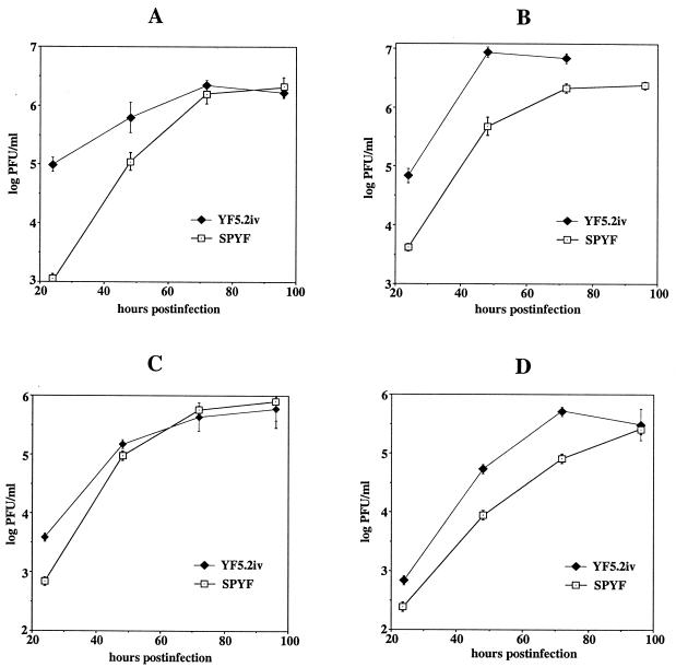
FIG. 4.
Growth curves of SPYF and YF5.2iv viruses in cell culture. Experiments were conducted as described in Materials and Methods, using multiplicities of infection of 0.03 PFU/cell. Samples were obtained in triplicate and titrated on Vero cells, and values indicate mean log PFU/ml ± standard deviation. (A) BHK cells. (B) SW-13 cells. (C) Vero cells. (D) C6/36 cells.
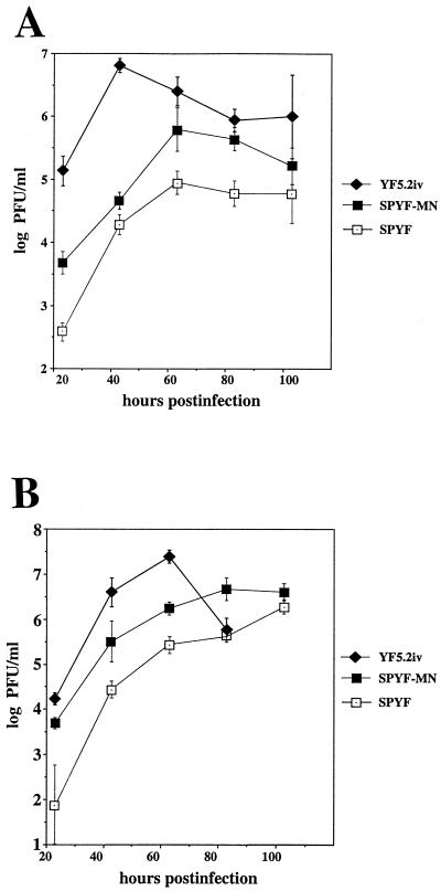
FIG. 6.
Growth curve analysis of SPYF, SPYF-MN and YF5.2iv viruses in BHK and SW-13 cells. Experimental procedures were as for Fig. 4, except that the multiplicity of infection in this experiment was 0.003 PFU per cell. (A) BHK cells. (B) SW-13 cells.
RESULTS
Properties of the neuroadapted SPYF virus.
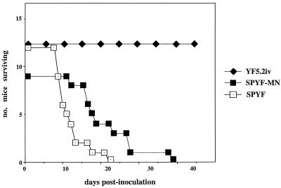
Serial passage of YF17D virus in mouse brain is known to result in enhanced neurovirulence for young adult mice relative to nonpassaged virus. In a previous study, when such neuroadapted virus was tested by inoculation of the olfactory bulb, it replicated more rapidly, generated a higher CNS virus burden, and caused earlier death than the nonadapted virus (55). In that study it was shown that there was genetic heterogeneity in the E protein of the neuroadapted strain, from which clones were derived directly from the original suckling mouse brain virus preparation. Hence we subsequently generated plaque-purified virus isolates from this stock and verified the neurovirulence properties prior to molecular cloning of the full-length SPYF genome. The original PYF virus (55) was passaged in adult SCID mice, followed by plaque purification of the brain-associated virus, amplification in mouse brain, and final growth in cell culture to provide a working virus stock free of mouse brain substances. This preparation, referred to as SPYF virus, was tested for virulence in SCID/ICR mice using YF5.2iv virus (derived from YF17D infectious clone), which had undergone the same plaque purification steps except for the passages in mice, as a control. Previous studies with YF5.2iv virus derived from this clone indicate that it has properties which generally resemble the YF17D vaccine (17, 33, 43, 55), although there are a few sequence differences between the two viruses (see below). Figure 1 shows the survival data for SCID/ICR mice subjected to i.p. inoculation with the SPYF and YF5.2iv viruses. SPYF caused a fatal encephalitis with an average incubation period of approximately 11 days (range, 8 to 21 days), whereas death associated with the YF5.2iv virus required considerably longer (average survival time, 20 weeks; minimum survival time, 46 days; range, 46 to 259 days [12 mice analyzed]). In some cases, YF5.2iv-inoculated mice have survived for prolonged periods (beyond 1 year) and no virus was recovered from the CNS at the time of death. In Fig. 1, SPYF-MN refers to the engineered cDNA clone of SPYF which is discussed below.
FIG. 1.
Mortality data for SCID/ICR mice inoculated with 106 PFU of SPYF, YF5.2iv, or SPYF-MN viruses by the i.p. route. Experimental procedures were carried out as described in Materials and Methods. The mortality curves were constructed using data from three separate experiments in which similar survival times were recorded within groups of mice inoculated with the same viruses.
Virus distribution in tissues of SCID mice.
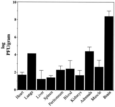
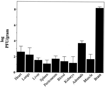
Experiments were next done to determine the distribution and content of SPYF virus in the SCID/ICR mice at the time of fatal encephalitis using plaque titration of tissue homogenates. This method was found to be sensitive for detection of even low levels of infectious virus (approximately 1 log PFU/g of tissue) from these animals. Results are shown in Fig. 2. High levels of SPYF virus were measured in the brains. The average content was approximately 8.2 log PFU/g at the time of onset of a moribund condition in such animals. A range of peripheral tissues was also tested for virus content. Almost all tissues tested from several individual mice contained measurable amounts of virus; however, there was variability in the levels detected. Moderate levels were found in lungs and adrenal glands (approximately 4 log PFU/g). Somewhat lower levels of virus were found in the hindlimb muscle, the kidneys, and the peritoneal membrane at the site of virus inoculation. The lowest levels were found in the livers and spleens (approximately 1 log PFU/g). Virus was detectable in the blood of these mice at approximately 2 log PFU/ml. Some of the tissues that did not yield direct plaque titers were found to contain infectious virus by culture of the tissue extract in liquid medium on Vero cells and plaque titration of the medium from the subculture, indicating that they harbored virus below the limit detectable by plaque assay.
FIG. 2.
Content of SPYF virus in tissues of SCID/ICR mice which had been inoculated by the i.p. route as for Fig. 1. Procedures were performed as described in Materials and Methods. Virus content is shown as mean PFU per gram of infected tissue, plus or minus the standard deviation, based on titrations done on three to five mice for each group, except for lung tissue content (average of two samples). Plaque assays were performed on Vero cells. Tissues were harvested on days 9, 10, 11, 12, and 15 after inoculation, when individual mice showed signs of encephalitis.
Virulence properties of SPYF in normal mice.
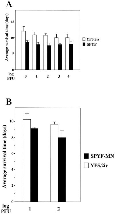
As described in a previous investigation (55), the parental PYF virus exhibits high neurovirulence in normal mice by i.c. inoculation, relative to nonneuroadapted YF5.2iv virus. To determine if SPYF exhibited a similar increased virulence for normal mice, a graded dose i.c. challenge was conducted in comparison to the YF5.2iv virus. Five-week-old ICR mice (Harlan) were used in this experiment. Both viruses caused mortality in all mice tested of this age, even at a dose of 1.0 PFU (Fig. 3A). However, average survival times for the SPYF virus were significantly shorter than for YF5.2iv for all doses tested (P < 0.05; two-sided Wilcoxon test). This suggested more rapid replication of the SPYF strain in the brains of these mice, which is consistent with the studies of neuroadapted virus production in infected mouse brain (55). To determine if any fatal neuroinvasiveness occured with the SPYF virus, normal ICR mice (Taconic; same ICR background as the SCID/ICR lineage) were tested by i.p. inoculation. In experiments with weanling mice inoculated with the same doses of virus used for the SCID/ICR experiments, six of eight of these mice succumbed to peripheral inoculation with SPYF (average survival time, 9.33 days), whereas zero of eight succumbed after inoculation with YF5.2iv (P < 0.05) (Table 1). To determine if the susceptibility to SPYF was age dependent, 5-week-old mice were also tested. In this case, SPYF still caused substantial mortality (4 of 10 mice; average survival time, 10.5 days) (Table 1). The quantities of SPYF virus in the brains were 7.18 and 7.32 log PFU/g (two mice in the 3-week-old group) and 6.9 and 7.32 log PFU/g (two mice in the 5-week-old group). These results were somewhat surprising because the original PYF virus preparation does not exhibit this degree of neuroinvasiveness in adult mice (data not shown). More subtle measures of increased virulence, such as the occurrence of subclinical neuroinvasion with either SPYF or the YF5.2iv virus in mice which did not succumb to infection, were not analyzed in these experiments. The ICR mice which succumbed to infection with SPYF virus in these experiments were also tested for viremia using a direct plaque assay as shown in Fig. 2. No viremia was detectable in these samples, despite the progression to lethal encephalitis in these animals.
FIG. 3.
Average survival times of ICR mice (Harlan) subjected to graded dose i.c. inoculations. (A) SPYF compared to YF5.2iv virus. (B) SPYF-MN compared to YF5.2iv virus. Groups of five to six mice at 5 weeks of age were used for these experiments. Average survival times in days (means ± standard deviations) are shown. Differences between SPYF and YF5.2iv were significant for all doses (see text). Differences between SPYF-MN and YF5.2iv were significant for both doses (see text).
TABLE 1.
i.p. inoculation of ICR mice
| Virus | Age (wks) | Dose (log10 PFU) | Mortality (no. of deaths/total [%])a | Average survival time (days) |
|---|---|---|---|---|
| SPYF | 3 | 6 | 6/8 (75) | 9.33 |
| 5 | 6 | 4/10 (40) | 10.5 | |
| SPYF-MN | 3 | 6 | 0/8 (0) | NAb |
| YF5.2iv | 3 | 6 | 0/8 (0) | NA |
P value = 0.003 for comparison of mortality rates of 3-week-old SPYF mice to the other viruses.
NA, not applicable.
Growth efficiency of SPYF in cell culture.
To determine if the neurovirulence phenotypes of the SPYF and YF5.2iv viruses correlated with any differences in replication in cell culture, the two viruses were compared for growth efficiency in several different cell lines (Fig. 4). In all mammalian cell lines tested, SPYF exhibited less efficient replication, as indicated by the rate of virus production over the first 24 to 48 h. This was most pronounced in BHK cells (panel A), where virus yields at 24 h differed by approximately 2 logs. Differences of 0.5 to 1.0 log were observed in the other cell lines at 24 h (panels B, C, and D). Peak titers of virus occurred between 48 and 72 h for both viruses in BHK and Vero cells, reaching approximately 6 log of virus per ml. On SW-13 and C6/36 cells, SPYF exhibited lower titers than YF5.2iv over the interval examined. The plaque efficiency of the two viruses also differed. SPYF formed more distinct plaques on Vero cells than YF5.2iv did, but they were slightly smaller in size (0.9 versus 0.8 mm). In contrast, on SW-13 cells, SPYF formed small indistinct plaques of <0.5 mm, whereas YF5.2iv formed larger distinct plaques (2 to 3 mm) (data not shown).
Nucleotide sequence analysis of the SPYF virus.
In order to define the extent of nucleotide substitutions which differentiated the genome of the SPYF virus from nonneuroadapted YF5.2iv, the entire SPYF genome was sequenced for duplicate reverse transcription-PCR-derived clones from the plaque-purified virus isolate described above. A total of 29 nucleotide substitutions were identified in the SPYF sequence relative to that of YF5.2iv. The differences were distributed throughout many regions of the viral genome (Fig. 5 and Table 2). One of the two clones contained a silent substitution (an A-to-T transversion at nt 4150), but otherwise all nucleotide and predicted amino acid substitutions were the same for the two clones. The E protein contained the most amino acid differences of all the proteins. These occurred at amino acid positions 52, 173, 305, 326, and 380 (numbering relative to the amino terminus of E). A single amino acid substitution was present in the NS1 protein. The NS2A protein contained three substitutions, the NS4A and NS4B proteins each contained one substitution, and the NS5 protein contained two predicted substitutions. Two substitutions were identified in the 3′ untranslated region. To gain insight into whether any of the SPYF substitutions were likely to represent virulence determinants, the sequence of SPYF was compared with those of other YFV strains known to exhibit a high level of virulence (Table 2). Comparison of these nucleotide and amino acid substitutions revealed that nine of the nucleotide substitutions of SPYF were identical to those of the Asibi, FNV, and FVV viruses, resulting in seven amino acid identities (positions 52, 173, and 380 of the E protein, position 173 of NS2A, position 107 of NS4A, position 232 of NS4B, and position 657 of NS5). Two other SPYF substitutions occurred at positions where the virulent strains differ from YF5.2iv, but these involved replacement with a different residue (position 305 of E and position 169 of NS2A). At one position (NS2A residue 17), SPYF contained the residue found in the FNV strain. Nucleotide sequence changes previously reported in the prM-through-NS2A region of the original PYF viral genome (55) were also identified in the SPYF sequence. It is important to point out that the sequence of the YF5.2iv virus differs from the consensus sequence of YF17D (17D-204) (50) at three nucleotide positions (nt 8212, C to T [silent]; nt 4025, G to A [methionine to valine at NS2A residue 173]; and nt 5641, G to A [silent]) (51). Although SPYF contains valine at NS2A residue 173, a subclone of the YF17D-204 strain also contains this residue (50), suggesting that it is not a critical virulence determinant.
FIG. 5.
Schematic of the YFV genome with its open reading frame, 5′ and 3′ untranslated regions, and positions of nucleotide and amino acid substitutions between SPYF and YF5.2iv viruses. Amino acid substitutions are shown above the genome. Silent nucleotide changes and those in the 3′ untranslated region are shown below the genome. Residues or nucleotides which are common to SPYF, Asibi, FNV, and/or FVV are marked with dark shading. Open circles and squares indicate changes unique to SPYF. Diagonal shading indicates the position where differences occur among YF5.2iv, SPYF, and the virulent YFV strains (Asibi, FNV, and FVV). Horizontal shading indicates identity with FNV (residue 17; NS2A) or with Asibi and FVV (nucleotide 1432).
TABLE 2.
Comparison of YFV genomes
| Name of region | Position of substitution
|
Substitution (nucleotide/amino acid) in indicated virus
|
|||||
|---|---|---|---|---|---|---|---|
| Nucleotide | Amino acid | YF5.2iva | SPYF | Asibib | FNVc | FVVc | |
| E | 1127 | 52 | a/Arg | g/Gly | g/Gly | g/Gly | g/Gly |
| 1432 | 153 | u/Asn | c/Asn | c/Asn | a/Lys | c/Asn | |
| 1491 | 173 | u/Ile | c/Thr | c/Thr | c/Thr | c/Thr | |
| 1886 | 305 | u/Phe | g/Val | u/Ser | u/Ser | u/Ser | |
| 1949 | 326 | a/Lys | g/Glu | a/Lys | a/Lys | a/Lys | |
| 2112 | 380 | g/Arg | c/Thr | c/Thr | c/Thr | c/Thr | |
| 2431 | 486 | u/Phe | c/Phe | u/Phe | u/Phe | u/Phe | |
| NS1 | 2554 | 34 | c/Tyr | u/Tyr | c/Tyr | c/Tyr | c/Tyr |
| 3135 | 228 | u/Leu | a/Gln | u/Leu | u/Leu | c/Pro | |
| NS2A | 3557 | 17 | a/Met | u/Leu | a/Met | c/Leu | a/Met |
| 4014 | 169 | u/Phe | c/Ser | u/Leu | u/Leu | u/Phe | |
| 4025 | 173 | a/Met | g/Val | g/Val | g/Val | g/Val | |
| 4054 | 182 | u/Asn | c/Asn | c/Asn | c/Asn | c/Asn | |
| 4150 | 214 | a/Ala | u/Ala | a/Ala | a/Ala | a/Ala | |
| NS3 | 5338 | 256 | c/Gly | u/Gly | c/Gly | c/Gly | c/Gly |
| 5554 | 328 | u/Asp | c/Asp | u/Asp | u/Asp | u/Asp | |
| NS4A | 6529 | 30 | c/Phe | u/Phe | u/Phe | u/Leu | u/Phe |
| 6664 | 75 | a/Lys | g/Lys | a/Lys | a/Lys | a/Lys | |
| 6758 | 107 | g/Val | a/Ile | a/Ile | a/Ile | a/Ile | |
| NS4B | 7492 | 202 | u/Ala | c/Ala | u/Ala | u/Ala | u/Ala |
| 7580 | 232 | c/His | u/Tyr | u/Tyr | u/Tyr | u/Tyr | |
| NS5 | 7993 | 119 | g/Leu | a/Leu | g/Leu | g/Leu | g/Leu |
| 8212 | 192 | u/Leu | c/Leu | c/Leu | c/Leu | c/Leu | |
| 9151 | 505 | a/Gly | g/Gly | a/Gly | a/Gly | a/Gly | |
| 9605 | 657 | g/Asp | a/Asn | a/Asn | a/Asn | a/Asn | |
| 9719 | 695 | a/Ile | c/Leu | a/Ile | a/Ile | a/Ile | |
| 10243 | 869 | a/Leu | g/Leu | g/Leu | g/Leu | g/Leu | |
| 3′ untranslated region | 10555 | a | g | a | a | a | |
| 10631 | t | c | t | t | t | ||
Taken together, these results suggest that the identified substitutions in SPYF most likely represent consensus sequence elements among virulent YF viruses and not random errors introduced by the cloning procedures during derivation of SPYF. However, residue 326 of the E protein may represent a clonal substitution unique to the plaque isolate of SPYF derived in these experiments, since it is not present in the other virulent YFV strains. The glutamate substitution is also not found in the parental PYF preparation, which has a lysine residue at this position (55). Silent nucleotide changes between SPYF and YF5.2iv were found at 14 positions and were of two types. Nine of these were unique to SPYF, and five were common to Asibi and other virulent strains. The two mutations in the 3′ terminus of the genome were unique to the SPYF virus.
Construction and properties of the SPYF-MN infectious clone.
To assess the importance of the multiple nucleotide substitutions identified in the SPYF virus for the neurovirulence phenotype, they were introduced into the YF5.2iv infectious clone to evaluate whether the resulting virus would exhibit the properties of the parental SPYF virus. All nucleotide substitutions except for two silent changes (nt 5338 and 8212) were introduced. Transfection of Vero cell monolayers with RNA transcripts derived from the template yielded infectious virus within approximately 5 days. The plaque size of this virus (hereafter referred to as SPYF-MN) on SW-13 cells was small (1 mm), resembling that of the parental SPYF virus, but was larger in size (2.5 mm) and more distinct on Vero cells. This presumably reflected the clonal nature of the virus derived from the neuroadapted PYF strain, which does exhibit some heterogeneity in plaque size. Further characterization of the SPYF-MN virus included growth curve studies in BHK and SW-13 cells in comparison to the YF5.2iv and SPYF viruses (Fig. 6). These experiments revealed that SPYF-MN was impaired in replication efficiency in both cell lines relative to YF5.2iv, particularly in BHK cells. However, it replicated more efficiently than the parental SPYF virus, with levels of virus production intermediate between SPYF and YF5.2iv.
Virulence properties of the SPYF-MN virus.
To determine if the virulence properties of the engineered SPYF-MN virus resembled those of its parental SPYF virus, SCID/ICR mice were tested for their susceptibility using the experimental design shown in Fig. 1. The average time to fatal encephalitis caused by the SPYF-MN virus in SCID/ICR mice was 20 days (range, 11 to 35 days). This was similar to the average survival time of 11 days for the SPYF parent, but the difference was significant (Fig. 1) (P < 0.05; Wilcoxon test). However, the difference in average survival time between SPYF-MN and the YF5.2iv virus (20 weeks) was highly significant (P < 0.01). Thus, the SPYF-MN virus was not exactly identical to the parental SPYF virus in its virulence properties for SCID/ICR mice but could be classified as highly virulent relative to the YF5.2iv virus. To determine if the neuroinvasiveness of SPYF-MN was restricted to SCID/ICR mice, normal ICR mice (Taconic) at 3 weeks of age received the same i.p. dose as was used in the SCID/ICR experiments. In this case, SPYF-MN did not exhibit any apparent neuroinvasiveness, with all mice appearing healthy during the observation period (Table 1). This was in contrast to the results with the SPYF virus, which as stated earlier was neuroinvasive in both 3- and 5-week-old mice, with mortalities of 75 and 40% in these respective groups.
To further compare the virulence properties of the SPYF-MN virus with its SPYF parent, tissue titration studies were performed on the SCID/ICR mice which had succumbed to infection in order to determine the distribution and extent of virus burden at the time of fatal encephalitis (Fig. 7). The virus content in tissues of these mice was distributed similarly to that in mice infected with SPYF (Fig. 2), but in some cases the magnitude differed. Titers of brain-associated virus were essentially the same as those observed with SPYF (8.2 log PFU/g). Virus was isolated in similar amounts from adrenal glands (approximately 4.0 log PFU/g). Most other tissues contained quantities of titratable virus similar to those seen with the SPYF virus, except lower levels were found in the lungs, and the viremia was between 0.5 and 1.0 log PFU/ml lower than with the SPYF virus. Taken together, these results suggest that some or all of the genetic determinants introduced into the SPYF-MN virus govern replication efficiency in the peripheral tissues of SCID mice, in conjunction with their effects on inducing fatal encephalitis in this host.
FIG. 7.
Content of SPYF-MN virus in tissues recovered from SCID/ICR mice after i.p. inoculation as in Fig. 1. Experimental procedures were as described for Fig. 2. Values (mean log PFU/g ± standard deviation) were determined from between four and seven mice in these experiments. Tissues were harvested on days 15 (two mice), 17, 20, 23, 24, and 33, when individual mice showed signs of encephalitis.
To further investigate the relationship between replication of YFV in peripheral tissues and the occurrence of encephalitis, experiments were done to see if the YF5.2iv virus generated a virus burden in SCID mouse tissues similar to those seen with the SPYF and SPYF-MN viruses. Three SCID/ICR mice infected with YF5.2iv were sacrificed at the same time that mice inoculated with SPYF-MN were succumbing to encephalitis (between 2 and 3 weeks postinfection), and virus contents were determined in the tissue homogenates as for Fig. 2 and 7. The peripheral tissues and brains of these mice were devoid of any detectable virus based on plaque assay, except for a single sample of peritoneal membrane from one mouse which yielded 2 log PFU/g. Thus, a lower virus burden is present in mice infected with the YF5.2iv virus at the time that mice infected with SPYF-MN have accumulated their peak virus burdens, despite similar doses of the two viruses being administered. This suggests that YF5.2iv is either less infectious or replicates less efficiently in mouse tissues than SPYF and SPYF-MN.
Because of the large difference in survival time between SCID/ICR mice infected with YF5.2iv virus by the i.p. route and those infected with the SPYF and SPYF-MN viruses, experiments were done to determine if the incubation time for fatal encephalitis after i.c. inoculation differed among the three viruses. Although this seemed an unlikely possibility, it was not known how the absence of an immune system in SCID mice would influence the behavior of virus after entry into the CNS. For these experiments, dose ranging was done at low doses (0.5 to 100 PFU per mouse), which was predicted to mimic the amount of virus entering the brain during neuroinvasion following peripheral infection (based on the viremia data shown in Fig. 2 and 7). As shown in Table 3, 100% mortality was observed with as little as 5 PFU of SPYF virus, but this dose was sublethal for both SPYF-MN and YF5.2iv. Differences between the average survival times for SPYF and either SPYF-MN or YF5.2iv at this dose were significant (P = 0.025), but the difference between SPYF-MN and YF5.2iv was not. A dose of 100 PFU caused 100% mortality with both the SPYF-MN and YF5.2iv viruses; however, the difference between the average survival times at this dose was significant (11 versus 23 days; range, 10 to 12 days versus 10 to 56 days, respectively). Although the average survival time appeared longer for mice receiving 100 PFU of YF5.2iv virus than for those receiving 5 PFU, it is important to note that only three of five mice died in the latter group. All mice inoculated with 0.5 PFU of virus survived, except for one recipient of SPYF-MN which died 11 days postinoculation. The brain content of virus at the time of onset of the moribund condition was determined for mice receiving the SPYF-MN and YF5.2iv viruses. Values of 7.64 and 8.14 log PFU/g were obtained for SPYF-MN (two mice tested). Values of 7.19, 7.43, and 7.27 log PFU/g were obtained for YF5.2iv (three mice tested).
TABLE 3.
i.c. inoculation of SCID/ICR mice
| Virus | Dose (PFU) | Mortality (no. of deaths/total [%]) | Average survival time (days) |
|---|---|---|---|
| SPYF | 100 | NT | NAe |
| 5 | 6/6 (100)a | 9.6c | |
| 0.5 | 0/5 (0)a | NA | |
| SPYF-MN | 100 | 6/6 (100)b | 11d |
| 5 | 3/5 (60) | 10.66a | |
| 0.5 | 1/5 (20)b | 11 | |
| YF5.2iv | 100 | 6/6 (100) | 23d |
| 5 | 3/5 (60) | 12.33c | |
| 0.5 | 0/5 (0) | NA |
P value for comparison of mortality rates and average survival times among groups is 0.002.
P = 0.017.
P = 0.025 (one-sided).
P < 0.025 (one-sided).
NA, not applicable.
The neurovirulence of the SPYF-MN and YF5.2iv viruses was also compared in normal ICR mice (Harlan) (Fig. 3B). At two doses, 10 and 100 PFU delivered by i.c. inoculation, mortality was 100% for both viruses. Average (± standard deviation) survival times were 9.14 ± 1.0 and 8.0 ± 0.8 days for these doses of SPYF-MN versus 10.16 ± 0.75 and 9.66 ± 0.5 days for YF5.2iv, respectively, and were significantly different between the two viruses in both cases (P < 0.05). Taken together, the data from these various experiments indicate that SPYF is the most virulent of these viruses and that SPYF-MN is more virulent than YF5.2iv for SCID and normal ICR mice, based on mortality rates and average survival times. The failure of the YF5.2iv virus to cause rapidly fatal infection in SCID mice did not result from an inability to replicate in the CNS of these mice, since lethal virus burdens could be generated in some cases if a sufficient quantity of virus was introduced (between 5 and 100 PFU). The average incubation times for fatal encephalitis with the three viruses after i.c. inoculation in SCID mice ranged from 9 to 23 days and differences were statististically significant among the groups. It is also notable that the neuroadapted viruses were more virulent for ICR mice (Harlan) than for SCID/ICR mice of the same age, both in terms of dose required for 100% mortality and average survival time (compare Fig. 3 and Table 3). This could reflect differences in susceptibility to YFV among outbred mice of different lineages, as observed previously (4), or some differential effect associated with the SCID mutation itself on viral pathogenesis.
DISCUSSION
YF17D, similar to other flaviviruses, exhibits a high level of neurovirulence when introduced into the CNS of mice but usually does not cause fatal encephalitis following infection of immunocompetent adult mice by a peripheral route of inoculation. It has been observed that the incubation time for the encephalitis can be measurably shortened by serial passage of the virus in mouse brain, and this process is associated with selection for viruses with enhanced replication efficiency in neural tissue (37, 56, 59). Direct comparison of the replication efficiencies and virulence properties in mouse brain of genetically defined strains of neuroadapted and nonneuroadapted flaviviruses is therefore an approach to defining the virus-specific factors which govern this process and is a first step towards understanding the mechanisms involved in the pathogenesis of encephalitis in this model.
The E protein is a major virulence factor for flaviviruses, with numerous studies demonstrating that determinants within this protein affect virulence in the mouse model (reviewed in reference 36; see also references 10, 32, 45, 46, 52, and 54). It is becoming evident, however, that other molecular determinants involved in the functions of either nonstructural proteins or the 5′ and 3′ untranslated regions also influence the virulence phenotypes of flaviviruses (7, 14, 31, 38, 43, 47). To gain insight into this process, we determined the genomic sequence of a neurovirulent plaque isolate selected from the mouse-neuroadapted PYF strain. This study differs from various other investigations using engineered virus mutants because the substitutions we identified are directly related to the process of neuroadaptation and their presence in other highly virulent strains such as Asibi and FVV suggests a strong association with the virulence phenotype. The multiple sequence differences detected in the SPYF virus compared to nonneuroadapted virus were unlikely to represent deleterious mutations accumulated during plaque purification or molecular cloning, since virus engineered to contain the substitutions displayed cell culture properties that were generally similar to the original SPYF virus. In studies of virulence, this SPYF-MN molecular clone was also similar to the SPYF parent based on neuroinvasiveness and neurovirulence for SCID/ICR mice. In normal ICR mice, however, SPYF-MN differed from SPYF in being nonneuroinvasive but had neurovirulence properties which were of a higher grade than the YF5.2iv virus (based on average survival times; Fig. 3B). Thus, the SCID mouse was a more sensitive model than normal mice to discriminate the genetic basis for virulence determinants of YFV. This failure to reconstruct a full virulence phenotype using cDNA-derived virus clones has been observed with other neurotropic flaviviruses (9). In the present case, this may result from clonal differences in neurovirulence determinants among viruses in the heterogeneous SPYF pool. Alternatively, mutations introduced by SP6 transcription during synthesis of infectious RNA transcripts could confer properties on the virus which reduce the effects of neurovirulence determinants. However, the results do indicate that one or more of the nucleotide sequence differences identified between the YF5.2iv and SPYF-MN viruses largely governs the virulence of SPYF for SCID/ICR mice. Since some of the substitutions observed in SPYF were common to the Asibi, FVV, and FNV viruses, it is not likely that they are sequence artifacts unrelated to the neurovirulence phenotype.
With respect to the E protein, it is believed that molecular determinants distributed throughout domains I, II, and III, as well as the stem-anchor region, are critical for the structural changes in this protein which are required for virus entry (1, 2, 21, 57). Mutations at numerous positions are in turn capable of modulating the virulence properties conferred by this protein (49). This has generally been the case for encephalitic flaviviruses which have been characterized in mice (36). Consistent with this hypothesis, predicted amino acid substitutions were found in domains I, II, and III in the E protein of the SPYF virus. Mutation at residue 52 has been associated with neurovirulence differences and alterations of virus-cell interactions for Japanese encephalitis (JE) virus (20). Position 173 has been identified as a site which defines a YFV wild-type epitope and is associated with altered neurovirulence of YF17D virus (52). Positions 326 and 380 lie within the putative receptor binding domain at two distinct regions which have been proposed as critical for the function of this portion of the E protein (6). A YF17D substrain-specific epitope is known to involve the adjacent residue 325, whose substitution is associated with alteration in mouse neurovirulence (54). Residue 380 is included within the RGD motif of mosquito-borne flaviviruses, in which mutations have been shown to affect the efficiency of virus spread in cell culture and neuroinvasion in mice (29, 60). Collectively, these observations suggest that the substitutions in the SPYF E protein are likely to functionally alter its properties during pathogenesis of encephalitis in the mouse model. However, unlike cases where as few as two residues govern the neurovirulence of a flavivirus (23), the presence of five substitutions in the E protein of SPYF suggests that neurovirulence may depend on multiple genetic determinants, as has been shown in studies of the JE virus E protein (4). Fine mapping of these candidate virulence determinants within the SPYF E protein using the SCID/ICR mouse model will allow identification of the critical residues and may give insight into potential mechanisms underlying the neuroinvasive and neurovirulence properties of this virus. Since the effects of substitutions in the nonstructural region on neurovirulence have not been determined, it is premature to conclude that the E protein is the only protein responsible for the enhanced neurovirulence of SPYF. Studies on the attenuation of the Asibi strain of YFV suggest a selection for mutations in the E protein as well as nonstructural proteins, even after limited passages in cell substrates (14, 18). In particular, substitutions in NS1, NS2A, NS4B, and NS5 have been noted. It is conceivable that such substitutions enhance neurovirulence by increasing the efficiency of RNA synthesis, leading to higher virus burdens and induction of apoptotic cell death (13). In addition, comparison of the sequences of SPYF-MN, Asibi, FVV, and FNV viruses reveals that some silent nucleotide substitutions are common to these viruses. This could reflect selection for RNA structures which impart higher replication efficiency or confer host range effects which are important during pathogenesis. Although this is not a well-explored area, it is increasingly being realized that effects of mutations on RNA structures are likely to influence flavivirus replication (48).
The pattern of disease occurring in the SCID mouse subjected to infection with neuroadapted virus is characterized by a high virus burden in the CNS, together with lower quantities of virus in extraneural tissues and a relatively low-level viremia. This level of viremia is typical of that generated by attenuated virus strains in other rodent models of flavivirus neuropathogenesis (40, 44, 58). The presence and magnitude of viremia has generally been regarded as an important factor for neuroinvasion (19, 41), but the consequences of a given level may depend to some extent on the host immune response to circulating virus or the associated virus burden in the peripheral tissues of infected animals. The low viremia observed in this SCID/ICR model may result in part from YFV being inherently less efficient than other encephalitic flaviviruses for replication in mice because of its evolution and adaptation towards lymphoid tissue of primates (42). In addition, clearance by nonspecific immune defenses such as macrophages and/or natural killer cells may be a mechanism which reduces circulating virus to relatively low levels in this model (39, 62). The higher virus burden in SCID/ICR mice inoculated with SPYF-MN than those inoculated with YF5.2iv at 2 to 3 weeks postinfection suggests that genetic determinants within SPYF-MN promote more efficient replication in peripheral tissues in this host. YF17D vaccine strains have been observed to be less infectious for mice than virulent strains, in an age-dependent fashion (15). The failure of YF5.2iv to generate much virus burden in SCID/ICR mice may therefore reflect a low efficiency of infection even at the doses used in our experiments. Alternatively, resistance of SPYF to clearance by the innate immune response has not been eliminated as an explanation for the observed differences in virus burden. For instance, evidence exists from studies of other flavivirus infections in SCID mice that alpha and beta interferons play important roles in host defense against this family of viruses (24, 27), but there is little information available on virus-host interactions at this level (12).
The difference between SPYF and SPYF-MN with respect to mortality of normal ICR mice inoculated by the peripheral route is also notable. This difference could be based on a highly neuroinvasive quasispecies within the SPYF population which is not represented by SPYF-MN. Alternatively, if a certain threshold of viremia is required for neuroinvasion, SPYF may be more efficient in achieving this prior to induction of sufficient neutralizing antibody activity and/or cytotoxic T lymphocytes which then act to eliminate cirtculating virus. In contrast to what was observed in SCID/ICR mice, the absence of detectable viremia in ICR mice at the time of fatal encephalitis is consistent with the clearance of circulating virus by such mechanisms (41).
The average incubation time to onset of fatal encephalitis after i.c. inoculation of SCID/ICR mice with YF5.2iv virus was somewhat longer than for the SPYF and SPYF-MN viruses (Table 3); however, the time interval between peripheral inoculation and death was considerably longer (Fig. 1). A difference was also observed in the amount of virus required to cause lethal encephalitis after i.c. inoculation of these mice with the three viruses, with SPYF-MN and YF5.2iv being sublethal at doses at or below 5 PFU (Table 3). These results suggest that the time required for neuroinvasion is the principle variable which accounts for the prolonged delay before onset of encephalitis with YF5.2iv virus. However, entry of only small amounts of YF5.2iv virus (<5 PFU) may not always be sufficient to generate a lethal infection in the CNS. Hence, differences in neuroinvasiveness and neurovirulence may both be involved in the virulence properties of SPYF compared to YF5.2iv in SCID/ICR mice. The long incubation time of the nonneuroadapted virus presumably reflects a requirement for accumulation of a virus burden in peripheral tissues. In this regard, neuroadaptation of YF17D virus may reflect a generalized adaptation to mouse tissue which is the basis for the more efficient replication of SPYF than of YF5.2iv virus in all extraneural tissues examined. It is possible that nonspecific immune defenses may be more efficient in clearing virus from peripheral tissues than from the CNS, which could also contribute to the long interval needed for accumulation of YF5.2iv virus prior to neuroinvasion. Clearly, the level of virus burden appears to be related to the occurrence of neuroinvasion. It is not known whether neuroinvasion results from a critical level of circulating virus or requires participation of inflammatory responses from nonspecific host defenses such as activated macrophages and natural killer cells, as suggested by other models (30). In any case, due to the multiple number of nucleotide substitutions between the SPYF-MN and YF5.2iv viruses, further studies with engineered mutants are needed to establish whether the determinants which enhance growth of virus in the periphery and those which promote neuroinvasion also govern replication efficiency in the CNS. It will also be of interest to establish whether there is a redundancy in the effects of multiple independent mutations on the virulence phenotype or whether certain residues have dominant effects. The mechanism of enhanced neurovirulence of neuroadapted YFV has been proposed to involve the generation of a high virus burden in the CNS resulting from an enhanced replication efficiency and leading to death of target cells (55). Other mechanisms could also contribute to neuronal death, including deleterious effects of the virus-specific inflammatory response. In the SCID model this is certainly not the case, although a detailed analysis of the CNS inflammatory response is needed to determine the role of nonspecific host immune responses, which have been implicated in the pathogenesis of Murray Valley encephalitis virus (3).
Finally, it remains undetermined whether any mutations occur in the YF5.2iv virus during the long incubation period preceeding the onset of encephalitis which occurs in some mice infected with this nonneuroadapted virus. If so, this might reflect emergence of viruses with an increase in neurovirulence. Evolution of virulent viruses during persistent infection of SCID mice with Sindbis virus has been described as a virus-specific mechanism of pathogenesis (26). This question is under investigation for YFV using brain-associated virus recovered from mice which have succumbed to infection with YF5.2iv virus after prolonged time intervals.
ACKNOWLEDGMENTS
This work was supported by grants from the NIH (AI-37646) and the Edward Mallinckrodt, Jr., Foundation.
REFERENCES
- 1.Allison S L, Schalich J, Stiasny K, Mandl C W, Kunz C, Heinz F X. Oligomeric rearrangement of tick-borne encephalitis virus envelope proteins induced by an acidic pH. J Virol. 1995;69:695–700. doi: 10.1128/jvi.69.2.695-700.1995. [DOI] [PMC free article] [PubMed] [Google Scholar]
- 2.Allison S L, Stiasny K, Stadler K, Mandl C W, Heinz F X. Mapping of functional elements in the stem-anchor region of tick-borne encephalitis virus envelope protein E. J Virol. 1999;73:5605–5612. doi: 10.1128/jvi.73.7.5605-5612.1999. [DOI] [PMC free article] [PubMed] [Google Scholar]
- 3.Andrews D M, Matthews V B, Sammels L M, Carrello A C, McMinn P C. The severity of Murray Valley encephalitis in mice is linked to neutrophil infiltration and inducible nitric oxide synthase activity in the central nervous system. J Virol. 1999;73:8781–8790. doi: 10.1128/jvi.73.10.8781-8790.1999. [DOI] [PMC free article] [PubMed] [Google Scholar]
- 4.Arroyo J, Guirakhoo F, Fenner S, Zhang Z-X, Monath T P, Chambers T J. Molecular basis for attenuation of neurovirulence of a yellow fever virus/Japanese encephalitis virus chimera vaccine (ChimeriVax-JE) J Virol. 2001;75:934–942. doi: 10.1128/JVI.75.2.934-942.2001. [DOI] [PMC free article] [PubMed] [Google Scholar]
- 5.Barrett A D T, Gould E A. Comparison of neurovirulence of different strains of yellow fever virus in mice. J Gen Virol. 1986;67:631–637. doi: 10.1099/0022-1317-67-4-631. [DOI] [PubMed] [Google Scholar]
- 6.Bhardwaj S, Holbrook M, Shope R E, Barrett A D T, Watowich S J. Biophysical characterization and vector-specific antagonist activity of domain III of the tick-borne flavivirus envelope protein. J Virol. 2001;75:4002–4007. doi: 10.1128/JVI.75.8.4002-4007.2001. [DOI] [PMC free article] [PubMed] [Google Scholar]
- 7.Bray M, Men R, Tokimatsu I, Lai C-J. Genetic determinants responsible for acquisition of dengue type 2 virus mouse neurovirulence. J Virol. 1998;72:1647–1651. doi: 10.1128/jvi.72.2.1647-1651.1998. [DOI] [PMC free article] [PubMed] [Google Scholar]
- 8.Butrapet S, Huang C Y-H, Pierro D J, Bhamarapravati N, Gubler D J, Kinney R M. Attenuation markers of a candidate dengue type 2 vaccine virus, strain 16681 (PDK-53), are defined by mutations in the 5′ noncoding region and nonstructural proteins 1 and 3. J Virol. 2000;74:3011–3019. doi: 10.1128/jvi.74.7.3011-3019.2000. [DOI] [PMC free article] [PubMed] [Google Scholar]
- 9.Campbell M S, Pletnev A G. Infectious clones of Langat tick-borne flavivirus that differ from their parent in peripheral neurovirulence. Virology. 2000;269:225–237. doi: 10.1006/viro.2000.0220. [DOI] [PubMed] [Google Scholar]
- 10.Chambers T J, Nestorowicz A, Mason P W, Rice C M. Yellow fever/Japanese encephalitis chimeric viruses: construction and biological properties. J Virol. 1999;73:3095–3101. doi: 10.1128/jvi.73.4.3095-3101.1999. [DOI] [PMC free article] [PubMed] [Google Scholar]
- 11.Despres P, Frenkiel M-P, Ceccaldi P-E, Duarte dos Santos C, Deubel V. Apoptosis in the mouse central nervous system in response to infection with mouse-neurovirulent dengue viruses. J Virol. 1998;72:823–829. doi: 10.1128/jvi.72.1.823-829.1998. [DOI] [PMC free article] [PubMed] [Google Scholar]
- 12.Diamond M S, Roberts T G, Edgil D, Lu B, Ernst J, Harris E. Modulation of dengue virus infection in human cells by alpha, beta, and gamma interferons. J Virol. 2000;74:4957–4966. doi: 10.1128/jvi.74.11.4957-4966.2000. [DOI] [PMC free article] [PubMed] [Google Scholar]
- 13.Duarte dos Santos C N, Frenkiel M-P, Courageot M-P, Rocha C F S, Vazeille-Falcoz M-C, Wien M W, Rey F A, Deubel V, Despres P. Determinants in the envelope E protein and viral RNA helicase NS3 that influence the induction of apoptosis in response to infection with dengue type 1 virus. Virology. 2000;274:292–308. doi: 10.1006/viro.2000.0457. [DOI] [PubMed] [Google Scholar]
- 14.Dunster L M, Wang H, Ryman K D, Miller B R, Watowich S J, Minor P D, Barrett A D T. Molecular and biological changes associated with HeLa cell attenuation of wild-type yellow fever virus. Virology. 1999;261:309–318. doi: 10.1006/viro.1999.9873. [DOI] [PubMed] [Google Scholar]
- 15.Fitzgeorge R, Bradish C J. The in vivo differentiation of strains of yellow fever virus in mice. J Gen Virol. 1980;46:1–13. doi: 10.1099/0022-1317-46-1-1. [DOI] [PubMed] [Google Scholar]
- 16.Freestone D S. Yellow fever vaccine. In: Plotkin S A, Mortimer E M, editors. Vaccines. 2nd ed. Philadelphia, Pa: W. B. Saunders; 1994. p. 741-779. [Google Scholar]
- 17.Guirakhoo F, Zhang Z-X, Chambers T J, Delagrave S, Arroyo J, Barrett A D T, Monath T P. Immunogenicity, genetic stability, and protective efficacy of a recombinant, chimeric yellow fever-Japanese encephalitis virus (ChimeriVax-JE) as a live, attenuated vaccine candidate against Japanese encephalitis. Virology. 1999;257:363–372. doi: 10.1006/viro.1999.9695. [DOI] [PubMed] [Google Scholar]
- 18.Hahn C S, Dalrymple J M, Strauss J H, Rice C M. Comparison of the virulent Asibi strain of yellow fever virus with the 17D vaccine strain derived from it. Proc Natl Acad Sci USA. 1987;84:2019–2023. doi: 10.1073/pnas.84.7.2019. [DOI] [PMC free article] [PubMed] [Google Scholar]
- 19.Halevy M, Akov Y, Ben-Nathan D, Kobiler D, Lachmi B, Lustig S. Loss of active neuroinvasiveness in attenuated strains of West Nile virus: pathogenicity in immunocompetent and SCID mice. Arch Virol. 1994;137:355–370. doi: 10.1007/BF01309481. [DOI] [PubMed] [Google Scholar]
- 20.Hasegawa H, Yoshida M, Shiosaka T, Fujita S, Kobayashi Y. Mutations in the envelope protein of Japanese encephalitis virus affect entry into cultured cells and virulence in mice. Virology. 1992;191:158–165. doi: 10.1016/0042-6822(92)90177-q. [DOI] [PubMed] [Google Scholar]
- 21.Heinz F X, Stiasny K, Puschner-Auer G, Holtzmann H, Allison S L, Mandl C W, Kunz C. Structural changes and functional control of the tick-borne encephalitis virus glycoprotein E by the heterodimeric association with protein prM. Virology. 1994;198:109–117. doi: 10.1006/viro.1994.1013. [DOI] [PubMed] [Google Scholar]
- 22.Jennings A D, Whitby J E, Minor P D, Barrett A D T. Comparison of the nucleotide and deduced amino acid sequences of the envelope protein genes of the wild-type French viscerotropic strain of yellow fever virus and the live vaccine strain, French neurotropic vaccine, derived from it. Virology. 1993;192:692–695. doi: 10.1006/viro.1993.1090. [DOI] [PubMed] [Google Scholar]
- 23.Jennings A D, Gibson C A, Miller B R, Mathews J H, Mitchell C J, Roehrig J T, Wood D J, Taffs F, Sil B K, Whitby S N, Whitby J E, Monath T P, Minor P D, Sanders P G, Barrett A D T. Analysis of a yellow fever virus isolated from a fatal case of vaccine-associated human encephalitis. J Infect Dis. 1994;169:512–518. doi: 10.1093/infdis/169.3.512. [DOI] [PubMed] [Google Scholar]
- 24.Johnson A J, Roehrig J T. New mouse model for dengue virus vaccine testing. J Virol. 1999;73:783–786. doi: 10.1128/jvi.73.1.783-786.1999. [DOI] [PMC free article] [PubMed] [Google Scholar]
- 25.Kawano H, Rostapshov V, Rosen L, Lai C-J. Genetic determinants of dengue type 4 virus neurovirulence for mice. J Virol. 1993;67:6567–6575. doi: 10.1128/jvi.67.11.6567-6575.1993. [DOI] [PMC free article] [PubMed] [Google Scholar]
- 26.Levine B, Griffin D E. Molecular analysis of neurovirulent strains of Sindbis virus that evolve during persistent infection of scid mice. J Virol. 1993;67:6872–6875. doi: 10.1128/jvi.67.11.6872-6875.1993. [DOI] [PMC free article] [PubMed] [Google Scholar]
- 27.Leyssen P, Van Lommel A, Drosten C, Schmitz H, De Clercq E, Neyts J. A novel model for the study of the therapy of flavivirus infections using the Modoc virus. Virology. 2001;279:27–37. doi: 10.1006/viro.2000.0723. [DOI] [PubMed] [Google Scholar]
- 28.Liprandi F. Isolation of plaque variants differing in virulence from the 17D strain of yellow fever virus. J Gen Virol. 1981;56:363–370. doi: 10.1099/0022-1317-56-2-363. [DOI] [PubMed] [Google Scholar]
- 29.Lobigs M, Usha R, Nestorowicz A, Marshall I D, Weir R C, Dalgarno L. Host cell selection of Murray Valley encephalitis virus variants altered at an RGD sequence in the envelope protein and in mouse virulence. Virology. 1990;176:587–595. doi: 10.1016/0042-6822(90)90029-q. [DOI] [PubMed] [Google Scholar]
- 30.Lustig S, Danenberg H D, Kafri Y, Kobiler D, Ben-Nathan D. Viral neuroinvasion and encephalitis induced by lipopolysaccharide and its mediators. J Exp Med. 1992;176:707–712. doi: 10.1084/jem.176.3.707. [DOI] [PMC free article] [PubMed] [Google Scholar]
- 31.Mandl C W, Holzmann H, Meixner T, Rauscher S, Stadler P F, Allison S L, Heinz F X. Spontaneous and engineered deletions in the 3′ noncoding region of tick-borne encephalitis virus: construction of highly attenuated mutants of a flavivirus. J Virol. 1998;72:2132–2140. doi: 10.1128/jvi.72.3.2132-2140.1998. [DOI] [PMC free article] [PubMed] [Google Scholar]
- 32.Mandl C W, Allison S L, Holzmann H, Meixner T, Heinz F X. Attenuation of tick-borne encephalitis virus by structure-based site-specific mutagenesis of a putative flavivirus receptor binding site. J Virol. 2000;74:9601–9609. doi: 10.1128/jvi.74.20.9601-9609.2000. [DOI] [PMC free article] [PubMed] [Google Scholar]
- 33.Marchevsky R S, Mariano J, Ferreira V S, Almeida E, Cerqueira M J, Carvalho R, Pissurno J W, Travassos Da Rosa A P A, Simoes M C, Santos C N D, Ferreira I I, Muylaert I R, Mann G F, Rice C M, Galler R. Phenotypic analysis of yellow fever virus derived from cDNA. Am J Trop Med Hyg. 1995;52:75–80. doi: 10.4269/ajtmh.1995.52.75. [DOI] [PubMed] [Google Scholar]
- 34.McMinn P C, Lee E, Hartley S, Roehrig J T, Dalgarno L, Weir R C. Murray Valley encephalitis virus envelope protein antigenic variants with altered hemagglutination properties and reduced neuroinvasiveness in mice. Virology. 1995;211:10–20. doi: 10.1006/viro.1995.1374. [DOI] [PubMed] [Google Scholar]
- 35.McMinn P C, Weir R C, Dalgarno L. A mouse-attenuated envelope protein variant of Murray Valley encephalitis virus with altered fusion activity. J Gen Virol. 1996;77:2085–2088. doi: 10.1099/0022-1317-77-9-2085. [DOI] [PubMed] [Google Scholar]
- 36.McMinn P C. The molecular basis of virulence of the encephalitogenic flaviviruses. J Gen Virol. 1997;78:2711–2722. doi: 10.1099/0022-1317-78-11-2711. [DOI] [PubMed] [Google Scholar]
- 37.Meers P D. Adaptation of the 17D yellow fever virus to mouse brain by serial passage. Trans R Soc Trop Med Hyg. 1959;53:445–457. [Google Scholar]
- 38.Men R, Bray M, Clark D, Chanock R M, Lai C-J. Dengue type 4 virus mutants containing deletions in the 3′ noncoding region of the RNA genome: analysis of growth restriction in cell culture and altered viremia pattern and immunogenicity in rhesus monkeys. J Virol. 1996;70:3930–3937. doi: 10.1128/jvi.70.6.3930-3937.1996. [DOI] [PMC free article] [PubMed] [Google Scholar]
- 39.Monath T P C, Borden E C. Effects of thorotrast on humoral antibody, viral multiplication and interferon during infection with St. Louis encephalitis virus in mice. J Infect Dis. 1971;123:297–300. doi: 10.1093/infdis/123.3.297. [DOI] [PubMed] [Google Scholar]
- 40.Monath T P, Cropp C B, Bowen G S, Kemp G E, Mitchell C J, Gardner J J. Variation in virulence for mice and rhesus monkeys among St. Louis encephalitis virus strains of different origin. Am J Trop Med Hyg. 1980;29:948–962. doi: 10.4269/ajtmh.1980.29.948. [DOI] [PubMed] [Google Scholar]
- 41.Monath T P. Pathobiology of the flaviviruses. In: Schlesinger S, Schlesinger M J, editors. The togaviridae and the flaviviridae. New York, N.Y: Plenum Press; 1986. pp. 375–440. [Google Scholar]
- 42.Monath T P. Yellow fever and dengue: the interactions of virus, vector and host in the re-emergence of epidemic disease. Semin Virol. 1994;5:133–143. [Google Scholar]
- 43.Muylaert I R, Chambers T J, Galler R, Rice C M. Mutagenesis of the N-linked glycosylation sites of the yellow fever virus NS1 protein: effects on virus replication and mouse neurovirulence. Virology. 1996;222:159–168. doi: 10.1006/viro.1996.0406. [DOI] [PubMed] [Google Scholar]
- 44.Ni H, Barrett A D T. Molecular differences between wild-type Japanese encephalitis virus strains of high and low mouse neuroinvasiveness. J Gen Virol. 1996;77:1449–1455. doi: 10.1099/0022-1317-77-7-1449. [DOI] [PubMed] [Google Scholar]
- 45.Ni H, Barrett A D T. Attenuation of Japanese encephalitis virus by selection of its mouse brain membrane receptor preparation escape variants. Virology. 1998;241:30–36. doi: 10.1006/viro.1997.8956. [DOI] [PubMed] [Google Scholar]
- 46.Ni H, Ryman K D, Wang H, Saeed M F, Hull R, Wood D, Minor P D, Watowich S J, Barrett A D T. Interaction of yellow fever virus French neurotropic vaccine strain with monkey brain: characterization of monkey brain membrane receptor escape variants. J Virol. 2000;74:2903–2906. doi: 10.1128/jvi.74.6.2903-2906.2000. [DOI] [PMC free article] [PubMed] [Google Scholar]
- 47.Pletnev A G, Bray M, Lai C-J. Chimeric tick-borne encephalitis and dengue type 4 viruses: effects of mutations on neurovirulence in mice. J Virol. 1993;67:4956–4963. doi: 10.1128/jvi.67.8.4956-4963.1993. [DOI] [PMC free article] [PubMed] [Google Scholar]
- 48.Proutski V, Gaunt M W, Gould E A, Holmes E C. Secondary structure of the 3′-untranslated region of yellow fever virus: implications for virulence, attenuation and vaccine development. J Gen Virol. 1997;78:1543–1549. doi: 10.1099/0022-1317-78-7-1543. [DOI] [PubMed] [Google Scholar]
- 49.Rey F A, Heinz F X, Mandl C, Kunz C, Harrison S C. The envelope glycoprotein from tick-borne encephalitis virus at 2 angstrom resolution. Nature. 1995;375:291–298. doi: 10.1038/375291a0. [DOI] [PubMed] [Google Scholar]
- 50.Rice C M, Lenches E M, Eddy S R, Shin S J, Sheets R I, Strauss J H. Nucleotide sequence of yellow fever virus: implications for flavivirus gene expression and evolution. Science. 1985;229:726–733. doi: 10.1126/science.4023707. [DOI] [PubMed] [Google Scholar]
- 51.Rice C M, Grakoui A, Galler R, Chambers T J. Transcription of infectious yellow fever virus RNA from full-length cDNA templates produced by in vitro ligation. New Biol. 1989;1:285–296. [PubMed] [Google Scholar]
- 52.Ryman K D, Xie H, Ledger T N, Campbell G A, Barrett A D T. Antigenic variants of yellow fever virus with an altered neurovirulence phenotype in mice. Virology. 1997;230:376–380. doi: 10.1006/viro.1997.8496. [DOI] [PubMed] [Google Scholar]
- 53.Ryman K D, Ledger T N, Weir R C, Schlesinger J J, Barrett A D T. Yellow fever virus envelope protein has two discrete type-specific neutralizing epitopes. J Gen Virol. 1997;78:1353–1356. doi: 10.1099/0022-1317-78-6-1353. [DOI] [PubMed] [Google Scholar]
- 54.Ryman K D, Ledger T N, Campbell G A, Watowich S J, Barrett A D T. Mutation in a 17D-204 vaccine substrain-specific envelope protein epitope alters the pathogenesis of yellow fever virus in mice. Virology. 1998;244:59–65. doi: 10.1006/viro.1998.9057. [DOI] [PubMed] [Google Scholar]
- 55.Schlesinger J J, Chapman S, Nestorowicz A, Rice C M, Ginocchio T E, Chambers T J. Replication of yellow fever virus in the mouse central nervous system: Comparison of neuroadapted and non-neuroadapted virus and partial sequence analysis of the neuroadapted strain. J Gen Virol. 1996;77:1277–1285. doi: 10.1099/0022-1317-77-6-1277. [DOI] [PubMed] [Google Scholar]
- 56.Schlesinger R W. Virus-host interactions in natural and experimental infections with alphaviruses and flaviviruses. In: Schlesinger R W, editor. The togaviruses. New York, N.Y: Academic Press; 1980. pp. 83–104. [Google Scholar]
- 57.Stiasny K, Allison S L, Marchler-Bauer A, Kunz C, Heinz F X. Structural requirements for low-pH-induced rearrangements in the envelope glycoprotein of tick-borne encephalitis virus. J Virol. 1996;70:8142–8147. doi: 10.1128/jvi.70.11.8142-8147.1996. [DOI] [PMC free article] [PubMed] [Google Scholar]
- 58.Sumiyoshi H, Tignor G H, Shope R E. Characterization of a highly attenuated Japanese encephalitis virus generated from molecularly cloned cDNA. J Infect Dis. 1995;171:1144–1151. doi: 10.1093/infdis/171.5.1144. [DOI] [PubMed] [Google Scholar]
- 59.Theiler M. The virus. In: Strode G K, editor. Yellow fever. New York, N.Y: McGraw-Hill; 1951. pp. 46–136. [Google Scholar]
- 60.Van der Most R G, Corver J, Strauss J H. Mutagenesis of the RGD motif in the yellow fever virus 17D envelope protein. Virology. 1999;265:83–95. doi: 10.1006/viro.1999.0026. [DOI] [PubMed] [Google Scholar]
- 61.Wang E, Ryman K D, Jennings A D, Wood D J, Taffs F, Minor P D, Sanders P G, Barrett A D T. Comparison of the genomes of the wild-type French viscerotropic strain of yellow fever virus with its vaccine derivative French neurotropic vaccine. J Gen Virol. 1995;76:2749–2755. doi: 10.1099/0022-1317-76-11-2749. [DOI] [PubMed] [Google Scholar]
- 62.Zisman B, Wheelock E F, Allison A C. Role of macrophages and antibody in resistance of mice against yellow fever virus. J Immunol. 1971;107:236–243. [PubMed] [Google Scholar]


