Abstract
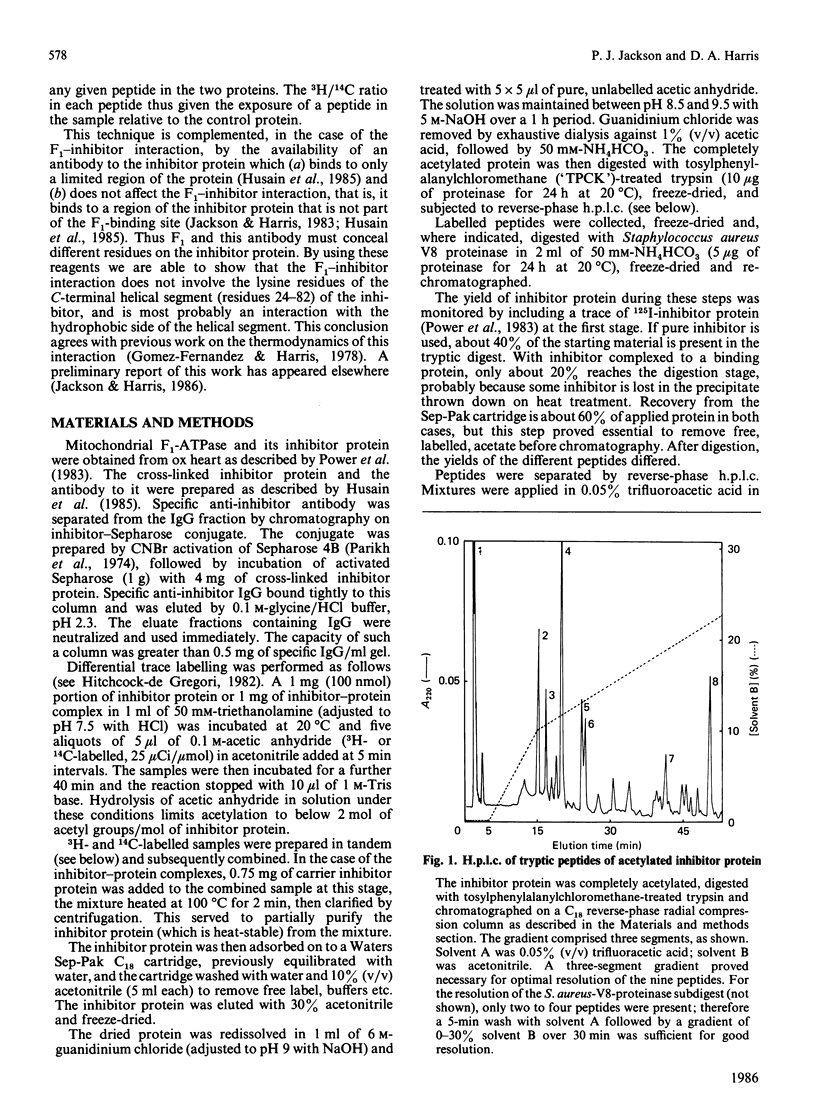
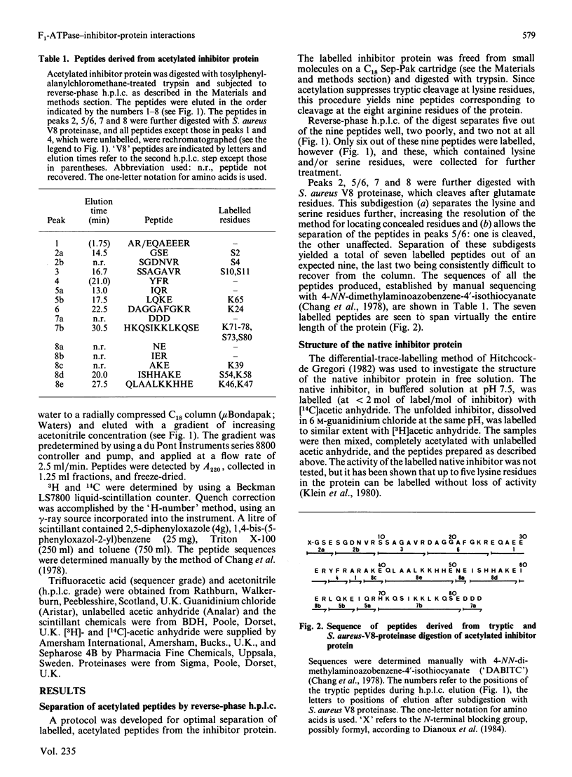
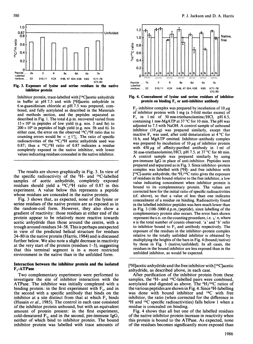
We have investigated the structure of the mitochondrial F1-ATPase inhibitor protein from ox heart by using a differential trace-labelling method. This method has also been used to determine sites on the inhibitor protein involved in binding to both the isolated mitochondrial ATPase (F1) and to a specific anti-inhibitor antibody. Native, free inhibitor was trace-labelled on its lysine and serine residues with [14C]acetic anhydride, and inhibitor protein unfolded in guanidinium chloride or specifically bound to another protein, with [3H]acetic anhydride. Exposure/concealment of residues was deduced from the 14C/3H ratios of the peptides in a proteolytic digest of the inhibitor, after separation by h.p.l.c. None of the lysine or serine residues in the native inhibitor are as exposed as in the unfolded form. There is a gradient of reactivity, with residues 54-58 being most concealed and exposure increasing towards either end of the protein. A slight decrease in reactivity is noted in residues 1-3, suggesting that the N-terminus may be in a fairly restricted environment. These findings are discussed in the light of the predicted structure of the inhibitor protein. All but one of the labelled residues increases in reactivity when inhibitor protein binds to F1. The exception, Lys-24, is only slightly concealed. Hence, F1 binding appears not to involve the lysine or serine residues directly. This finding is consistent with the view that the F1-inhibitor interaction is hydrophobic in nature. Complementary information was provided using an anti-inhibitor antibody that binds to a site on the inhibitor different from that at which F1 binds. Binding of this antibody conceals residues 54, 58, and 65 considerably. This confirms that F1 does not interact with these hydrophilic residues on the inhibitor protein.
Full text
PDF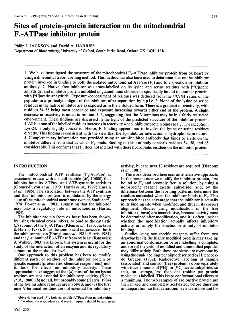
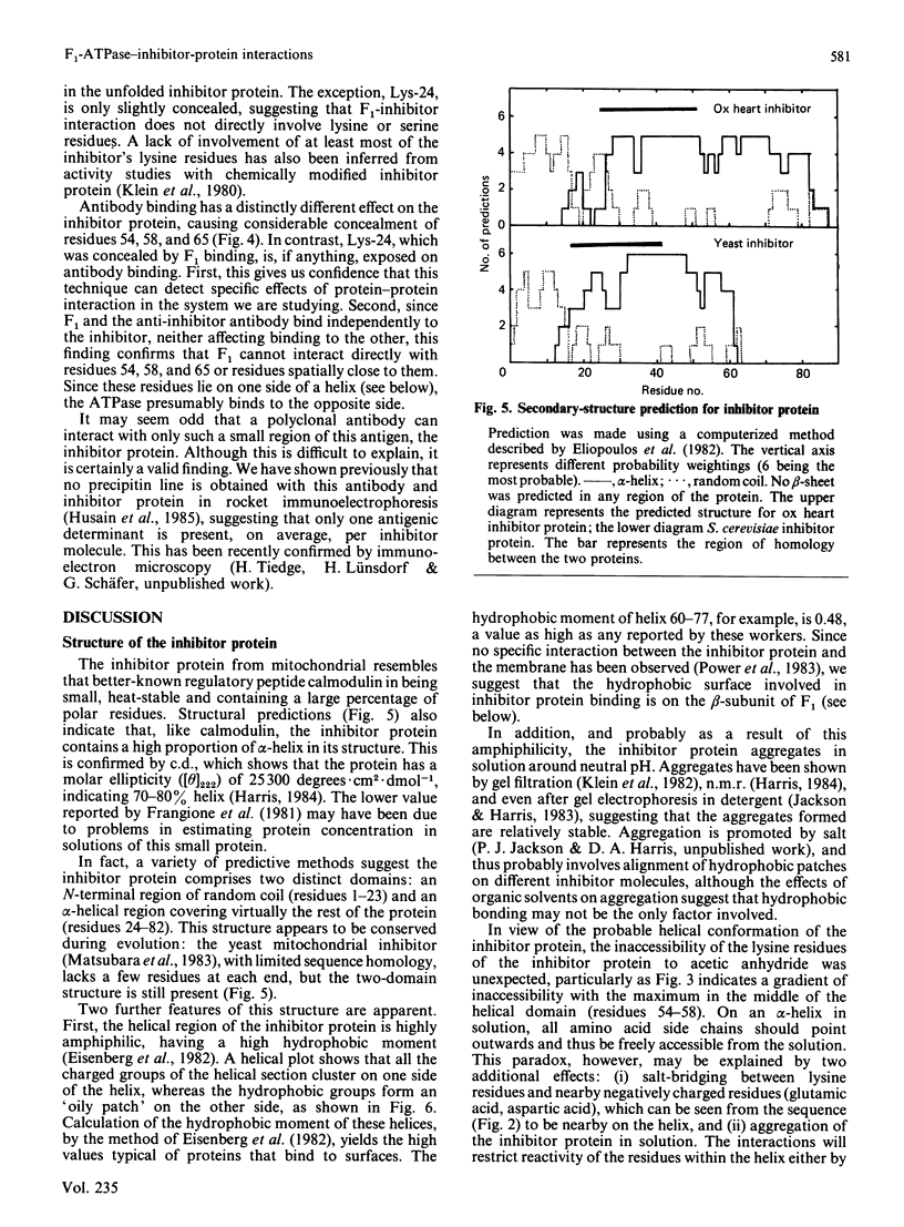
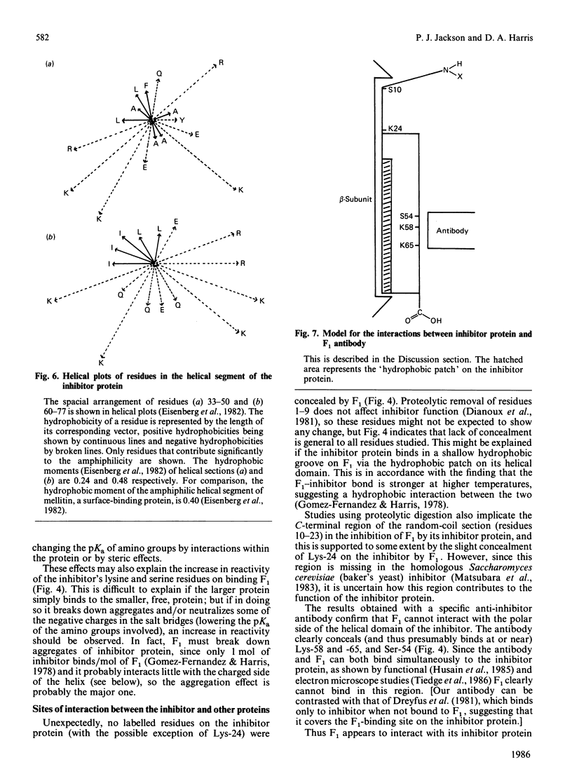
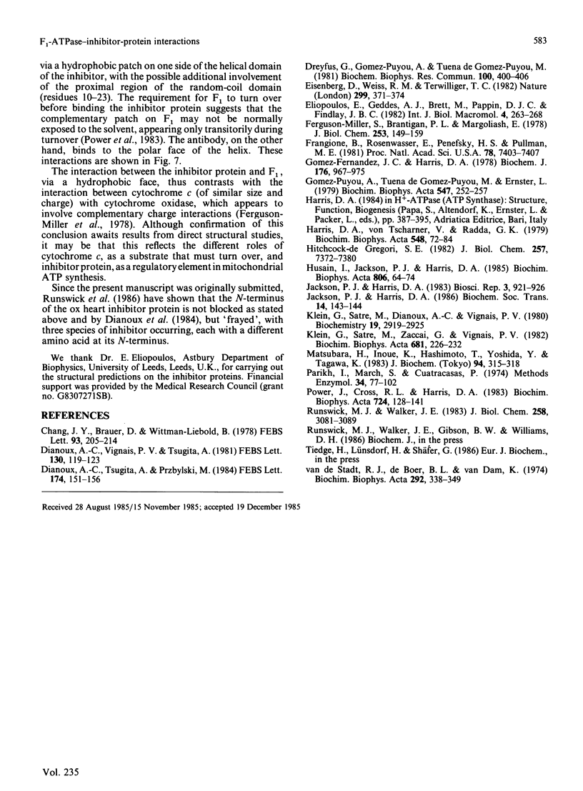
Selected References
These references are in PubMed. This may not be the complete list of references from this article.
- Dianoux A. C., Tsugita A., Przybylski M. Mass spectral identification of the blocked N-terminal tryptic peptide of the ATPase inhibitor from beef heart mitochondria. FEBS Lett. 1984 Aug 20;174(1):151–156. doi: 10.1016/0014-5793(84)81095-9. [DOI] [PubMed] [Google Scholar]
- Dianoux A. C., Vignais P. V., Tsugita A. Partial proteolysis of the natural ATPase inhibitor from beef heart mitochondria. Isolation and characterization of an active cleavage product. FEBS Lett. 1981 Jul 20;130(1):119–123. doi: 10.1016/0014-5793(81)80678-3. [DOI] [PubMed] [Google Scholar]
- Dreyfus G., Gómez-Puyou A., Iuena de Gómez-Puyou M. Electrochemical gradient induced displacement of the natural ATPase inhibitor protein from mitochondrial ATPase as directed by antibodies against the inhibitor protein. Biochem Biophys Res Commun. 1981 May 15;100(1):400–406. doi: 10.1016/s0006-291x(81)80110-6. [DOI] [PubMed] [Google Scholar]
- Eisenberg D., Weiss R. M., Terwilliger T. C. The helical hydrophobic moment: a measure of the amphiphilicity of a helix. Nature. 1982 Sep 23;299(5881):371–374. doi: 10.1038/299371a0. [DOI] [PubMed] [Google Scholar]
- Ferguson-Miller S., Brautigan D. L., Margoliash E. Definition of cytochrome c binding domains by chemical modification. III. Kinetics of reaction of carboxydinitrophenyl cytochromes c with cytochrome c oxidase. J Biol Chem. 1978 Jan 10;253(1):149–159. [PubMed] [Google Scholar]
- Frangione B., Rosenwasser E., Penefsky H. S., Pullman M. E. Amino acid sequence of the protein inhibitor of mitochondrial adenosine triphosphatase. Proc Natl Acad Sci U S A. 1981 Dec;78(12):7403–7407. doi: 10.1073/pnas.78.12.7403. [DOI] [PMC free article] [PubMed] [Google Scholar]
- Gomez-Fernandez J. C., Harris D. A. A thermodynamic analysis of the interaction between the mitochondrial coupling adenosine triphosphatase and its naturally occurring inhibitor protein. Biochem J. 1978 Dec 15;176(3):967–975. doi: 10.1042/bj1760967. [DOI] [PMC free article] [PubMed] [Google Scholar]
- Gómez-Puyou A., de Gómez-Puyou M. T., Ernster L. Inactive to active transitions of the mitochondrial ATPase complex as controlled by the ATPase inhibitor. Biochim Biophys Acta. 1979 Aug 14;547(2):252–257. doi: 10.1016/0005-2728(79)90008-2. [DOI] [PubMed] [Google Scholar]
- Harris D. A., von Tscharner V., Radda G. K. The ATPase inhibitor protein in oxidative phosphorylation. The rate-limiting factor to phosphorylation in submitochondrial particles. Biochim Biophys Acta. 1979 Oct 10;548(1):72–84. doi: 10.1016/0005-2728(79)90188-9. [DOI] [PubMed] [Google Scholar]
- Hitchcock-De Gregori S. E. Study of the structure of troponin-I by measuring the relative reactivities of lysines with acetic anhydride. J Biol Chem. 1982 Jul 10;257(13):7372–7380. [PubMed] [Google Scholar]
- Husain I., Jackson P. J., Harris D. A. Interaction between F1-ATPase and its naturally occurring inhibitor protein. Studies using a specific anti-inhibitor antibody. Biochim Biophys Acta. 1985 Jan 23;806(1):64–74. doi: 10.1016/0005-2728(85)90082-9. [DOI] [PubMed] [Google Scholar]
- Jackson P. J., Harris D. A. Binding of mitochondrial ATPase from ox heart to its naturally occurring inhibitor protein: localization by antibody binding. Biosci Rep. 1983 Oct;3(10):921–926. doi: 10.1007/BF01140661. [DOI] [PubMed] [Google Scholar]
- Klein G., Satre M., Dianoux A. C., Vignais P. V. Radiolabeling of natural adenosine triphosphatase inhibitor with phenyl (14C)isothiocyanate and study of its interaction with mitochondrial adenosine triphosphatase. Localization of inhibitor binding sites and stoichiometry of binding. Biochemistry. 1980 Jun 24;19(13):2919–2925. doi: 10.1021/bi00554a016. [DOI] [PubMed] [Google Scholar]
- Klein G., Satre M., Zaccai G., Vignais P. V. Spontaneous aggregation of the mitochondrial natural ATPase inhibitor in salt solutions as demonstrated by gel filtration and neutron scattering. Application to the concomitant purification of the ATPase inhibitor and F1-ATPase. Biochim Biophys Acta. 1982 Aug 20;681(2):226–232. doi: 10.1016/0005-2728(82)90026-3. [DOI] [PubMed] [Google Scholar]
- Matsubara H., Inoue K., Hashimoto T., Yoshida Y., Tagawa K. A stabilizing factor of yeast mitochondrial F1F0-ATPase-inhibitor complex: common amino acid sequence with yeast ATPase inhibitor and E. coli epsilon and bovine delta subunits. J Biochem. 1983 Jul;94(1):315–318. doi: 10.1093/oxfordjournals.jbchem.a134347. [DOI] [PubMed] [Google Scholar]
- Parikh I., March S., Cuatercasas P. Topics in the methodology of substitution reactions with agarose. Methods Enzymol. 1974;34:77–102. doi: 10.1016/s0076-6879(74)34009-8. [DOI] [PubMed] [Google Scholar]
- Power J., Cross R. L., Harris D. A. Interaction of F1-ATPase, from ox heart mitochondria with its naturally occurring inhibitor protein. Studies using radio-iodinated inhibitor protein. Biochim Biophys Acta. 1983 Jul 29;724(1):128–141. doi: 10.1016/0005-2728(83)90034-8. [DOI] [PubMed] [Google Scholar]
- Runswick M. J., Walker J. E. The amino acid sequence of the beta-subunit of ATP synthase from bovine heart mitochondria. J Biol Chem. 1983 Mar 10;258(5):3081–3089. [PubMed] [Google Scholar]
- van de Stadt R. J., de Boer B. L., van Dam K. The interaction between the mitochondrial ATPase (F 1 ) and the ATPase inhibitor. Biochim Biophys Acta. 1973 Feb 22;292(2):338–349. doi: 10.1016/0005-2728(73)90040-6. [DOI] [PubMed] [Google Scholar]


