

Abstract
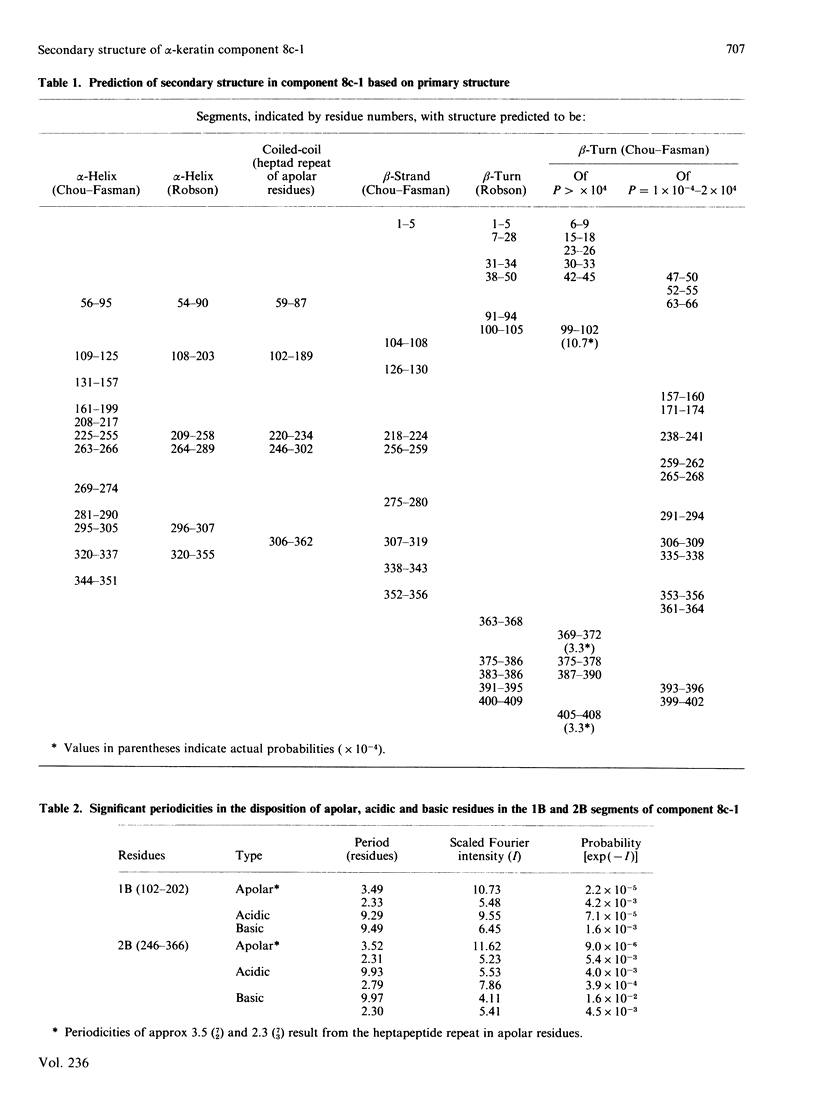
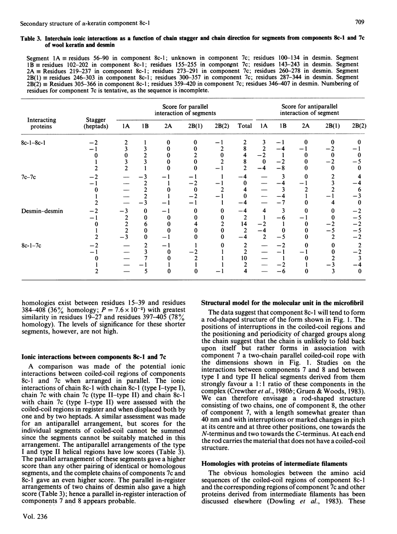
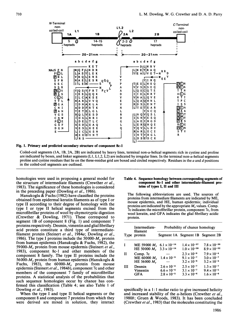
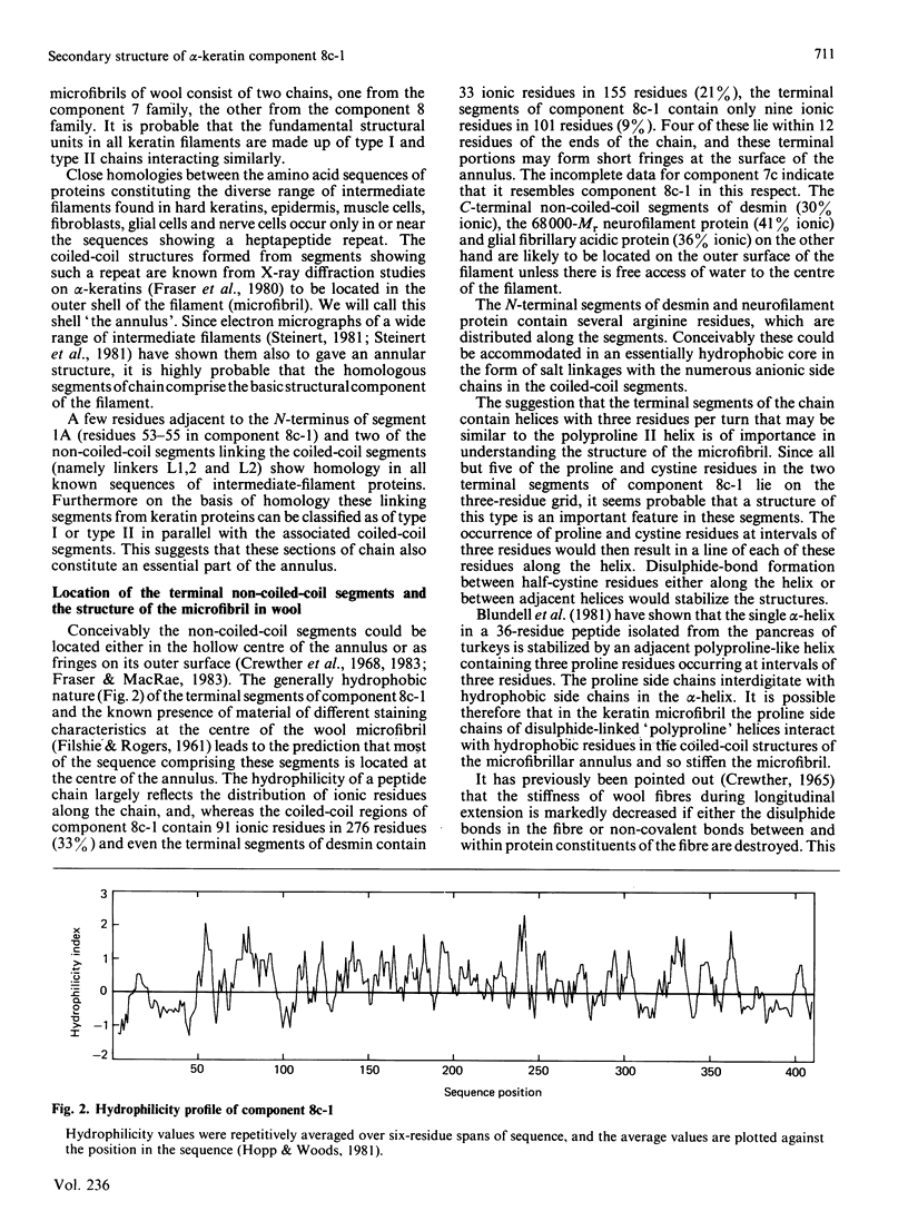
The amino acid sequence of component 8c-1 from alpha-keratin was analysed by using secondary-structure prediction techniques, homology search methods, fast Fourier-transform techniques to detect regularities in the linear disposition of amino acids, interaction counts to assess possible modes of chain aggregation and assessment of hydrophilicity distribution. The analyses show the following. The molecule has two lengths of coiled-coil structure, each about 20 nm long, one from residues 56-202 with a discontinuity from about residue 91 to residue 101, and the other from residues 219-366 with discontinuities from about residue 238 to residue 245 and at about residue 306. The acidic and basic residues in the coiled-coil segment between residues 102 and 202 show a 9,4-residue structural period in their linear disposition, whereas between residues 246 and 366 a period of 9.9 residues is observed in the positioning of ionic residues. Acidic and basic residues are out of phase by 180 degrees. Similar repeats occur in corresponding regions of other intermediate-filament proteins. The overall mean values for the repeats are 9.55 residues in the N-terminal region and 9.85 residues in the C-terminal region. The regions at each end of the protein chain (residues 1-55 and 367-412) are not alpha-helical and contain many potential beta-bends. The regions specified in have a significant degree of homology mainly due to a semi-regular disposition of proline and half-cystine residues on a three-residue grid; this is especially apparent in the C-terminal segment, in which short (Pro-Cys-Xaa)n regions occur. The coiled-coil segments of component 8c-1 bear a striking similarity to corresponding segments of other intermediate-filament proteins as regards sequence homology, structural periodicity of ionic residues and secondary/tertiary-structure predictions. The assessments of the probabilities that these homologies occurred by chance indicate that there are two populations of keratin filament proteins. The non-coiled-coil regions at each end of the chain are less hydrophilic than the coiled-coil regions. Ionic interactions between the heptad regions of components 8c-1 and 7c from the microfibrils of alpha-keratin are optimized when a coiled-coil structure is formed with the heptad regions of the constituent chains both parallel and in register.
Full text
PDF
Selected References
These references are in PubMed. This may not be the complete list of references from this article.
- Blundell T. L., Pitts J. E., Tickle I. J., Wood S. P., Wu C. W. X-ray analysis (1. 4-A resolution) of avian pancreatic polypeptide: Small globular protein hormone. Proc Natl Acad Sci U S A. 1981 Jul;78(7):4175–4179. doi: 10.1073/pnas.78.7.4175. [DOI] [PMC free article] [PubMed] [Google Scholar]
- Chou P. Y., Fasman G. D. Conformational parameters for amino acids in helical, beta-sheet, and random coil regions calculated from proteins. Biochemistry. 1974 Jan 15;13(2):211–222. doi: 10.1021/bi00699a001. [DOI] [PubMed] [Google Scholar]
- Chou P. Y., Fasman G. D. Prediction of protein conformation. Biochemistry. 1974 Jan 15;13(2):222–245. doi: 10.1021/bi00699a002. [DOI] [PubMed] [Google Scholar]
- Doolittle R. F., Cassman K. G., Cottrell B. A., Friezner S. J., Takagi T. Amino acid sequence studies on the alpha chain of human fibrinogen. Covalent structure of the alpha-chain portion of fragment D. Biochemistry. 1977 Apr 19;16(8):1710–1715. doi: 10.1021/bi00627a029. [DOI] [PubMed] [Google Scholar]
- Dowling L. M., Crewther W. G., Inglis A. S. The primary structure of component 8c-1, a subunit protein of intermediate filaments in wool keratin. Relationships with proteins from other intermediate filaments. Biochem J. 1986 Jun 15;236(3):695–703. doi: 10.1042/bj2360695. [DOI] [PMC free article] [PubMed] [Google Scholar]
- Dowling L. M., Parry D. A., Sparrow L. G. Structural homology between hard alpha-keratin and the intermediate filament proteins desmin and vimentin. Biosci Rep. 1983 Jan;3(1):73–78. doi: 10.1007/BF01121573. [DOI] [PubMed] [Google Scholar]
- FILSHIE B. K., ROGERS G. E. The fine structure of alpha-keratin. J Mol Biol. 1961 Dec;3:784–786. doi: 10.1016/s0022-2836(61)80084-3. [DOI] [PubMed] [Google Scholar]
- Fraser R. D., MacRae T. P., Suzuki E. Structure of the alpha-keratin microfibril. J Mol Biol. 1976 Dec;108(2):435–452. doi: 10.1016/s0022-2836(76)80129-5. [DOI] [PubMed] [Google Scholar]
- Fraser R. D., MacRae T. P. The structure of the alpha-keratin microfibril. Biosci Rep. 1983 Jun;3(6):517–525. doi: 10.1007/BF01120695. [DOI] [PubMed] [Google Scholar]
- Garnier J., Osguthorpe D. J., Robson B. Analysis of the accuracy and implications of simple methods for predicting the secondary structure of globular proteins. J Mol Biol. 1978 Mar 25;120(1):97–120. doi: 10.1016/0022-2836(78)90297-8. [DOI] [PubMed] [Google Scholar]
- Geisler N., Weber K. Amino acid sequence data on glial fibrillary acidic protein (GFA); implications for the subdivision of intermediate filaments into epithelial and non-epithelial members. EMBO J. 1983;2(11):2059–2063. doi: 10.1002/j.1460-2075.1983.tb01700.x. [DOI] [PMC free article] [PubMed] [Google Scholar]
- Geisler N., Weber K. The amino acid sequence of chicken muscle desmin provides a common structural model for intermediate filament proteins. EMBO J. 1982;1(12):1649–1656. doi: 10.1002/j.1460-2075.1982.tb01368.x. [DOI] [PMC free article] [PubMed] [Google Scholar]
- Gibbs A. J., McIntyre G. A. The diagram, a method for comparing sequences. Its use with amino acid and nucleotide sequences. Eur J Biochem. 1970 Sep;16(1):1–11. doi: 10.1111/j.1432-1033.1970.tb01046.x. [DOI] [PubMed] [Google Scholar]
- Gough K. H., Inglis A. S., Crewther W. G. Amino acid sequences of alpha-helical segments from S-carbosymethylkerateine-A. Complete sequence of a type-I segment. Biochem J. 1978 Aug 1;173(2):373–385. doi: 10.1042/bj1730373. [DOI] [PMC free article] [PubMed] [Google Scholar]
- Gruen L. C., Woods E. F. Structural studies on the microfibrillar proteins of wool. Interaction between alpha-helical segments and reassembly of a four-chain structure. Biochem J. 1983 Mar 1;209(3):587–595. doi: 10.1042/bj2090587. [DOI] [PMC free article] [PubMed] [Google Scholar]
- Hanukoglu I., Fuchs E. The cDNA sequence of a Type II cytoskeletal keratin reveals constant and variable structural domains among keratins. Cell. 1983 Jul;33(3):915–924. doi: 10.1016/0092-8674(83)90034-x. [DOI] [PubMed] [Google Scholar]
- Hanukoglu I., Fuchs E. The cDNA sequence of a human epidermal keratin: divergence of sequence but conservation of structure among intermediate filament proteins. Cell. 1982 Nov;31(1):243–252. doi: 10.1016/0092-8674(82)90424-x. [DOI] [PubMed] [Google Scholar]
- Hopp T. P., Woods K. R. Prediction of protein antigenic determinants from amino acid sequences. Proc Natl Acad Sci U S A. 1981 Jun;78(6):3824–3828. doi: 10.1073/pnas.78.6.3824. [DOI] [PMC free article] [PubMed] [Google Scholar]
- McLachlan A. D., Karn J. Periodic charge distributions in the myosin rod amino acid sequence match cross-bridge spacings in muscle. Nature. 1982 Sep 16;299(5880):226–231. doi: 10.1038/299226a0. [DOI] [PubMed] [Google Scholar]
- McLachlan A. D., Stewart M. The 14-fold periodicity in alpha-tropomyosin and the interaction with actin. J Mol Biol. 1976 May 15;103(2):271–298. doi: 10.1016/0022-2836(76)90313-2. [DOI] [PubMed] [Google Scholar]
- McLachlan A. D., Stewart M. Tropomyosin coiled-coil interactions: evidence for an unstaggered structure. J Mol Biol. 1975 Oct 25;98(2):293–304. doi: 10.1016/s0022-2836(75)80119-7. [DOI] [PubMed] [Google Scholar]
- Nozaki Y., Tanford C. The solubility of amino acids and two glycine peptides in aqueous ethanol and dioxane solutions. Establishment of a hydrophobicity scale. J Biol Chem. 1971 Apr 10;246(7):2211–2217. [PubMed] [Google Scholar]
- Parby D. A. Fibrinogen: a preliminary analysis of the amino acid sequences of the portion of the alpha, beta and gamma-chains postulated to form the interdomainal link between globular regions of the molecule. J Mol Biol. 1978 Apr 25;120(4):545–551. doi: 10.1016/0022-2836(78)90353-4. [DOI] [PubMed] [Google Scholar]
- Parry D. A. Analysis of the amino acid sequence of a tropomyosin-binding fragment from troponin-T. J Mol Biol. 1981 Feb 25;146(2):259–263. doi: 10.1016/0022-2836(81)90435-6. [DOI] [PubMed] [Google Scholar]
- Parry D. A. Analysis of the primary sequence of alpha-tropomyosin from rabbit skeletal muscle. J Mol Biol. 1975 Nov 5;98(3):519–535. doi: 10.1016/s0022-2836(75)80084-2. [DOI] [PubMed] [Google Scholar]
- Parry D. A., Crewther W. G., Fraser R. D., MacRae T. P. Structure of alpha-keratin: structural implication of the amino acid sequences of the type I and type II chain segments. J Mol Biol. 1977 Jun 25;113(2):449–454. doi: 10.1016/0022-2836(77)90153-x. [DOI] [PubMed] [Google Scholar]
- Steinert P. M., Parry D. A., Racoosin E. L., Idler W. W., Steven A. C., Trus B. L., Roop D. R. The complete cDNA and deduced amino acid sequence of a type II mouse epidermal keratin of 60,000 Da: analysis of sequence differences between type I and type II keratins. Proc Natl Acad Sci U S A. 1984 Sep;81(18):5709–5713. doi: 10.1073/pnas.81.18.5709. [DOI] [PMC free article] [PubMed] [Google Scholar]
- Steinert P. M., Rice R. H., Roop D. R., Trus B. L., Steven A. C. Complete amino acid sequence of a mouse epidermal keratin subunit and implications for the structure of intermediate filaments. Nature. 1983 Apr 28;302(5911):794–800. doi: 10.1038/302794a0. [DOI] [PubMed] [Google Scholar]
- Stewart M., McLachlan A. D. Fourteen actin-binding sites on tropomyosin? Nature. 1975 Sep 25;257(5524):331–333. doi: 10.1038/257331a0. [DOI] [PubMed] [Google Scholar]