Abstract
The 1957 human pandemic strain of influenza A virus contained an avian virus hemagglutinin (HA) and neuraminidase (NA), both of which acquired specificity for the human receptor, N-acetylneuraminic acid linked to galactose of cellular glycoconjugates via an α2-6 bond (NeuAcα2-6Gal). Although the NA retained considerable specificity for NeuAcα2-3Gal, its original substrate in ducks, it lost the ability to support viral growth in the duck intestine, suggesting a growth-restrictive change other than a shift in substrate specificity. To test this possibility, we generated a panel of reassortant viruses that expressed the NA genes of human H2N2 viruses isolated from 1957 to 1968 with all other genes from the avian virus A/duck/Hong Kong/278/78 (H9N2). Only the NA of A/Singapore/1/57 supported efficient viral growth in the intestines of orally inoculated ducks. The growth-supporting capacity of the NA correlated with a high level of enzymatic activity, comparable to that found to be associated with avian virus NAs. The specific activities of the A/Ann Arbor/6/60 and A/England/12/62 NAs, which showed greatly restricted abilities to support viral growth in ducks, were only 8 and 5%, respectively, of the NA specific activity for A/Singapore/1/57. Using chimeric constructs based on A/Singapore/1/57 and A/England/12/62 NAs, we localized the determinants of high specific NA activity to a region containing six amino acid substitutions in A/England/12/62: Ser331→Arg, Asp339→Asn, Asn367→Ser, Ser370→Leu, Asn400→Ser, and Pro431→Glu. Five of these six residues (excluding Asn400) were required and sufficient for the full specific activity of the A/Singapore/1/57 NA. Thus, in addition to a change in substrate specificity, a reduction in high specific activity may be required for the adaptation of avian virus NAs to growth in humans. This change is likely needed to maintain an optimal balance between NA activity and the lower affinity shown by human virus HAs for their cellular receptor.
During replication, influenza A viruses require the activities of two viral surface glycoproteins, hemagglutinin (HA), responsible for binding to terminal sialic acid on cell surface glycoconjugates, and neuraminidase (NA), with an associated enzymatic activity that removes sialic acid from host cell glycoconjugates as well as newly synthesized viral proteins to facilitate the budding of progeny virions from cells (16). The primordial reservoir for all subtypes of influenza A viruses is believed to be wild aquatic birds (26), and the activities of these two proteins in avian viruses are highly adapted to the specific recognition of N-acetylneuraminic acid (NeuAc) attached via an α2-3 linkage to neighboring galactose (α2-3Gal). NeuAcα2-3Gal is important for the replication of influenza viruses in the intestines of aquatic birds, the natural site of virus replication in these hosts (7, 8, 18, 19). In addition, the opposing activities of HA and NA must be balanced (4, 6, 13, 14) to ensure that NA possesses sufficient enzymatic activity to remove sialic acids from infected cells but does not reduce the efficiency of infection by removing sialic acid from uninfected cells before virus attachment occurs.
Occasionally viruses are transmitted to other host species, resulting in severe outbreaks of disease and often the long-term establishment of a new viral strain in the nonavian host (reviewed in reference 26). In the last century, at least two outbreaks, in 1957 and 1968, associated with transmission of avian viruses or the introduction of avian viral genes into human viruses led to serious pandemics. After the appearance of these viruses in humans, HA variants were selected that had a higher specificity for the major species of sialic acid, NeuAc-α2-6-galactose (NeuAcα2-6Gal), on cells of the upper respiratory tract in humans than for the original receptor, NeuAcα2-3Gal, in wild aquatic birds (2, 3, 18, 19). The N2 subtype of NA first appeared in human viruses in the 1957 outbreak of H2N2 virus and over several years gradually acquired an increased substrate specificity for NeuAcα2-6Gal while maintaining significant specificity for the original target of its activity in birds, NeuAcα2-3Gal (1, 10).
However, even though the human virus N2 NA maintained substantial specificity for NeuAcα2-3Gal, it became altered in its ability to support viral growth in ducks. Orally inoculated avian viruses can replicate to a high titer in the lower intestines of ducks. A reassortant virus, containing the N2 NA gene of human virus A/Udorn/307/72 (H3N2) and all other genes from an avian virus, A/mallard/New York/6750/72, was not able to replicate in the lower intestines of orally inoculated ducks (5). This difference in growth of the parent avian virus versus the reassortant virus stemmed from differences between the avian and human NAs that occurred as a consequence of adaptation of the NA to growth in humans.
Because the human virus NA has retained appreciable specificity for NeuAcα2-3Gal despite acquiring specificity for NeuAcα2-6Gal, we asked why it has lost the ability to support viral replication in ducks. Apparently, NA substrate specificity, while an important determinant of the ability of an influenza virus to replicate in any host, is not sufficient to explain the differences in host range properties between avian and human viruses. To better understand the role of NA in host range restriction, we generated a series of reassortants in which the NA of an avian virus, A/duck/Hong Kong/278/78 (H2N9), was replaced with the N2 NAs from a series of human H2N2 viruses isolated between 1957 and 1968. The ability of each reassortant to grow in ducks was assessed to determine when the human virus NA lost the ability to support virus growth in these hosts. Differences in the enzymatic activities of the NAs were also assessed to identify differences in the NAs that might be related to the restriction of growth in ducks.
MATERIALS AND METHODS
Viruses, cells, and antibodies.
Two avian influenza viruses, A/duck/Hong Kong/278/78 (H2N9) and A/duck/Hong Kong/7/75 (H3N2), and four H2N2 human viruses, A/Singapore/1/57, A/Ann Arbor/6/60, A/England/12/62, and A/Korea/426/68, were obtained from the repository at St. Jude Children's Research Hospital. Madin-Darby canine kidney (MDCK) cells were cultured in Eagle's minimum essential medium with 5% newborn calf serum, and 293T cells were cultured in Dulbecco's modified Eagle medium supplemented with 10% fetal calf serum. For selection of reassortant viruses expressing N2 NAs, a pool of several monoclonal antibodies specific for the N9 NA were used. This antibody pool and a pool of anti-N2 monoclonal antibodies were used for inhibition assays.
Generation of reassortant viruses.
Reassortant viruses were generated with all genes except that for NA derived from avian virus A/duck/Hong Kong/278/78 (H2N9) by using a previously described procedure (24). The NA genes of the reassortants were derived from the human H2N2 viruses A/Singapore/1/57, A/Ann Arbor/6/60, A/England/12/62, and A/Korea/426/68, and the reassortants were subsequently designated Dk78/Sing57N2, Dk78/AA60N2, Dk78/Eng62N2, and Dk78/Korea68N2, respectively. After identifying the viruses that expressed the N2 NA, we extracted viral RNAs from infection supernatants by phenol-chloroform extraction and ethanol-sodium acetate precipitation. cDNA was generated for each viral genome segment by using UNI12, a primer complementary to the 12-base sequence at the 3′ end of the viral RNA that is identical among all the segments, and Moloney murine leukemia virus reverse transcriptase (Promega, Madison, Wis.). The genotypes of the internal gene segments were determined by PCR with a primer pair that is specific for the avian virus gene segment and another for the human virus gene segment for all of the internal genes. In cases where PCR was not diagnostic (i.e., both primer pairs for a given gene yielded an amplification product), the PCR products were further analyzed by restriction enzyme digestion with enzymes specific for only one of the gene sequences. The identity of the H2 HA incorporated into the reassortants was also determined by PCR amplification of a portion of the gene, followed by digestion with restriction enzymes specific for the avian and human H2 HAs. The sequences of all primers are available upon request. Only reassortants expressing the N2 gene from the human viruses and all other genes from the avian viruses were used for growth analysis in ducks. Each reassortant was plaque purified three times on MDCK cells and then inoculated into 11-day-old embryonated chicken eggs. Allantoic fluid stocks were kept at −70°C.
Growth of reassortant viruses in ducks.
Pekin ducks (Ridgeway Hatcheries, LaRue, Ohio) were orally inoculated with equal titers (median egg infectious dose, 6.75) of each reassortant virus. Three days postinfection, the ducks were sacrificed and a 6-cm-long portion of the colon was removed. The tissue was homogenized and resuspended in phosphate-buffered saline (PBS) containing antibiotics, and the virus in the PBS suspension was titrated in 11-day-old embryonated chicken eggs. Allantoic fluid harvested from each egg was tested for virus by hemagglutination assay with chicken red blood cells.
NA enzymatic activity assays.
NA enzymatic activity was determined by using 2′-(4-methylumbelliferyl)-α-d-N-acetylneuraminic acid (MU-NANA) (Sigma), and sialyllactose containing NeuAc bound to the galactose of lactose through an α2-3 ketosidic linkage (NeuAcα2-3Gal) (Sigma), as previously described (10). NA activities were determined with cell-expressed NAs and concentrated virus preparations as previously described (10). In all assays, a time course analysis with several dilutions of cell-expressed NA or virus was performed to determine the optimal conditions for comparison of NA activities. Assay conditions were selected to determine NA activities within the linear range of the sialidase activity-versus-time plot for all of the NAs. The data reported are the means of duplicate reactions for each sample and are representative of two or three separate assays.
Immunoprecipitation of NA.
Twenty microliters of virus was diluted 1:10 in RIPA buffer (150 mM NaCl, 50 mM Tris-HCl [pH 7.4], 5 mM EDTA, 1% Triton X-100, 1% sodium deoxycholate, 1% sodium dodecyl sulfate [SDS]) and incubated for 15 min at 37°C. One microliter of the anti-N2 monoclonal antibody pool was added to the disrupted virus, and the mixture was incubated at room temperature for 2 h. Next, 80 μl of a 50% suspension of protein A beads (RepliGen, Needham, Mass.), previously washed three times in RIPA buffer, was added and incubated at room temperature for 2 h with gentle agitation. The beads were collected by centrifugation and washed three times with RIPA buffer and then once with 50 mM Tris-HCl (pH 6.8) in a clean tube. The beads were resuspended in SDS-polyacrylamide gel electrophoresis sample buffer (nonreducing) in preparation for electrophoresis.
Determination of relative specific activities of NA in reassortant virions.
One 175-cm2 flask of MDCK cells at 90% confluency was infected with Dk78/Sing57N2, Dk78/AA60N2, or Dk78/Eng62N2 at a multiplicity of infection of 1 and maintained in minimum essential medium-bovine serum albumin. Similarly, one flask of cells was infected with Dk78/75N2, a virus previously generated by reverse genetics that contains the H2 HA gene and all internal genes from A/duck/Hong Kong/278/78 and the N2 NA gene from A/duck/Hong Kong/7/75 (9). This virus grows efficiently in the duck intestine and provided an avian N2 NA for comparison with the specific activities of the human N2 NAs. Four hours postinfection, the medium was replaced with methionine- and cysteine-deficient RPMI 1640 (ICN, Costa Mesa, Calif.) supplemented with N-tosyl-l-phenylalanine chloromethyl ketone-treated trypsin for 1 h to deplete the endogenous reserves of methionine and cysteine. Then, 250 μCi of methionine-cysteine Trans35S-Label (ICN) was added to each flask. The supernatants were harvested at 24 h postinfection, following lysis of 100% of the cells in each flask. The supernatant fluids were centrifuged at 5,000 × g for 30 min in an SW28 rotor to pellet cell debris, and the virus was pelleted by centrifugation at 65,000 × g for 1.5 h. Each pellet was resuspended in 200 μl of PBS, divided into aliquots, and stored at −20°C.
The enzymatic activity of each concentrated virus was determined as described above in equal volumes of virus preparation, thus providing a comparison of NA activity per unit volume of virus stock. Next, the viral proteins from an equal volume of each virus preparation were separated by electrophoresis in an SDS–9% polyacrylamide gel under nonreducing conditions. To identify NA bands on the gel, immunoprecipitation of the NA from each of the virus preparations was performed as described above and resolved on the same gel as the virus preparations. The viral proteins were detected by autoradiography. Each immunoprecipitated NA resolved as a high-molecular-weight double band that corresponded to a double band in an identical position in each whole-virus lane. The relative NA protein content in each virus preparation was determined from the combined optical densities of both NA bands, as previously described (9). The NA activity (determined as described above) divided by the NA protein content provided the relative specific activity of each NA, reported here as a normalized value relative to that of Dk78/75N2.
Cloning of viral NA genes and generation of chimeric NA constructs.
The full-length NA genes from Dk78/Sing57N2 and Dk78/Eng62N2 were cloned into pUC19 and the plasmid expression vector pCAGGS/MCS, as previously described (10, 15), generating the constructs pUC/Sing57N2, pCA/Sing57N2, pUC/Eng62N2, and pCA/Eng62N2. The sequences of these NAs were confirmed to be identical to those of the original parent viruses, A/Singapore/1/57 and A/England/12/62. To provide a common antibody recognition sequence for protein quantitation of cell-expressed NA, we inserted the coding sequence for the FLAG epitope tag (DYKDDDDK) (17) in frame into the region of each NA gene coding for the stalk after Pro45. Mutagenesis was performed with the oligonucleotide 5′-GCATTACTTGGTTGCTCGCCCTTGTCATCGTCGTCCTTGTAGT-3′ and the Clontech Transformer site-directed mutagenesis kit (number K1600-1), according to the manufacturer's instructions. Sequence analysis confirmed insertion of the FLAG sequence, whose presence did not affect the enzymatic activities of the NAs (data not shown).
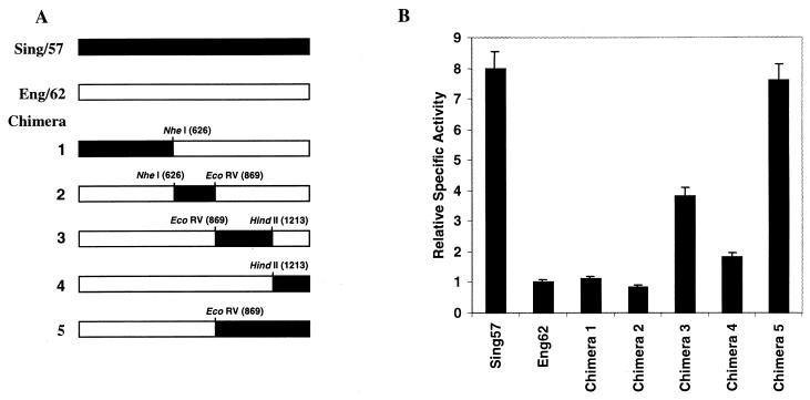
Five chimeric constructs of A/Singapore/1/57 and A/England/12/62 were generated as described previously (10) (see Fig. 2A).
FIG. 2.
Identification of the NA region associated with high specific activity. (A) The sequence coding for the FLAG epitope tag (DYKDDDDK) was inserted, in frame, into the NA genes of A/Singapore/1/57 (Sing/57) and A/England/12/62 (Eng/62), and chimeras 1 to 5 were generated by using shared restriction sites. All genes were subcloned into the plasmid expression vector pCAGGS/MCS. (B) Two 6-cm2 wells of 293T cells were transfected with 2 μg of each NA-expressing plasmid/well. 48 h later, the cell-associated NA activity was determined by using equal numbers of cells transfected with each NA and 0.1 mM NeuAcα2-3Gal sialyllactose substrate. NA protein expression levels were determined by isolation of cell membranes and measurement of NA by chemiluminescent Western blot analysis with the anti-FLAG antibody. Enzymatic activities were normalized to protein expression for each NA and are reported relative to that of A/England/12/62. The error bars indicate standard deviations.
Site-specific mutagenesis of A/England/12/62 NA amino acid residues.
To study the contributions of particular amino acid residues to NA specific activity, we replaced certain residues of the A/England/12/62 NA with those of A/Singapore/1/57. Mutations were generated in the coding sequences of amino acid residues 331, 339, 367, 370, 400, and 431 individually and/or in groups of two or more. Mutagenesis of individual residues at positions 367(Ser→Asn) and 370 (Leu→Ser) was performed with the oligonucleotides 5′-GCGCAAATCCTTGTTGATCGTTCTTCCC-3′ and 5′-CCTGTTAACCTGAGCGTGAATCCTTGCTGATCG-3′, respectively, and the Clontech Transformer site-directed mutagenesis kit, as described above.
Residue 400 (Ser→Asn) was mutated by amplification with the mutagenic PCR primer 5′-GTCATAGTTGACAACAATAATTGGTCAGG-3′, which contains the HindII site of the NA gene at nucleotide position 1213, and the reverse primer 5′-ATGACCATGATTACGCCAAGCTTGC-3′, which binds nucleotides 445 to 469 of the pUC19 MCS (10). The Glu431→Pro mutation in pUC/Eng62N2 was introduced by PCR amplification of construct pUC/Sing57N2 with a mutagenic primer, 5′-GTCATAGTTGACAGCAATAATTGGTCTGG-3′, which binds in the same position as the above mutagenic forward primer, and the reverse primer listed above. Within this region there are only two nucleotide differences that result in amino acid differences between the A/England/12/62 and A/Singapore/1/57 NAs. Mutagenesis altered A/Singapore/1/57 residue Asn400 to A/England/12/62 residue Ser400, and when this fragment was subcloned into pUC/Eng62N2, the net effect was a shift from Gln431 to Pro431. All mutants were confirmed by sequencing and subcloned into pCAGGS/MCS.
Amino acids found in the A/Singapore/1/57 NA were introduced into the A/England/12/62 NA in groups of two or more by subcloning appropriate fragments from suitable constructs, using the shared NheI, EcoRV, FokI, and HindII restriction enzyme sites at positions 626, 869, 1105, and 1213, respectively, and Asp718 in the pUC19 MCS.
Determination of relative specific activities of expressed NAs.
The pCAGGS/MCS expression plasmid for each wild-type, chimeric, or mutant NA was expressed in 293T cells (two 10-cm2 wells per construct) as previously described (10). NA enzymatic activity was determined with the NeuAcα2-3Gal sialyllactose and MU-NANA substrates, as described above, in dilutions of equal volumes of each cell suspension. After determination of the cell surface-expressed NA activity, the cell membranes were isolated for quantitation of the membrane-associated NA protein. Cells were pelleted by centrifugation at 500 × g for 30 s, resuspended in 1 ml of ice-cold Dounce buffer (10 mM Tris-Cl [pH 7.5] and 0.5 mM MgCl2, with the protease inhibitors leupeptin [10 μg/ml], aprotinin [10 μg/ml], phenylmethylsulfonyl fluoride [1 mM], and iodoacetamide [1.8 mg/ml]), and incubated on ice for 10 min. The cells were lysed by 30 strokes in a Dounce homogenizer with the tight (type B) pestle, and tonicity restoration buffer (10 mM Tris-Cl [pH 7.6], 0.5 mM MgCl2, and 0.6 M NaCl, plus protease inhibitors as in Dounce buffer) was immediately added to bring the solution to a final concentration of 150 mM NaCl to prevent disruption of the nuclei. The nuclei were pelleted by centrifugation at 4°C for 5 min at 500 × g. To the supernatant, 0.5 M EDTA was added to a final concentration of 5 mM, and the membrane suspensions were centrifuged at 4°C for 45 min at 150,000 × g. Each membrane preparation was solubilized in 200 μl of resuspension buffer (300 mM NaCl, 50 mM Tris-Cl [pH 7.6], and 0.2% SDS, plus protease inhibitors).
To measure the membrane-associated NA protein content, we diluted each membrane preparation in dilution buffer (300 mM NaCl, 50 mM Tris-Cl [pH 7.5], 0.1% SDS) and spotted 1 μl of the dilutions in duplicate onto prewetted Immun-Blot polyvinylidene difluoride membranes (Bio-Rad, Hercules, Calif.). Several dilutions of each membrane were tested to ensure that attempts to detect the bound protein were within the linear detection range of the assay. The protein was allowed to bind for 30 min, after which the membranes were incubated for 1 h at room temperature in blocking buffer (PBS containing 2% nonfat milk and 0.05% Tween 20) with gentle agitation. After being blocked and between subsequent steps, the membranes were washed extensively with PBS containing 0.1% Tween 20. NA protein was detected by incubating the blots with a 1:5,000 dilution of the anti-FLAG M2 antibody (Kodak IB13010) in blocking buffer at room temperature for 1 h. The blots were then incubated for 1 h at room temperature with a 1:10,000 dilution of anti-mouse horseradish peroxidase-conjugated antibody (Sigma number A-9044) in blocking buffer. The bound horseradish peroxidase activity was detected by chemiluminescence with the ECL Western blotting kit (Amersham, Piscataway, N.J.) according to the manufacturer's protocol. The optical densities of the spots were determined with the Kodak KDS1D imaging software. The previously determined cell-associated NA activities were then normalized to the relative level of protein expression for each construct. The relative specific activity for each construct normalized to that of the pCA/Eng62N2 is reported in Results.
RESULTS
Growth of reassortant viruses in ducks.
The N2 NA, originally derived from an avian virus, first appeared in human viruses in 1957. To determine when the NA lost its ability to support the growth of virus in ducks after its introduction into the human population, we generated reassortants containing N2 NA genes from human H2N2 viruses isolated between 1957 and 1968 in the background of all other genes from avian virus A/duck/Hong Kong/278/78. Although none of the reassortants differed from the parent avian virus in the ability to grow in MDCK cell culture or in the allantoic cavity of embryonated chicken eggs (data not shown), there were marked differences in their replication in the duck intestine (Table 1). Both A/duck/Hong Kong/278/78 and Dk78/Sing57N2 grew well in this tissue, while reassortant Dk78/AA60N2 grew poorly and Dk78/Eng62N2 and Dk78/Korea68N2 did not grow to detectable levels. Thus, by 1962 the human virus N2 had lost the ability to support productive virus replication in orally inoculated ducks.
TABLE 1.
Growth of reassortant viruses in duck intestinea
| Virus | NA source | No. of ducks positive for virus/no. inoculated |
|---|---|---|
| Dk78/Sing57N2 | A/Singapore/1/57 | 6/6 |
| Dk78/AA60N2 | A/Ann Arbor/6/60 | 1/6 |
| Dk78/Eng62N2 | A/England/12/62 | 0/6 |
| Dk78/Korea68N2 | A/Korea/426/68 | 0/6 |
Pekin ducks were orally inoculated with virus (median egg infectious dose, 6.75). Three days postinfection, viral growth in the intestines was assayed as described in Materials and Methods.
N2 NA enzymatic activity in reassortant viruses.
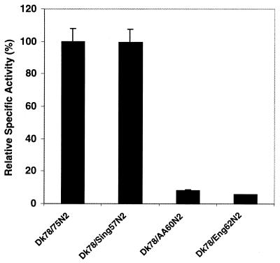
In previous analyses of the enzymatic activities of avian and human influenza A virus NAs, we had observed that in preparations of similar virus concentration, avian viruses generally possessed higher levels of activity than human viruses (unpublished observations). To measure differences in the enzymatic properties of the human virus NAs that might account for their differing abilities to support viral replication in ducks, we measured the relative specific activities of the NAs in the reassortants Dk78/Sing57N2, Dk78/AA60N2, and Dk78/Eng62N2 and compared them with the result for the N2 NA in Dk78/75N2, which was derived from an avian virus, A/duck/Hong Kong/7/75 (H3N2), in the background of A/duck/Hong Kong/278/78. Dk78/75N2 was chosen for this analysis because it replicates efficiently in the duck intestine and its N2 subtype is more closely related in sequence to the human virus NAs than the N9 NA of A/duck/Hong Kong/278/78. The avian and A/Singapore/1/57 NAs both possessed high levels of specific enzymatic activity (Fig. 1), in contrast to the A/Ann Arbor/6/60 and A/England/12/62 NAs, whose activities were only about 8 and 5%, respectively, of that of the high-activity NAs. The same trend in activity was observed when this assay was performed using NeuAcα2,3-lactose as the substrate (data not shown). Thus, an appreciable reduction in the enzymatic activity of NA during its adaptation in humans correlates with its reduced ability to support viral growth in ducks.
FIG. 1.
Relative specific enzymatic activities of avian and human virus NAs. Enzymatic activity was determined with the MU-NANA substrate, as described in Materials and Methods. Relative specific activity was calculated by normalizing NA activity to the level of NA protein in virions, as determined by nonreducing polyacrylamide gel electrophoresis of 35S-labeled virions, followed by autoradiography and densitometric analysis of the NA bands. The results are shown in relation to the activity of the avian NA of Dk78/75N2, set at 100%. The error bars indicate standard deviations.
NA region associated with high specific activity.
To identify the region of the NA molecule that contained the molecular determinants for high specific activity, we constructed a series of chimeras in which regions of the A/England/12/62 NA, the earliest human NA not able to support viral growth in ducks, were replaced with corresponding regions of the A/Singapore/1/57 NA (Fig. 2), whose ability to support replication in ducks parallels that of avian virus NAs. The constructs were expressed in cell culture, and the cell-associated NA activities and protein expression levels were used to calculate the relative specific activity for each construct.
The cell-expressed A/Singapore/1/57 NA had about fivefold-higher specific activity than the A/England/12/62 NA. Substitution in the A/England/12/62 NA with the A/Singapore/1/57 residues in chimeras 1 and 2 had little effect on enzymatic activity. Chimera 3 had about 3-fold-higher and chimera 4 about 1.5-fold-higher activity than did the A/England/12/62 NA. When the regions represented by chimeras 3 and 4 were combined in chimera 5, the full activity of the A/Singapore/1/57 NA was generated in the A/England/12/62 NA, indicating that multiple residues are involved in determining the level of NA specific activity. Residues from other portions of the molecule, such as those represented in chimeras 1 and 2, probably do not contribute to this function.
Molecular determinants of high specific activity in NA.
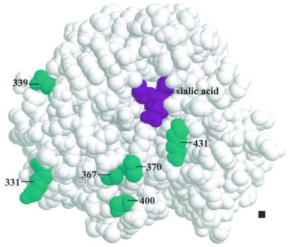
There are a total of six amino acid differences between the A/Singapore/1/57 and A/England/12/62 NAs in the region represented by chimera 5 (Table 2). To assess the contribution of these residues to high specific activity, we first examined the effect of replacing each of the four residues at positions 367, 370, 400, and 431 in A/England/12/62, all of which lie close to the enzymatic active site (Fig. 3), with the corresponding residue from A/Singapore/1/57. Mutants containing combinations of two, three, or all four of these residues were also evaluated. Because residues 331 and 339 were distant from the enzymatic active site, their contribution to specific activity was assessed as a pair in combination with the other residues under examination. All mutants were evaluated with the α2,3-sialyllactose substrate.
TABLE 2.
Amino acid sequence comparison of the N2 NAs of avian and early human H2N2 viruses in the region represented by chimera 5 (nucleotides 869 to 1467)
| Virus | Amino acid at positiona:
|
||||||||
|---|---|---|---|---|---|---|---|---|---|
| 331 | 334 | 339 | 367 | 370 | 392 | 400 | 431 | 463 | |
| A/Singapore/1/57 | S | N | D | N | S | I | N | P | N |
| A/Ann Arbor/6/60 | S | L | V | ||||||
| A/England/12/62 | R | N | S | L | S | Q | |||
| A/duck/Hong Kong/7/75 | S | S | V | D | |||||
Only the amino acids that differ from those of A/Singapore/1/57 are shown.
FIG. 3.
Location of amino acid differences between A/Singapore/1/57 and A/England/12/62 in the region of NA exchanged in chimera 5. The positions of the amino acid residues (cyan) and the enzymatic active site, indicated by the position of the bound sialic acid (purple), are shown on the NA structure of A/Tokyo/3/67 (H3N2) (23). The fourfold-symmetry axis (▪) that generates the tetrameric head of NA is perpendicular to the plane of the figure.
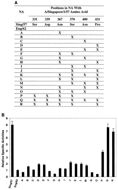
Individual amino acid mutations at residues 370 (mutant B) and 431 (mutant D) led to increased NA activity: 156 and 191% of the value for A/England/12/62 (Fig. 4). The change in mutant A at position 367 caused a decrease of 33% in the activity of A/England/12/62 NA, while the change at residue 400 in mutant C did not appreciably affect activity. Mutant D, with the highest activity of any of these altered NAs, was still far less active than the A/Singapore/1/57 NA. The variable effects of these mutations suggest that the individual residues do not make a straightforward contribution to NA specific activity but rather interact in a complex manner such that residue combinations, perhaps including all residues in chimera 5, cooperate to produce the high activity of the A/Singapore/1/57 NA.
FIG. 4.
Determination of the contributions of individual amino acid residues to high specific activity. (A) Mutants were generated in which amino acid residues in the A/England/12/62 (Eng/62) NA were replaced (X) individually and in groups with the corresponding A/Singapore/1/57 (Sing/57) residues. (B) The relative specific activities of the mutants were determined as described in the legend to Fig. 2. The error bars indicate standard deviations.
Mutation of residues 400 and 431 as a pair in mutant E, equivalent in sequence to chimera 4, had no additional effect on activity compared with the effect of residue 431 (mutant D) alone (Fig. 4). The paired mutation of residues 367 and 370 in mutant F did not substantially affect NA activity compared with that of wild-type A/England/12/62. When the residue 367 mutation was paired with either the residue 400 or 431 mutation (mutants G and H, respectively), it decreased NA activity compared to results with either residue alone. The combination of residues 370 and 400 (mutant I) yielded an intermediate NA activity (between those observed with either residue alone), while the addition of residue 370 to 431 in mutant J slightly increased the NA activity relative to that of the 431 mutant (D). Thus, when associated in pairs, none of these residues can account for the high specific activity of the A/Singapore/1/57 NA.
The analysis was expanded by generating additional combinations of three or four amino acid mutations. Changes at positions 367, 370, 400, and 431 in mutant K resulted in an NA with about the same activity as mutant D (Fig. 4). Although, it was possible to change residue 400 back to the A/England/12/62 identity (mutant L) without affecting the activity of the NA, similar reversions involving other residues produced variable results. For example, mutant M, with the A/England/12/62 identity at residue 367, had slightly lower activity than mutant K, whereas reversions at residue 370 in mutant N and 431 in mutant O to the A/England/12/62 identity dramatically lowered the respective NA activities to just 65 and 39% of the A/England/12/62 activity. When residues 331 and 339 were introduced in combination with residues 367 and 370 to generate mutant P, equivalent in sequence to chimera 3, we observed a 3.8-fold increase in activity compared with that of the wild-type A/England/12/62 NA. This suggests that the high specific activity of the A/Singapore/1/57 NA, while partially dependent on residues 367, 370, and 431, also depends on the identities of residues 331 and 339, despite their distance from the enzyme's active site.
Finally, a mutant construct containing residues 331 and 339 together with residues 367, 370, 400, and 431 (mutant Q), equivalent to chimera 5, had the full specific activity of the A/Singapore/1/57 NA (Fig. 4). Again, the mutation at position 400 (mutant R) was dispensable in generating a construct with full activity. Thus, the combination of residues at positions 331, 339, 367, 370, and 431 appears adequate to restore high levels of specific NA activity to A/England/12/62. By analogy, the high specific activity of the A/Singapore/1/57 NA can be viewed as requiring the interplay of several amino acid residues and cannot be attributed to the identity of the amino acid at any single position.
DISCUSSION
Understanding the molecular changes that accompany the adaptation of influenza A virus to a new host can yield important insight into the requirements for productive viral replication and cross-species transmission. Here, we show that the N2 NA from a human virus, A/Singapore/1/57, can support the replication of an avian reassortant virus with all other genes from A/duck/Hong Kong/278/78 in the duck intestine following oral inoculation. In similar reassortants, the N2 NA of A/Ann Arbor/6/60 could support virus growth, but at appreciably reduced efficiency, while the N2 NA of A/England/12/62 and a later human virus could not. Because all reassortants contained the same avian genes, with the exception of the NA gene, and the original avian virus was replication competent in orally inoculated ducks, we reasoned that clues to the molecular determinants(s) of viral growth resided in differences among the human NAs. Subsequent analyses revealed high levels of specific enzymatic activity in avian virus NAs and a human virus NA isolated in 1957, with sharp reductions in activity for viruses isolated after 1957. Thus, the loss of ability of most human viral NAs to support efficient viral replication in the duck intestine can be associated with reductions in the specific enzymatic activity of this viral surface glycoprotein.
Hinshaw et al. (5) were the first to note the inability of human viral NA to support replication in the duck intestine following oral inoculation. This finding was made with a human-avian reassortant possessing the N2 NA gene of A/Udorn/307/72 (H3N2), a virus isolated 15 years after the appearance of the N2 subtype of NA in humans, with all other genes from the avian virus A/mallard/New York/6750/78 (H2N2). A/Singapore/1/57, in which the HA and NA were both derived from an avian virus, represents the first appearance of the pandemic human H2N2 viruses in 1957 (20, 21). While the NA of this earliest human H2N2 virus appears to have retained properties of its avian ancestor, ongoing changes in later virus isolates, likely due to adaptation to growth in humans, greatly reduced its ability to support viral replication in ducks. Thus, by 1962, the human virus N2 NA in the reassortant Dk78/Eng62N2 was incapable of supporting viral replication in orally inoculated ducks, while Dk78/AA60N2, with the NA of human virus A/Ann Arbor/6/60, could replicate in some but not all ducks. In our analysis, the specific activity of the A/England/12/62 NA was slightly lower than that of A/Ann Arbor/6/60, but the reduction is too small to account for the differences in growth among the reassortants. It is possible that the ability of NA to support infection is dependent on high specific activity and one or more unknown additional properties, retained in the A/Ann Arbor/6/60 NA to some extent, that disappeared by 1962. We suggest that the NA of A/Ann Arbor/6/60 may represent an intermediate stage in the adaptation of NA to viral growth requirements in humans.
For practical reasons, not all possible combinations of the six residues at positions 331, 339, 367, 370, 400, and 431 were generated in this study. However, residues 331 and 339 appear indispensable (but not sufficient by themselves) for high specific NA activity. Although, the NA of A/Ann Arbor/6/60 has the same amino acid residues at positions 331, 339, 400, and 431 as does the A/Singapore/1/57 NA, it possesses low specific activity similar to that of A/England/12/62. The A/Ann Arbor/6/60 NA has serine at position 367 and valine at 392, similar to the high-activity NA of A/duck/Hong Kong/7/75, but has leucine at 370 like the A/England/12/62 NA. Because Leu370 represents the only amino acid difference between the A/Ann Arbor/6/60 NA and high-activity NAs and it lies in the NA region that determines high specific activity, residue 370 may be an important determinant of activity. Residue 367, on the other hand, differs in the NAs of A/Singapore/1/57 and A/England/12/62 but is the same in both the high-activity A/duck/Hong Kong/7/75 NA and the low-activity NAs of A/Ann Arbor/6/60 and A/England/12/62, suggesting that its identity is not particularly important for determining high specific NA activity.
The identity of residue 431 makes a significant contribution to high specific activity. Interestingly, this amino acid was also found to be important for recognition of the N-glycolylneuraminic species of sialic acid (10), an important receptor for influenza virus replication in the duck intestine (8). As is evident from the crystallographic structure of the A/Tokyo/3/67 N2 NA (22, 23), the position of residue 431 is too far from the sialic acid binding pocket of the enzymatic site to directly interact with the sialic acid species in glycoconjugates, suggesting that it interacts with a portion of the glycoconjugate farther up the chain (10); however, this prediction is not consistent with the fact that changes at position 431 can affect enzymatic activity even with small substrates, such as MU-NANA, which lacks neighboring sugar moieties. Thus, it could be that proline at position 431 results in conformational changes in the structure of the NA, permitting residue 431 or other residues to directly affect binding and/or activity for sialoglycoconjugates.
The amino acids analyzed in this study showed very high levels of conservation among avian influenza viruses: all 10 avian viruses with the N2 subtype shared Ser331, Asp339, Ser367, Ser370, Asn400, and Pro431 (Table 3). These residues were also found in the NAs of A/Singapore/1/57 and A/Ann Arbor/6/60, with the exception of Ser367→Asn in A/Singapore/1/57 and Ser370→Leu in A/Ann Arbor/6/60. The high degree of conservation of the residues at these six positions in avian viruses indicates the importance of their contribution to NA function. Most human viruses isolated after 1960 and all swine viruses display similar substitutions in four of the six residues of interest. Most notably, in the sequences of 25 human and 13 swine N2 viruses, residue 339 was always asparagine and residue 400 was always serine (except for one human isolate with arginine). Numerous replacements were observed at position 431 in human and swine virus NAs, including Gln, Arg, Lys, Glu, and Gly, but none of the isolates had the avian proline residue. Residue 331 was also highly conserved among human viruses isolated after 1962 and in the swine viruses; all had arginine at position 331 except for a human isolate with lysine and another with serine. The positions of residues 331 and 339 in the NA structure would not permit their direct interaction with a substrate, and they do not participate in the interaction between NA subunits in the active tetramer form of the enzyme (Fig. 3). Conceivably, these residues could contribute to high specific activity through long-range conformational effects. Residues at positions 367 and 370 were more heterogeneous in identity, displaying overlap between the amino acid identities in avian viruses and other amino acids found only in the human and swine viruses. It was also noted that residues 367, 370, and 400, conserved in high-activity NAs, are also important for NA hemadsorption activity, a conserved property of all NA subtypes in avian viruses that is lost in human virus NAs (9, 11, 25). However, the biological significance of NA hemadsorption activity for viral replication has not been determined (9).
TABLE 3.
Sequence comparison of avian, swine, and human N2 NAs
| Host species (no. of isolates) | Amino acid at positiona:
|
|||||
|---|---|---|---|---|---|---|
| 331 | 339 | 367 | 370 | 400 | 431 | |
| Avian (10) | Ser | Asp | Ser | Ser | Asn | Pro |
| Human 1957–1960 (2) | Ser | Asp | Asn | Leu | Asn | Pro |
| Ser | Ser | |||||
| Human 1962–1989 (22) | Arg (20) | Asn (22) | Asn (6) | Leu (18) | Arg (1) | Arg (1) |
| Lys (1) | Gly (6) | Ser (4) | Ser (21) | Gln (3) | ||
| Ser (1) | Ser (10) | Glu (8) | ||||
| Lys (10) | ||||||
| Swine (14) | Arg (13) | Asn (14) | Gly (1) | Leu (6) | Ser (14) | Arg (3) |
| Gly (1) | Ser (13) | Ser (8) | Glu (6) | |||
| Gly (2) | ||||||
| Lys (3) | ||||||
The numbers in parentheses indicate the number of isolates with that amino acid in the indicated position.
We postulate that the different levels of specific activity of the NAs from human and avian viruses stem from different viral growth requirements in the two hosts. Aquatic birds are believed to be the primordial reservoir for all subtypes of influenza viruses, and the NA of avian viruses is well adapted to support viral growth in these hosts. It is likely that high NA specific activity is required for maintenance of the HA affinity-NA activity balance needed for efficient growth in the duck intestine. The importance of this balance has been illustrated in several examples in which NA activity was inhibited by drugs, reduced by mutagenesis, or removed from virions (4, 6, 13, 14). In these studies, compensatory mutations that reduced HA affinity for its cellular receptor were observed, and they reduced viral dependence on NA activity during the budding of progeny virions from infected cells. Soon after H2N2 viruses were transmitted to humans, amino acid mutations in the HA increased its affinity for NeuAcα2-6Gal and reduced its affinity for NeuAcα2-3Gal. Since the affinity of human virus HAs for NeuAcα2-6Gal is generally less than that shown by avian virus HAs for NeuAcα2-3Gal (12), human virus NAs would be expected to require correspondingly less NA activity for budding of progeny virions. Alternatively, the reduction in human NA specific activity may simply be related to the removal of selective pressure on growth in an avian host or even to growth factors in the human respiratory tract that we do not yet understand.
Our findings help to define the unique requirements for influenza virus growth among different hosts and the process by which influenza viruses adapt to new hosts and establish stable lineages. Although H2N2 viruses had disappeared from humans by about 1968, the adaptation of the avian-virus-derived N2 NA resulted in the persistence of a human-adapted NA in H3N2 viruses, which continues to circulate worldwide. Expanding our understanding of the factors that influence cross-species transmission and adaptation of influenza viruses may aid in identifying viruses with the potential to cause pandemic outbreaks and thus in implementing timely and effective countermeasures.
ACKNOWLEDGMENTS
We thank Robert G. Webster for providing the antibodies to the N2 and N9 NAs. We are also grateful to Clayton Naeve and the St. Jude Children's Research Hospital Center for Biotechnology for preparing oligonucleotides and for computer support. We also thank John Gilbert for helpful suggestions and for editing the manuscript.
This work was supported by National Institute of Allergy and Infectious Diseases Public Health Service research grants, Cancer Center Support (CORE) grant CA-21765, and the American Lebanese Syrian Associated Charities (ALSAC).
REFERENCES
- 1.Baum L G, Paulson J C. The N2 neuraminidase of human influenza virus has acquired a substrate specificity complementary to the hemagglutinin receptor specificity. Virology. 1991;180:10–15. doi: 10.1016/0042-6822(91)90003-t. [DOI] [PubMed] [Google Scholar]
- 2.Connor R J, Kawaoka Y, Webster R G, Paulson J C. Receptor specificity in human, avian, and equine H2 and H3 influenza virus isolates. Virology. 1994;205:17–23. doi: 10.1006/viro.1994.1615. [DOI] [PubMed] [Google Scholar]
- 3.Couceiro J N, Paulson J C, Baum L G. Influenza virus strains selectively recognize sialyloligosaccharides on human respiratory epithelium: the role of the host cell in selection of hemagglutinin receptor specificity. Virus Res. 1993;29:155–165. doi: 10.1016/0168-1702(93)90056-s. [DOI] [PubMed] [Google Scholar]
- 4.Gubareva L V, Bethell R, Hart G J, Murti K G, Penn C R, Webster R G. Characterization of mutants of influenza A virus selected with the neuraminidase inhibitor 4-guanidino-Neu5Ac2en. J Virol. 1996;70:1818–1827. doi: 10.1128/jvi.70.3.1818-1827.1996. [DOI] [PMC free article] [PubMed] [Google Scholar]
- 5.Hinshaw V S, Webster R G, Naeve C W, Murphy B R. Altered tissue tropism of human-avian reassortant influenza viruses. Virology. 1983;128:260–263. doi: 10.1016/0042-6822(83)90337-9. [DOI] [PubMed] [Google Scholar]
- 6.Hughes M T, Matrosovich M, Rodgers M E, McGregor M, Kawaoka Y. Influenza A viruses lacking sialidase activity can undergo multiple cycles of replication in cell culture, eggs, or mice. J Virol. 2000;74:5206–5212. doi: 10.1128/jvi.74.11.5206-5212.2000. [DOI] [PMC free article] [PubMed] [Google Scholar]
- 7.Ito T, Couceiro J N, Kelm S, Baum L G, Krauss S, Castrucci M R, Donatelli I, Kida H, Paulson J C, Webster R G, Kawaoka Y. Molecular basis for the generation in pigs of influenza A viruses with pandemic potential. J Virol. 1998;72:7367–7373. doi: 10.1128/jvi.72.9.7367-7373.1998. [DOI] [PMC free article] [PubMed] [Google Scholar]
- 8.Ito T, Suzuki Y, Suzuki T, Takada A, Horimoto T, Wells K, Kida H, Otsuki K, Kiso M, Ishida H, Kawaoka Y. Recognition of N-glycolylneuraminic acid linked to galactose by the α2,3 linkage is associated with intestinal replication of influenza A virus in ducks. J Virol. 2000;74:9300–9305. doi: 10.1128/jvi.74.19.9300-9305.2000. [DOI] [PMC free article] [PubMed] [Google Scholar]
- 9.Kobasa D, Rodgers M E, Wells K, Kawaoka Y. Neuraminidase hemadsorption activity, conserved in avian influenza A viruses, does not influence viral replication in ducks. J Virol. 1997;71:6706–6713. doi: 10.1128/jvi.71.9.6706-6713.1997. [DOI] [PMC free article] [PubMed] [Google Scholar]
- 10.Kobasa D, Kodihalli S, Luo M, Castrucci M R, Donatelli I, Suzuki Y, Suzuki T, Kawaoka Y. Amino acid residues contributing to the substrate specificity of the influenza A virus neuraminidase. J Virol. 1999;73:6743–6751. doi: 10.1128/jvi.73.8.6743-6751.1999. [DOI] [PMC free article] [PubMed] [Google Scholar]
- 11.Laver W G, Colman P M, Webster R G, Hinshaw V S, Air G M. Influenza virus neuraminidase with hemagglutinin activity. Virology. 1984;137:314–323. doi: 10.1016/0042-6822(84)90223-x. [DOI] [PubMed] [Google Scholar]
- 12.Matrosovich M, Tuzikov A, Bovin N, Gambaryan A, Klimov A, Castrucci M R, Donatelli I, Kawaoka Y. Early alterations of the receptor-binding properties of H1, H2, and H3 avian influenza virus hemagglutinins after their introduction into mammals. J Virol. 2000;74:8502–8512. doi: 10.1128/jvi.74.18.8502-8512.2000. [DOI] [PMC free article] [PubMed] [Google Scholar]
- 13.McKimm-Breschkin J L, Blick T J, Sahasrabudhe A, Tiong T, Marshall D, Hart G J, Bethell R C, Penn C R. Generation and characterization of variants of NWS/G70C influenza virus after in vitro passage in 4-amino-Neu5Ac2en and 4-guanidino-Neu5Ac2en. Antimicrob Agents Chemother. 1996;40:40–46. doi: 10.1128/aac.40.1.40. [DOI] [PMC free article] [PubMed] [Google Scholar]
- 14.Mitnaul L J, Matrosovich M N, Castrucci M R, Tuzikov A B, Bovin N V, Kobasa D, Kawaoka Y. Balanced hemagglutinin and neuraminidase activities are critical for efficient replication of influenza A virus. J Virol. 2000;74:6015–6020. doi: 10.1128/jvi.74.13.6015-6020.2000. [DOI] [PMC free article] [PubMed] [Google Scholar]
- 15.Niwa H, Yamamura K, Miyazaki J. Efficient selection for high-expression transfectants with a novel eukaryotic vector. Gene. 1991;108:193–200. doi: 10.1016/0378-1119(91)90434-d. [DOI] [PubMed] [Google Scholar]
- 16.Palese P, Tobita K, Ueda M, Compans R W. Characterization of temperature-sensitive influenza virus mutants defective in neuraminidase. Virology. 1974;61:397–410. doi: 10.1016/0042-6822(74)90276-1. [DOI] [PubMed] [Google Scholar]
- 17.Prickett K S, Amberg D C, Hopp T P. A calcium-dependent antibody for identification and purification of recombinant proteins. BioTechniques. 1989;7:580–589. [PubMed] [Google Scholar]
- 18.Rogers G N, Paulson J C. Receptor determinants of human influenza virus isolates: differences in receptor specificity of the H3 hemagglutinin based on species of origin. Virology. 1983;127:361–373. doi: 10.1016/0042-6822(83)90150-2. [DOI] [PubMed] [Google Scholar]
- 19.Rogers G N, D'Souza B L. Receptor binding properties of human and animal H1 influenza virus isolates. Virology. 1989;173:317–322. doi: 10.1016/0042-6822(89)90249-3. [DOI] [PubMed] [Google Scholar]
- 20.Schäfer J R, Kawaoka Y, Bean W J, Suss J, Senne D, Webster R G. Origin of the pandemic 1957 H2 influenza A virus and the persistence of its possible progenitors in the avian reservoir. Virology. 1993;194:781–788. doi: 10.1006/viro.1993.1319. [DOI] [PubMed] [Google Scholar]
- 21.Scholtissek C, Rohde W, Von Hoyningen V, Rott R. On the origin of the human influenza virus subtypes H2N2 and H3N2. Virology. 1978;87:13–20. doi: 10.1016/0042-6822(78)90153-8. [DOI] [PubMed] [Google Scholar]
- 22.Varghese J N, Colman P M. Three-dimensional structure of the neuraminidase of influenza virus A/Tokyo/3/67 at 2.2 Å resolution. J Mol Biol. 1991;221:473–486. doi: 10.1016/0022-2836(91)80068-6. [DOI] [PubMed] [Google Scholar]
- 23.Varghese J N, McKimm-Breschkin J L, Caldwell J B, Kortt A A, Colman P M. The structure of the complex between influenza virus neuraminidase and sialic acid, the viral receptor. Proteins. 1992;14:327–332. doi: 10.1002/prot.340140302. [DOI] [PubMed] [Google Scholar]
- 24.Webster R G. Antigenic hybrids of influenza A viruses with surface antigens to order. Virology. 1970;42:633–642. doi: 10.1016/0042-6822(70)90309-0. [DOI] [PubMed] [Google Scholar]
- 25.Webster R G, Air G M, Metzger D W, Colman P M, Varghese J N, Baker A T, Laver W G. Antigenic structure and variation in an influenza virus N9 neuraminidase. J Virol. 1987;61:2910–2916. doi: 10.1128/jvi.61.9.2910-2916.1987. [DOI] [PMC free article] [PubMed] [Google Scholar]
- 26.Webster R G, Bean W J, Gorman O T, Chambers T M, Kawaoka Y. Evolution and ecology of influenza A viruses. Microbiol Rev. 1992;56:152–179. doi: 10.1128/mr.56.1.152-179.1992. [DOI] [PMC free article] [PubMed] [Google Scholar]


