Abstract
A complete cDNA clone of the Newcastle disease virus (NDV) vaccine strain Hitchner B1 was constructed, and infectious recombinant virus expressing an influenza virus hemagglutinin was generated by reverse genetics. The rescued virus induces a strong humoral antibody response against influenza virus and provides complete protection against a lethal dose of influenza virus challenge in mice, demonstrating the potential of recombinant NDV as a vaccine vector.
Newcastle disease virus (NDV) is a member of the Rubulavirus genus in the Paramyxoviridae family and is categorized into three pathotypes depending on the severity of the disease that it causes in birds: lentogenic, mesogenic, or velogenic (1, 33). The ability to genetically engineer negative-strand RNA viruses has led to extraordinary advances in understanding their biology. The first reports of NDV rescue from cDNA were published in 1999 (22, 28). Both groups used the lentogenic NDV strain LaSota. Recently, this strain was further attenuated by modifying the RNA editing site in the P gene, which resulted in low-level expression of the V protein (18). In addition, the generation of a chimeric recombinant NDV containing a hybrid HN gene with the N-terminal region derived from NDV and the external C-terminal region from avian paramyxovirus type 4 was reported (23). Finally, Krishnamurthy et al. (15) and Huang et al. (11) rescued recombinant NDVs (mesogenic strain Beaudette C and lentogenic strain LaSota, respectively) stably expressing the chloramphenicol acetyltransferase (CAT) reporter gene.
An important application of reverse-genetic techniques is the generation of recombinant viruses for use as vaccine vectors (reviewed in references 4, 8, 19, 21, and 26). A number of recombinant negative-strand RNA viruses expressing foreign proteins have been constructed. Recombinant vesicular stomatitis viruses (VSVs) are able to express the human CD4 protein (32), the influenza virus hemagglutinin (HA) and neuraminidase (14, 25, 27), the respiratory syncytial virus G and F proteins (12), the human immunodeficiency virus Gag and Env proteins (10), and the bovine viral diarrhea virus E2 protein (9). Their efficacies as vaccine vectors have been studied (reviewed in reference 26). Among paramyxoviruses, several chimeric measles viruses and Sendai viruses expressing foreign genes have been constructed (29, 35, 38), and a rinderpest virus expressing an influenza virus HA protein has been described (37). The latter recombinant was shown to induce humoral immunity in vaccinated cattle (37). In this paper, a recombinant NDV expressing the HA protein of influenza A virus was generated from the full-length cDNA of the avirulent strain Hitchner B1 (ATCC VR108), a widely used NDV live vaccine strain. The potential of the recombinant NDV as an effective vaccine vector was evaluated.
Rescue of recombinant viruses from cloned cDNAs.
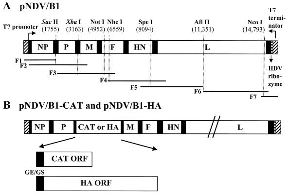
pNDV/B1, containing the full-length cDNA of the Hitchner B1 strain was constructed (Fig. 1), and two additional restriction enzyme sites (SacII, nucleotides [nt] 1755 to 1760; and XbaI, nt 3163 to 3168) were created as genetic tag sequences. The complete genome (15,186 nt) of pNDV/B1 was sequenced using a model ABI 3700 sequencer (Applied Biosystems). We chose the newly introduced XbaI site, located between the P and M genes to insert the CAT gene (24) or the HA gene of influenza A/WSN/33 virus (7). These constructs (pNDV/B1-CAT and pNDV/B1-HA) should retain the normal ratio of NP to P expression, which appears to be critical for effective viral replication (2). The numbers of the inserted nucleotides in pNDV/B1-CAT and pNDV/B1-HA were 696 and 1,752, respectively, maintaining the genome length as a multiple of 6 (3). To generate recombinant NDVs from pNDV/B1, pNDV/B1-CAT, and pNDV/B1-HA, HEp-2 or A549 cells in a six-well plate were infected with MVA-T7 (kindly provided by B. Moss) at a multiplicity of infection of 1 and then transfected with each NDV full-length clone (1 μg) together with the following expression plasmids: pTM1-NP (nucleoprotein) at 0.4 μg, pTM1-P (phosphoprotein) at 0.2 μg, and pTM1-L (RNA-dependent RNA polymerase) at 0.2 μg. After overnight incubation, the transfected cells were cocultured with chicken embryo fibroblasts (CEF) to amplify the produced virus and then incubated for an additional 2 to 3 days. The supernatants of transfected cells were injected into the allantoic cavities of 9- or 10-day-old embryonated chicken eggs. Three to four days later, allantoic fluids were harvested and virus growth was confirmed by hemagglutination assays followed by hemagglutination inhibition (HI) testing. Viral genomic RNAs were isolated from the rescued viruses (rNDV/B1, rNDV/B1-CAT, and rNDV/B1-HA) and the tagged restriction enzyme sites, and the presence of the inserted foreign genes was confirmed by reverse transcription-PCR–restriction enzyme digestion analysis (data not shown).
FIG. 1.
Schematic representation of pNDV/B1, pNDV/B1-CAT, and pNDV/B1-HA cDNA constructs. (A) pNDV/B1 was generated by seven PCR fragments spanning the following nucleotide positions: F1, T7 promoter, nt 1755 (SacII); F2, nt 1 to 3321; F3, nt 1755 (SacII) to 6580; F4, nt 6151 to 10,210; F5, nt 7381 to 11,351; F6, nt 11,351 to 14,995; and F7, nt 14,701 to 15,186. These sequences were followed by the hepatitis delta virus (HDV) ribozyme and the T7 terminator. The cDNA fragments were joined at shared restriction sites and assembled in plasmid pSL1180 (Amersham Pharmacia Biotech). SacII and XbaI are shown in italics to indicate that they are genomic tag sequences. (B) The pNDV/B1-CAT and pNDV/B1-HA constructs were made by inserting the CAT and influenza virus A/WSN/33 HA open reading frames (ORF), respectively, into the unique XbaI cloning site (nt 3163) located between the P and M genes of the pNDV/B1 clone. The inserted gene contains the gene end (GE; 5′-TTAGAAAAAA-3′), intercistronic nucleotide (T), and the gene start sequence (GS; 5′-ACGGGTAGAA-3′). In addition, seven nucleotides (5′-CGCCACC-3′) were inserted upstream of the initiation site to introduce an optimal Kozak sequence (13). In the case of pNDV/B1-HA, the gene start sequence is followed by the 5′ untranslated region (26 nt) of the HA gene.
rNDV/B1-HA virus stably expresses HA protein on the cell surface.
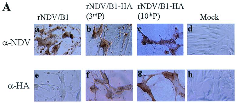
The expression of HA protein was analyzed by infection of 35-mm-diameter dishes of confluent CEF cells with either rNDV/B1 or rNDV/B1-HA at a multiplicity of infection of 1. Cells were fixed at 1 and 2 days postinfection using 1% paraformaldehyde. NDV proteins and HA protein were visualized using a mouse anti-NDV polyclonal serum and a monoclonal antibody (2G9) against influenza virus HA, respectively, followed by incubation with peroxidase-conjugated anti-mouse immunoglobulins (Boehringer Mannheim). Extensive cytopathic effects and typical syncytia were observed in cells infected with rNDV/B1 or rNDV/B1-HA at 2 days postinfection (Fig. 2A, images a, b, c, e, f, and g) but not in mock-infected cells (Fig. 2A, images d and h). NDV infection was demonstrated by staining with an anti-NDV serum (Fig. 2A, images a, b, and c). In contrast, the HA-specific monoclonal antibody (2G9) reacted only with rNDV/B1-HA-infected cells and not with rNDV/B1- or mock-infected cells (Fig. 2A, images e, f, g, and h). These results suggested that the HA protein is efficiently expressed and transferred to the cell surfaces of rNDV/B1-HA-infected cells. A similar result was reported for recombinant VSV and rinderpest virus expressing the HA protein (14, 37). In addition, we demonstrated that the HA protein was stably expressed from rNDV/B1-HA following 10 passages in embryonated eggs at a low multiplicity of infection (10 to 100 PFU/egg) (Fig. 2A, image g). The stable expression of a foreign gene was also confirmed using rNDV/B1-CAT. CAT expression was not changed after 10 passages in embryonated eggs at a low multiplicity of infection (data not shown).
FIG. 2.
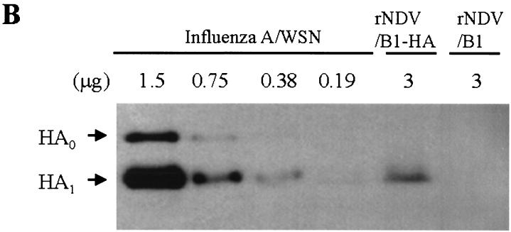
Detection of the HA protein on infected cells and in purified virions. (A) rNDV/B1- or rNDV/B1-HA-infected cells were fixed with 1% paraformaldehyde at day 2 postinfection, and cells were used for immunostaining analysis. NDV protein expression on the surfaces of cells infected with rNDV/B1 (a) or rNDV/B1-HA passaged in eggs at a low multiplicity of infection three times (3rdP) (b) or 10 times (10thP) (c) was analyzed by using mouse anti-NDV serum. HA expression on the cell surfaces of rNDV/B1 (e)- or rNDV/B1-HA (f and g)-infected cells was analyzed by using anti-HA monoclonal antibody (2G9). Mock-infected cells were also analyzed as a control (d and h). (B) rNDV/B1 and rNDV/B1-HA were purified from allantoic fluids of infected embryonated chicken eggs. Influenza A/WSN/33 virus was purified from MDBK cell culture supernatants as a control. Serial twofold dilutions of A/WSN/33 viral proteins (1.5 to 0.19 μg) and 3 μg of rNDV/B1-HA or rNDV/B1 viral proteins were separated on a sodium dodecyl sulfate–10% polyacrylamide gel. The gel was transferred to a nitrocellulose membrane, and the HA protein was detected by chemiluminescence using a mixture of anti-HA monoclonal antibodies and an anti-mouse IgG peroxidase-labeled antibody (DAKO). HA0 and HA1 indicate uncleaved and cleaved HA protein, respectively.
HA incorporation into virions.
Previous studies showed that the HA protein expressed from recombinant VSV was efficiently incorporated into virions (14). On the other hand, recombinant rinderpest virus did not incorporate the expressed HA protein into particles (37). Thus, we wished to determine whether the HA protein was incorporated into the virions of the recombinant NDV. For this purpose, virions of rNDV/B1-HA, rNDV/B1, or influenza A/WSN/33 virus were purified and concentrated using a two-step centrifugation method (7,000 × g for 30 min and 200, 000 × g for 90 min over a 30% sucrose cushion). The total amount of protein in purified virus preparations was measured using a Bradford assay kit (Bio-Rad). Viral proteins were electrophoresed on a sodium dodecyl sulfate–10% polyacrylamide gel, followed by immunoblotting with anti-HA monoclonal antibodies (H15-A13, B-8, B-9, B-15, B-17, C-10, C-12, and E-10, kindly provided by W. Gerhard). The result showed that the amount of HA protein in the rNDV/B1-HA virion was four- to eightfold lower than that in influenza A/WSN/33 virus, assuming that the same number of viral particles per microgram of viral proteins was present (Fig. 2B). The HA protein incorporated into rNDV/B1-HA particles appeared to be cleaved, indicating that the HA protein was accessible to proteolytic enzymes.
Virus growth kinetics and pathogenicity of rNDV/B1-HA.
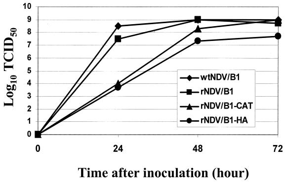
Embryonated chicken eggs were inoculated with the parent virus (wild-type NDV/B1 [wtNDV/B1]), rNDV/B1, rNDV/B1-CAT, or rNDV/B1-HA at 100 PFU per egg. Viral growth was analyzed at different time points after inoculation. A 50% tissue culture infective dose (TCID50) of each virus was determined by immunofluorescence assay. Ninety-six well plates of 80%-confluent CEF were infected with serial 10-fold dilutions of virus (four wells per dilution). Cells were incubated for 2 days and fixed with 2.5% formaldehyde containing 0.1% Triton X-100. Viral proteins were visualized using an anti-NDV rabbit serum followed by fluorescein isothiocyanate-conjugated anti-rabbit immunoglobulins (DAKO). wtNDV/B1, rNDV/B1, and rNDV/B1-CAT grew to similar titers (109 TCID50/ml), whereas the maximal viral titer achieved with rNDV/B1-HA was approximately 20-fold lower (5 × 10⁷ TCID50/ml) (Fig. 3). In order to test the virulence of rNDV/B1-HA, the mean time of death in eggs was determined. Serial 10-fold dilutions of infectious allantoic fluid (10−6 to 10−8) of wtNDV/B1, rNDV/B1, and rNDV/B1-HA viruses were inoculated into each of five embryonated eggs and the mean time in hours for the minimal lethal dose to kill embryos was determined. Embryos inoculated with the wtNDV/B1 and rNDV/B1 viruses died within 6 days. The mean times of death of the wild-type virus and rNDV/B1 were 108 and 113 h, respectively. In contrast, approximately 80% of embryos inoculated with rNDV/B1-HA survived beyond 8 days postinoculation. Similar results were obtained if higher amounts of viruses (10−3 to 10−5 dilutions) were inoculated. These results indicate that rNDV/B1-HA is attenuated in embryonated chicken eggs. Since NDV Hitchner B1 is routinely used to vaccinate poultry, rNDV/B1 expressing an avian influenza virus HA could be used as a vaccine against avian influenza viruses as well as NDV.
FIG. 3.
Growth curves of wtNDV/B1, rNDV/B1, rNDV/B1-CAT, and rNDV/B1-HA viruses in embryonated chicken eggs. Embryonated eggs were inoculated with 100 PFU of each virus, and allantoic fluids were harvested at different time points (24, 48, and 72 h postinoculation). Viral titers (TCID50) in CEF were determined by immunofluorescence assay using anti-NDV rabbit serum and an anti-rabbit IgG fluorescein isothiocyanate-labeled antibody (DAKO).
Immnunogenicity and pathogenicity of recombinant NDV in mice.
We demonstrated that rNDV/B1-HA efficiently expresses the HA protein on the surfaces of infected cells and that a significant amount of HA molecules is associated with the virus (Fig. 2). We next attempted to use this rNDV/B1-HA as a vaccine vector to protect mice against challenge with influenza virus. Previous studies showed that recombinant VSV and recombinant rinderpest virus expressing the HA protein induced a humoral antibody responses in vivo (25, 27, 37). BALB/c mice were inoculated with rNDV/B1-HA either intravenously or intraperitoneally at 3 × 10⁷ PFU per mouse. Phosphate-buffered saline (PBS) or the same amount of rNDV/B1 was administered intravenously as a control. No weight loss was observed in mice inoculated with rNDV/B1-HA or with rNDV/B1 (data not shown). Although growth levels of these viruses in mice were not determined, the lack of weight loss suggests that rNDV/B1 and rNDV/B1-HA have little or no toxicity in mice. Mice inoculated with rNDV/B1 or rNDV/B1-HA showed no measurable antibody response to either NDV or influenza virus 14 days after the initial inoculation (data not shown). One week after being given a booster injection (28 days after the initial inoculation), an antibody response against NDV was induced in mice inoculated with rNDV/B1 or rNDV/B1-HA by intravenous administration (Table 1). HI antibody titers against NDV in the two groups were similar (Table 1). Intraperitoneal administration of rNDV/B1-HA also induced a significant anti-NDV antibody response, but the HI titers were lower than those after the intravenous administration. As expected, specific anti-influenza virus antibody was detected only in mice inoculated with rNDV/B1-HA. Intravenous administration induced higher titers of antibody to influenza virus HA than intraperitoneal administration (Table 1).
TABLE 1.
Protection of mice immunized with rNDV/B1-HA following challenge with influenza A/WSN/33 virus
| Group | Immunization
|
HA antibody titera with:
|
No. of survivors (n = 5) after:
|
||||
|---|---|---|---|---|---|---|---|
| Virus | Dose (PFU) (107) | Administration | Anti-NDV | Anti-influenza A/WSN virus | Immunization | Challengeb | |
| A | rNDV/B1 | 3 | Intravenous | 1:480 | NDc | 5 | 0 |
| B | rNDV/B1-HA | 3 | Intravenous | 1:360 | 1:360 | 5 | 5 |
| C | rNDV/B1-HA | 3 | Intraperitoneal | 1:140 | 1:110 | 5 | 5 |
| D | PBS | 0 | Intravenous | ND | ND | 5 | 0 |
Sera were collected on day 28 (1 week after the booster injection) after the first inoculation. Values are averages of HA antibody titers of five mice.
Mice were challenged with 105 PFU (100 LD50) of influenza A/WSN/33 virus on day 35 after the first inoculation.
ND, not detectable.
Efficacy of recombinant NDV in generating protective immunity.
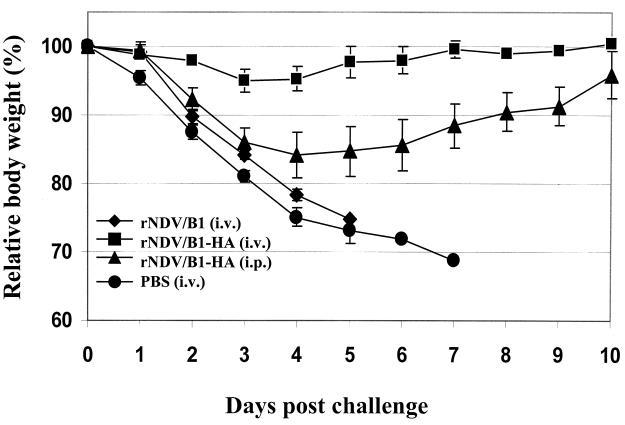
On day 35 after the initial inoculation (2 weeks after the booster injection), mice were challenged with 105 PFU (100 50% lethal doses [LD50]) of influenza A/WSN/33 virus by intranasal administration. As shown in Fig. 4 and Table 1, rNDV/B1-HA-immunized mice were protected against a lethal dose of influenza virus. In the case of intraperitoneal administration, although detectable weight loss was observed at first, mice fully recovered within 10 days (Fig. 4). In addition, no changes in physical activity and fur appearance were observed after influenza virus challenge in any of the vaccinated mice. In contrast, control mice inoculated with rNDV/B1 or PBS were not protected and died within 7 days of the influenza virus challenge (Fig. 4 and Table 1). Previous studies (6, 36) found that protection is related to HA antibody induction, and our investigation is consistent with these findings (Table 1). Little is known about the replication of lentogenic NDV in mice. Analysis of the growth kinetics and tissue distribution of NDV/B1 in mammalian animals remains to be done. However, it has been demonstrated that virulent NDV replicates in mice (17). Also, certain NDV strains (including pathogenic and avirulent strains) have been shown to possess oncolytic activity (reviewed in reference 34) and, most importantly, it has been reported that NDV can infect humans without inducing severe symptoms (5, 20, 30). Clinical trials using NDV as an antitumor agent have shown encouraging preliminary results for patients with a variety of cancers, and phase III trials are beginning in Europe (20). These results indicate the potential of using recombinant NDVs in humans. Recombinant NDVs expressing components of other human pathogens might be able to induce, without severe side effects, a protective immune response to these agents. A recombinant NDV-based vaccine against human immunodeficiency virus may thus be envisioned. In addition, NDV-infected cells have been shown to produce cytokines such as interferons and tumor necrosis factor, stimulating immune responses (16, 31, 34). This immune response-stimulatory activity of NDV may be the reason for the finding that patients developed a highly immunogenic response to their own tumor following administration of X-ray-irradiated NDV-infected autologous tumor cells (reviewed in reference 30).
FIG. 4.
Average body weights of vaccinated mice after influenza virus challenge. Vaccinated mice were challenged with a lethal dose (100 LD50) of influenza A/WSN/33 virus on day 35 (2 weeks after the booster injection). Relative average daily body weights (percentages) of mice vaccinated with rNDV/B1-HA intravenously (i.v.) (▪) or intraperitoneally (i.p.) (▴), with rNDV/B1 intravenously (♦), or with PBS (●) are shown. The vaccinating dose was 3 × 10⁷ PFU/ml in all cases. The error bars indicate ±0.5 times the standard deviation.
In conclusion, recombinant NDV expressing the influenza virus HA protein was generated from cDNA of the Hitchner B1 vaccine strain by reverse genetics. This recombinant virus stably expressed the HA protein in infected cells and induced a protective immune response against influenza virus in mice. Our studies suggest that recombinant NDV may be a safe and effective vaccine vector for possible use in mammalian and avian species.
Nucleotide sequence accession number.
The complete genome (15,186 nt) of pNDV/B1 was submitted to GenBank under accession number AF375823.
Acknowledgments
This work was partially supported by grants to A.G.-S. from the National Institutes of Health, by grants to P.P. from the National Institutes of Health, and by a grant to E.V. from the Direccion General de Ensenanza Superior of Spain (DGES PM97/0160). Y.N. was supported by an Uehara Memorial Bio-Medical Research Foundation fellowship.
A mixture of monoclonal antibodies to the influenza A/WSN/33 virus HA was kindly provided by Walter Gerhard. pTM1 and MVA-T7 were kindly provided by Bernard Moss.
REFERENCES
- 1.Alexander D J. Newcastle disease, Newcastle disease virus—an avian paramyxovirus. Dordrecht, The Netherlands: Kluwer Academic Publishers; 1988. pp. 1–22. [Google Scholar]
- 2.Baron M D, Foster-Cuevas M, Baron J, Barrett T. Expression in cattle of epitopes of a heterologous virus using a recombinant rinderpest virus. J Gen Virol. 1999;80:2031–2039. doi: 10.1099/0022-1317-80-8-2031. [DOI] [PubMed] [Google Scholar]
- 3.Calain P, Roux L. The rule of six, a basic feature for efficient replication of Sendai virus defective interfering RNA. J Virol. 1993;67:4822–4830. doi: 10.1128/jvi.67.8.4822-4830.1993. [DOI] [PMC free article] [PubMed] [Google Scholar]
- 4.Conzelmann K K. Nonsegmented negative-strand RNA viruses: genetics and manipulation of viral genomes. Annu Rev Genet. 1998;32:123–162. doi: 10.1146/annurev.genet.32.1.123. [DOI] [PubMed] [Google Scholar]
- 5.Csatary L K, Eckhardt S, Bukosza I, Czegledi F, Fenyvesi C, Gergely P, Bodey B, Csatary C M. Attenuated veterinary virus vaccine for the treatment of cancer. Cancer Detect Prev. 1993;17:619–627. [PubMed] [Google Scholar]
- 6.Deck R R, DeWitt C M, Donnelly J J, Liu M A, Ulmer J B. Characterization of humoral immune responses induced by an influenza hemagglutinin DNA vaccine. Vaccine. 1997;15:71–78. doi: 10.1016/s0264-410x(96)00101-6. [DOI] [PubMed] [Google Scholar]
- 7.Enami M, Palese P. High-efficiency formation of influenza virus transfectants. J Virol. 1991;65:2711–2713. doi: 10.1128/jvi.65.5.2711-2713.1991. [DOI] [PMC free article] [PubMed] [Google Scholar]
- 8.García-Sastre A. Negative-strand RNA viruses: applications to biotechnology. Trends Biotechnol. 1998;16:230–235. doi: 10.1016/s0167-7799(98)01192-5. [DOI] [PubMed] [Google Scholar]
- 9.Grigera P R, Marzocca M P, Capozzo A V, Buonocore L, Donis R O, Rose J K. Presence of bovine viral diarrhea virus (BVDV) E2 glycoprotein in VSV recombinant particles and induction of neutralizing BVDV antibodies in mice. Virus Res. 2000;69:3–15. doi: 10.1016/s0168-1702(00)00164-7. [DOI] [PubMed] [Google Scholar]
- 10.Haglund K, Forman J, Krausslich H G, Rose J K. Expression of human immunodeficiency virus type 1 Gag protein precursor and envelope proteins from a vesicular stomatitis virus recombinant: high-level production of virus-like particles containing HIV envelope. Virology. 2000;268:112–121. doi: 10.1006/viro.1999.0120. [DOI] [PubMed] [Google Scholar]
- 11.Huang Z, Krishnamurthy S, Panda A, Samal S K. High-level expression of a foreign gene from the most 3′-proximal locus of a recombinant Newcastle disease virus. J Gen Virol. 2001;82:1729–1736. doi: 10.1099/0022-1317-82-7-1729. [DOI] [PubMed] [Google Scholar]
- 12.Kahn J S, Schnell M J, Buonocore L, Rose J K. Recombinant vesicular stomatitis virus expressing respiratory syncytial virus (RSV) glycoproteins: RSV fusion protein can mediate infection and cell fusion. Virology. 1999;254:81–91. doi: 10.1006/viro.1998.9535. [DOI] [PubMed] [Google Scholar]
- 13.Kozak M. At least six nucleotides preceding the AUG initiator codon enhance translation in mammalian cells. J Mol Biol. 1987;196:947–950. doi: 10.1016/0022-2836(87)90418-9. [DOI] [PubMed] [Google Scholar]
- 14.Kretzschmar E, Buonocore L, Schnell M J, Rose J K. High-efficiency incorporation of functional influenza virus glycoproteins into recombinant vesicular stomatitis viruses. J Virol. 1997;71:5982–5989. doi: 10.1128/jvi.71.8.5982-5989.1997. [DOI] [PMC free article] [PubMed] [Google Scholar]
- 15.Krishnamurthy S, Huang Z, Samal S K. Recovery of a virulent strain of Newcastle disease virus from cloned cDNA: expression of a foreign gene results in growth retardation and attenuation. Virology. 2000;278:168–182. doi: 10.1006/viro.2000.0618. [DOI] [PubMed] [Google Scholar]
- 16.Le Bon A, Schiavoni G, D'Agostino G, Gresser I, Belardelli F, Tough D F. Type I interferons potently enhance humoral immunity and can promote isotype switching by stimulating dendritic cells in vivo. Immunity. 2001;14:461–470. doi: 10.1016/s1074-7613(01)00126-1. [DOI] [PubMed] [Google Scholar]
- 17.Lorence R M, Katubig B B, Reichard K W, Reyes H M, Phuangsab A, Sassetti M D, Walter R J, Peeples M E. Complete regression of human fibrosarcoma xenograft after local Newcastle disease virus therapy. Cancer Res. 1994;54:6017–6021. [PubMed] [Google Scholar]
- 18.Mebatsion T, Verstegen S, de Vaan L T, Römer-Oberdörfer A, Schrier C C. A recombinant Newcastle disease virus with low-level V protein expression is immunogenic and lacks pathogenicity for chicken embryos. J Virol. 2001;75:420–428. doi: 10.1128/JVI.75.1.420-428.2001. [DOI] [PMC free article] [PubMed] [Google Scholar]
- 19.Nagai Y, Kato A. Paramyxovirus reverse genetics is coming of age. Microbiol Immunol. 1999;43:613–624. doi: 10.1111/j.1348-0421.1999.tb02448.x. [DOI] [PubMed] [Google Scholar]
- 20.Nelson N J. Scientific interest in Newcastle disease virus is reviving. J Natl Cancer Inst. 1999;91:1708–1710. doi: 10.1093/jnci/91.20.1708. [DOI] [PubMed] [Google Scholar]
- 21.Palese P, Zheng H, Engelhardt O G, Pleschka S, García-Sastre A. Negative-strand RNA viruses: genetic engineering and applications. Proc Natl Acad Sci USA. 1996;93:11354–11358. doi: 10.1073/pnas.93.21.11354. [DOI] [PMC free article] [PubMed] [Google Scholar]
- 22.Peeters B P H, de Leeuw O S, Koch G, Gielkens A L J. Rescue of Newcastle disease virus from cloned cDNA: evidence that cleavability of the fusion protein is a major determinant for virulence. J Virol. 1999;73:5001–5009. doi: 10.1128/jvi.73.6.5001-5009.1999. [DOI] [PMC free article] [PubMed] [Google Scholar]
- 23.Peeters B P H, de Leeuw O S, Verstegan I, Koch G, Gielkens A L J. Generation of a recombinant chimeric Newcastle disease virus vaccine that allows serological differentiation between vaccinated and infected animals. Vaccine. 2001;19:1616–1627. doi: 10.1016/s0264-410x(00)00419-9. [DOI] [PubMed] [Google Scholar]
- 24.Pleschka S, Jaskunas R, Engelhardt O G, Zurcher T, Palese P, García-Sastre A. A plasmid-based reverse genetics system for influenza A virus. J Virol. 1996;70:4188–4192. doi: 10.1128/jvi.70.6.4188-4192.1996. [DOI] [PMC free article] [PubMed] [Google Scholar]
- 25.Roberts A, Kretzschmar E, Perkins A S, Forman J, Price R, Buonocore L, Kawaoka Y, Rose J K. Vaccination with a recombinant vesicular stomatitis virus expressing an influenza virus hemagglutinin provides complete protection from influenza virus challenge. J Virol. 1998;72:4704–4711. doi: 10.1128/jvi.72.6.4704-4711.1998. [DOI] [PMC free article] [PubMed] [Google Scholar]
- 26.Roberts A, Rose J K. Recovery of negative-strand RNA viruses from plasmid DNAs: a positive approach revitalizes a negative field. Virology. 1998;247:1–6. doi: 10.1006/viro.1998.9250. [DOI] [PubMed] [Google Scholar]
- 27.Roberts A, Buonocore L, Price R, Forman J, Rose J K. Attenuated vesicular stomatitis viruses as vaccine vectors. J Virol. 1999;73:3723–3732. doi: 10.1128/jvi.73.5.3723-3732.1999. [DOI] [PMC free article] [PubMed] [Google Scholar]
- 28.Römer-Oberdörfer A, Mundt E, Mebatsion T, Buchholz U, Mettenleiter T C. Generation of recombinant lentogenic Newcastle disease virus from cDNA. J Gen Virol. 1999;80:2987–2995. doi: 10.1099/0022-1317-80-11-2987. [DOI] [PubMed] [Google Scholar]
- 29.Sakai Y, Kiyotani K, Fukumura M, Asakawa M, Kato A, Shioda T, Yoshida T, Tanaka A, Hasegawa M, Nagai Y. Accommodation of foreign genes into the Sendai virus genome: sizes of inserted genes and viral replication. FEBS Lett. 1999;456:221–226. doi: 10.1016/s0014-5793(99)00960-6. [DOI] [PubMed] [Google Scholar]
- 30.Schirrmacher V, Ahlert T, Probstle T, Steiner H H, Herold-Mende C, Gerhards R, Hagmuller E, Steiner H H. Immunization with virus-modified tumor cells. Semin Oncol. 1998;25:677–696. [PubMed] [Google Scholar]
- 31.Schirrmacher V, Haas C, Bonifer R, Ahlert T, Gerhards R, Ertel C. Human tumor cell modification by virus infection: an efficient and safe way to produce cancer vaccine with pleiotropic immune stimulatory properties when using Newcastle disease virus. Gene Ther. 1999;6:63–73. doi: 10.1038/sj.gt.3300787. [DOI] [PubMed] [Google Scholar]
- 32.Schnell M J, Johnson J E, Buonocore L, Rose J K. Construction of a novel virus that targets HIV-1-infected cells and controls HIV-1 infection. Cell. 1997;90:849–857. doi: 10.1016/s0092-8674(00)80350-5. [DOI] [PubMed] [Google Scholar]
- 33.Seal B S, King D J, Sellers H S. The avian response to Newcastle disease virus. Dev Comp Immunol. 2000;24:257–268. doi: 10.1016/s0145-305x(99)00077-4. [DOI] [PubMed] [Google Scholar]
- 34.Sinkovics J G, Horvath J C. Newcastle disease virus (NDV): brief history of its oncolytic strains. J Clin Virol. 2000;16:1–15. doi: 10.1016/s1386-6532(99)00072-4. [DOI] [PubMed] [Google Scholar]
- 35.Spielhofer P, Bachi T, Fehr T, Christiansen G, Cattaneo R, Kaelin K, Billeter M A, Naim H Y. Chimeric measles viruses with a foreign envelope. J Virol. 1998;72:2150–2159. doi: 10.1128/jvi.72.3.2150-2159.1998. [DOI] [PMC free article] [PubMed] [Google Scholar]
- 36.Suarez D L, Scultz-Cherry S. Immunology of avian influenza virus: a review. Dev Comp Immunol. 2000;24:269–283. doi: 10.1016/s0145-305x(99)00078-6. [DOI] [PubMed] [Google Scholar]
- 37.Walsh E P, Baron M D, Rennie L F, Monaghan P, Anderson J, Barrett T. Recombinant rinderpest vaccines expressing membrane-anchored proteins as genetic markers: evidence of exclusion of marker protein from the virus envelope. J Virol. 2000;74:10165–10175. doi: 10.1128/jvi.74.21.10165-10175.2000. [DOI] [PMC free article] [PubMed] [Google Scholar]
- 38.Wang Z, Hangartner L, Cornu T I, Martin L R, Zuniga A, Billeter M A, Naim H Y. Recombinant measles viruses expressing heterologous antigens of mumps and simian immunodeficiency viruses. Vaccine. 2001;19:2329–2336. doi: 10.1016/s0264-410x(00)00523-5. [DOI] [PubMed] [Google Scholar]


