Abstract
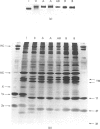
1. Combined histochemical and biochemical single-fibre analyses [Staron & Pette (1987) Biochem. J. 243, 687-693], were used to investigate the rabbit tibialis-anterior fibre population. 2. This muscle is composed of four histochemically defined fibre types (I, IIC, IIA and IIB). 3. Type I fibres contain slow myosin light chains LC1s and LC2 and the slow myosin heavy chain HCI, and types IIA and IIB contain the fast myosin light chains LC1f, LC2f and LC3f and the fast heavy chains HCIIa and HCIIb respectively. 4. A small fraction of fibres (IIAB), histochemically intermediate between types IIA and IIB, contain the fast light myosin chains but display a coexistence of HCIIa and HCIIb. 5. Similarly to the soleus muscle, C fibres in the tibialis anterior muscle contain both fast and slow myosin light chains and heavy chains. The IIC fibres show a predominance of the fast forms and the IC fibres (histochemically intermediate between types I and IIC) a predominance of the slow forms. 6. A total of 60 theoretical isomyosins can be derived from these findings on the distribution of fast and slow myosin light and heavy chains in the fibres of rabbit tibialis anterior muscle.
Full text
PDF


Images in this article
Selected References
These references are in PubMed. This may not be the complete list of references from this article.
- Andersen P., Henriksson J. Training induced changes in the subgroups of human type II skeletal muscle fibres. Acta Physiol Scand. 1977 Jan;99(1):123–125. doi: 10.1111/j.1748-1716.1977.tb10361.x. [DOI] [PubMed] [Google Scholar]
- Billeter R., Heizmann C. W., Howald H., Jenny E. Analysis of myosin light and heavy chain types in single human skeletal muscle fibers. Eur J Biochem. 1981 May 15;116(2):389–395. doi: 10.1111/j.1432-1033.1981.tb05347.x. [DOI] [PubMed] [Google Scholar]
- Billeter R., Weber H., Lutz H., Howald H., Eppenberger H. M., Jenny E. Myosin types in human skeletal muscle fibers. Histochemistry. 1980;65(3):249–259. doi: 10.1007/BF00493174. [DOI] [PubMed] [Google Scholar]
- Cleveland D. W., Fischer S. G., Kirschner M. W., Laemmli U. K. Peptide mapping by limited proteolysis in sodium dodecyl sulfate and analysis by gel electrophoresis. J Biol Chem. 1977 Feb 10;252(3):1102–1106. [PubMed] [Google Scholar]
- Edman K. A., Reggiani C., te Kronnie G. Differences in maximum velocity of shortening along single muscle fibres of the frog. J Physiol. 1985 Aug;365:147–163. doi: 10.1113/jphysiol.1985.sp015764. [DOI] [PMC free article] [PubMed] [Google Scholar]
- Gauthier G. F., Lowey S. Distribution of myosin isoenzymes among skeletal muscle fiber types. J Cell Biol. 1979 Apr;81(1):10–25. doi: 10.1083/jcb.81.1.10. [DOI] [PMC free article] [PubMed] [Google Scholar]
- Guth L., Yellin H. The dynamic nature of the so-called "fiber types" of nammalian skeletal muscle. Exp Neurol. 1971 May;31(2):227–300. doi: 10.1016/0014-4886(71)90196-8. [DOI] [PubMed] [Google Scholar]
- Hoh J. F., Yeoh G. P. Rabbit skeletal myosin isoenzymes from fetal, fast-twitch and slow-twitch muscles. Nature. 1979 Jul 26;280(5720):321–323. doi: 10.1038/280321a0. [DOI] [PubMed] [Google Scholar]
- Ingjer F. Effects of endurance training on muscle fibre ATP-ase activity, capillary supply and mitochondrial content in man. J Physiol. 1979 Sep;294:419–432. doi: 10.1113/jphysiol.1979.sp012938. [DOI] [PMC free article] [PubMed] [Google Scholar]
- Jansson E., Sjödin B., Tesch P. Changes in muscle fibre type distribution in man after physical training. A sign of fibre type transformation? Acta Physiol Scand. 1978 Oct;104(2):235–237. doi: 10.1111/j.1748-1716.1978.tb06272.x. [DOI] [PubMed] [Google Scholar]
- Lutz H., Weber H., Billeter R., Jenny E. Fast and slow myosin within single skeletal muscle fibres of adult rabbits. Nature. 1979 Sep 13;281(5727):142–144. doi: 10.1038/281142a0. [DOI] [PubMed] [Google Scholar]
- Miller D. M., 3rd, Ortiz I., Berliner G. C., Epstein H. F. Differential localization of two myosins within nematode thick filaments. Cell. 1983 Sep;34(2):477–490. doi: 10.1016/0092-8674(83)90381-1. [DOI] [PubMed] [Google Scholar]
- O'Farrell P. H. High resolution two-dimensional electrophoresis of proteins. J Biol Chem. 1975 May 25;250(10):4007–4021. [PMC free article] [PubMed] [Google Scholar]
- Oakley B. R., Kirsch D. R., Morris N. R. A simplified ultrasensitive silver stain for detecting proteins in polyacrylamide gels. Anal Biochem. 1980 Jul 1;105(2):361–363. doi: 10.1016/0003-2697(80)90470-4. [DOI] [PubMed] [Google Scholar]
- Pette D. J.B. Wolffe memorial lecture. Activity-induced fast to slow transitions in mammalian muscle. Med Sci Sports Exerc. 1984 Dec;16(6):517–528. [PubMed] [Google Scholar]
- Pette D. Metabolic heterogeneity of muscle fibres. J Exp Biol. 1985 Mar;115:179–189. doi: 10.1242/jeb.115.1.179. [DOI] [PubMed] [Google Scholar]
- Pette D., Spamer C. Metabolic properties of muscle fibers. Fed Proc. 1986 Dec;45(13):2910–2914. [PubMed] [Google Scholar]
- Pierobon-Bormioli S., Sartore S., Libera L. D., Vitadello M., Schiaffino S. "Fast" isomyosins and fiber types in mammalian skeletal muscle. J Histochem Cytochem. 1981 Oct;29(10):1179–1188. doi: 10.1177/29.10.7028858. [DOI] [PubMed] [Google Scholar]
- Pinter K., Mabuchi K., Sreter F. A. Isoenzymes of rabbit slow myosin. FEBS Lett. 1981 Jun 15;128(2):336–338. doi: 10.1016/0014-5793(81)80111-1. [DOI] [PubMed] [Google Scholar]
- Pullan L. M., Noltmann E. A. Purification and properties of pig muscle carbonic anhydrase III. Biochim Biophys Acta. 1985 Apr 17;839(2):147–154. doi: 10.1016/0304-4165(85)90031-5. [DOI] [PubMed] [Google Scholar]
- Riley D. A., Ellis S., Bain J. Carbonic anhydrase activity in skeletal muscle fiber types, axons, spindles, and capillaries of rat soleus and extensor digitorum longus muscles. J Histochem Cytochem. 1982 Dec;30(12):1275–1288. doi: 10.1177/30.12.6218195. [DOI] [PubMed] [Google Scholar]
- Shima K., Tashiro K., Hibi N., Tsukada Y., Hirai H. Carbonic anhydrase-III immunohistochemical localization in human skeletal muscle. Acta Neuropathol. 1983;59(3):237–239. doi: 10.1007/BF00703210. [DOI] [PubMed] [Google Scholar]
- Staron R. S., Pette D. Correlation between myofibrillar ATPase activity and myosin heavy chain composition in rabbit muscle fibers. Histochemistry. 1986;86(1):19–23. doi: 10.1007/BF00492341. [DOI] [PubMed] [Google Scholar]
- Staron R. S., Pette D. The multiplicity of combinations of myosin light chains and heavy chains in histochemically typed single fibres. Rabbit soleus muscle. Biochem J. 1987 May 1;243(3):687–693. doi: 10.1042/bj2430687. [DOI] [PMC free article] [PubMed] [Google Scholar]
- Vänänen H. K., Kumpulainen T., Korhonen L. K. Carbonic anhydrase in the type I skeletal muscle fibers of the rat. An immunohistochemical study. J Histochem Cytochem. 1982 Nov;30(11):1109–1113. doi: 10.1177/30.11.6216280. [DOI] [PubMed] [Google Scholar]
- d'Albis A., Janmot C., Bechet J. J. Comparison of myosins from the masseter muscle of adult rat, mouse and guinea-pig. Persistence of neonatal-type isoforms in the murine muscle. Eur J Biochem. 1986 Apr 15;156(2):291–296. doi: 10.1111/j.1432-1033.1986.tb09580.x. [DOI] [PubMed] [Google Scholar]
- d'Albis A., Pantaloni C., Bechet J. J. An electrophoretic study of native myosin isozymes and of their subunit content. Eur J Biochem. 1979 Sep;99(2):261–272. doi: 10.1111/j.1432-1033.1979.tb13253.x. [DOI] [PubMed] [Google Scholar]


