



Atrial fibrillation (AF) is the most common cause of cardioembolic stroke. Recognition is paramount since AF-related cardioembolic strokes are especially severe and oral anticoagulation (OAC) can prevent recurrent events.1 Optimal AF detection requires implantable loop recorders (ILR), which are resource-intensive with limited uptake.1 OAC after stroke without confirmed AF increases bleeding without improving outcomes. Efficient methods are needed to identify AF-related stroke, to guide intensive rhythm monitoring and OAC prescription.
Validated AF risk scores, such as Cohorts for Heart and Aging Research in Genomic Epidemiology-AF (CHARGE-AF),2 can discriminate cardioembolic stroke.1 However, scores are rarely employed due to cumbersome calculation and potential misclassification. Overlapping risk factors between cardioembolic and non-cardioembolic stroke also limits discriminative value.
ECG-AI is a validated deep learning model to estimate AF risk using the 12-lead electrocardiogram (ECG),3 an inexpensive test routinely performed after stroke. Since ECG-AI provides predictive utility beyond clinical risk,3 we hypothesized ECG-AI may be well-suited to discriminate cardioembolism at the time of stroke, and could provide a low-cost method for ILR prioritization.
We retrospectively analyzed 1,597 ischemic stroke patients from Massachusetts General Hospital and Brigham and Women’s Hospital undergoing prospective neurologist-adjudicated stroke subtyping using Trial of ORG 10172 in Acute Stroke Treatment (TOAST) or Causative Classification of Stroke (CCS) criteria.1 The Mass General Brigham Institutional Review Board approved the study. Informed consent was obtained or waived via protocol-specific allowance. Study data are available upon request.
We included patients with complete CHARGE-AF scores, ≥1 digital-format ECG obtained within 1 year before to 30 days after stroke, and no AF on the ECG used for analysis. Cardioembolism was defined using criteria favoring specificity (TOAST “definite cardioembolism” or CCS “evident or probable cardio-aortic embolism”), and adjudicators were unaware of ECG-AI results.1 We compared CHARGE-AF, ECG-AI and CH-AI (ECG-AI and CHARGE-AF) for discrimination of cardioembolism from other stroke subtypes using the area under the receiver operating characteristic curve (AUROC) and logistic regression. We assessed associations between ECG-AI and incident AF, defined using an electronic health record-based algorithm (positive predictive value 92%).4 In secondary analyses, we included ECGs demonstrating AF, and excluded individuals with AF diagnosed before or 30 days after stroke.
We analyzed 964 individuals with ischemic stroke (age 65±15, 40% women), of which 188 (19.5%) were cardioembolic. ECGs were performed median 1 (quartile-1: 0, quartile-3: 2) day from stroke. ECG-AI (AUROC 0.753 [95%CI 0.712–0.794]) and CH-AI (0.761 [0.722–0.800]) had greater discrimination of cardioembolic stroke than CHARGE-AF (0.675 [0.634–0.716], p<0.01 for both). Odds ratios for cardioembolism were larger for ECG-AI (2.72 per 1-SD increase [2.29–3.27]) and CH-AI (2.97 [2.44–3.65]) versus CHARGE-AF (2.00 [1.65–2.44]). ECG-AI stratified risk of longitudinal AF after stroke (10-year AF risk in top 10% of ECG-AI 60.6% [37.2–75.3], hazard ratio 2.50 [1.59–3.93] versus remainder; 10-year AF risk in bottom 10% of ECG-AI 14.4% [2.5–24.9], 0.34 [0.15–0.77] versus remainder) (Figure). ECG-AI remained favorable to CHARGE-AF when ECGs showing AF were included (n=1,069; AUROC ECG-AI 0.816 [0.785–0.848]; CHARGE-AF 0.683 [0.647–0.718]), and when all individuals with AF before or within 30 days after stroke were excluded (n=733; ECG-AI 0.641 [0.556–0.726]; CHARGE-AF 0.597 [0.504–0.690]).
Figure.
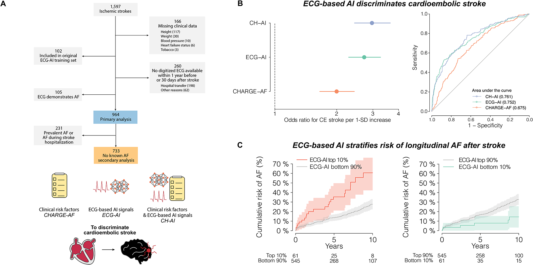
Summary of analysis and results. Panel A depicts an overview of the current study. A total of 964 individuals with acute stroke and paired ECG were included. A total of three AF risk estimation schemes were compared: a) CHARGE-AF, a validated clinical risk score, b) ECG-AI, a deep learning model using 12-lead ECG, and c) CH-AI a combination of CHARGE-AF and ECG-AI. Panel B depicts discrimination performance for each model, using univariable odds ratios on the left and receiver operating characteristic curves on the right. Panel C depicts the cumulative risk of AF among 606 individuals with no AF before or within 30 days after stroke and longitudinal follow-up available after stroke. The left panel depicts the cumulative risk of AF stratified by an ECG-AI score above the 90th percentile, and the right panel depicts the cumulative risk of AF stratified by an ECG-AI score below the 10th percentile. Shaded regions denote 95% confidence intervals.
Among ~1,000 acute stroke patients with neurologist-adjudicated stroke subtyping, we compared a validated AF risk score, ECG-based AI risk stratification tool, and their combination. AI-based models discriminated cardioembolic stroke favorably compared to clinical risk factors, and stratified risk of future AF.
Findings yield several implications. First, AI-enabled ECG analysis discriminates cardioembolic stroke. Although ECG-AI discrimination was favorable to CHARGE-AF, AUROC remained moderate and likely insufficient in isolation to classify cardioembolic stroke. Second, integrating clinical risk factors with ECG-based AI adds little value for identifying cardioembolism, likely due to common risk factors for cardioembolic and non-cardioembolic strokes. Third, ECG-AI may improve efficiency of ILR utilization after stroke. Nearly two-thirds of individuals with ECG-AI scores in the top 10% developed new AF within 10 years, identifying a population where ILR may be high-yield. Conversely, 10-year AF risk was 15% in the bottom 10% of ECG-AI, suggesting non-AF etiologies could be prioritized.
Individuals used to train ECG-AI were excluded, but patients are from the same healthcare system. Discrimination was lower when prevalent AF was excluded. Requiring digitized ECGs and complete CHARGE-AF scores introduces bias. Like ECG-AI, CHARGE-AF was designed to estimate AF risk rather than discriminate cardioembolic stroke. Even stroke neurologist-adjudicated subtyping is subject to misclassification. Incident AF detection after stroke depends on monitoring, which was left to the discretion of the clinical team. Monitor utilization is appreciable in our post-stroke population (6-month incidence 20–70% based on subtype5).
An AI-enabled AF risk model can discriminate cardioembolic stroke using one 12-lead ECG, performing favorably to a validated 11-component risk factor score. Future work is warranted to determine whether AI models specifically designed to identify cardioembolic stroke can achieve greater discrimination, and whether prospective application of ECG-AI can assist stroke subtype classification and prioritization of intensive rhythm monitoring after stroke.
Sources of Funding:
SAL was supported by National Institutes of Health (NIH) 1R01HL139731 and American Heart Association 18SFRN34250007. SKhurshid is supported by NIH K23HL169839 and AHA 23CDA1050571. SKany is supported by the Walter Benjamin Fellowship from the Deutsche Forschungsgemeinschaft (521832260). PTE is supported by grants from the National Institutes of Health (1RO1HL092577, 1R01HL157635, 5R01HL139731), from the American Heart Association (18SFRN34230127, 961045), and from the European Union (MAESTRIA 965286). CDA is supported by NIH R01NS103924, U01NS069673, AHA 18SFRN34250007 and 21SFRN812095, and the MGH McCance Center for Brain Health.
Disclosures:
SAL is employed at Novartis as of July 2022. SAL previously received sponsored research support from Bristol Myers Squibb, Pfizer, Boehringer Ingelheim, Fitbit, Medtronic, Premier, and IBM, and has consulted for Bristol Myers Squibb, Pfizer, Blackstone Life Sciences, and Invitae. PTE receives sponsored research support from Bayer AG, IBM Research, Bristol Myers Squibb, Pfizer and Novo Nordisk; he has also served on advisory boards or consulted for MyoKardia and Bayer AG. CDA has received sponsored research support from Bayer AG. The remaining authors have nothing to disclose.
References:
- 1.Khurshid S, Trinquart L, Weng L-C, Hulme OL, Guan W, Ko D, Schwab K, Rost NS, Al-Alusi MA, Benjamin EJ, et al. Atrial Fibrillation Risk and Discrimination of Cardioembolic From Noncardioembolic Stroke. Stroke. 2020;51:1396–1403. [DOI] [PMC free article] [PubMed] [Google Scholar]
- 2.Alonso A, Krijthe BP, Aspelund T, Stepas KA, Pencina MJ, Moser CB, Sinner MF, Sotoodehnia N, Fontes JD, Janssens ACJW, et al. Simple risk model predicts incidence of atrial fibrillation in a racially and geographically diverse population: the CHARGE-AF consortium. J Am Heart Assoc. 2013;2:e000102. [DOI] [PMC free article] [PubMed] [Google Scholar]
- 3.Khurshid S, Friedman S, Reeder C, Di Achille P, Diamant N, Singh P, Harrington LX, Wang X, Al-Alusi MA, Sarma G, et al. Electrocardiogram-based Deep Learning and Clinical Risk Factors to Predict Atrial Fibrillation. Circulation. 2021; [DOI] [PMC free article] [PubMed] [Google Scholar]
- 4.Khurshid S, Keaney J, Ellinor PT, Lubitz SA. A Simple and Portable Algorithm for Identifying Atrial Fibrillation in the Electronic Medical Record. Am J Cardiol. 2016;117:221–225. [DOI] [PMC free article] [PubMed] [Google Scholar]
- 5.Khurshid S, Li X, Ashburner JM, Lipsanopoulos ATT, Lee PR, Lin AK, Ko D, Ellinor PT, Schwamm LH, Benjamin EJ, et al. Usefulness of Rhythm Monitoring Following Acute Ischemic Stroke. Am J Cardiol. 2021;147:44–51. [DOI] [PubMed] [Google Scholar]