The authors regret any inconvenience caused due to the publication of identical images in Fig. 2C Clinical and Developmental Immunology study published in 2013 and Fig. 1K in the current manuscript.
Please note that the studies published in Clinical Immunology and those in Clinical and Developmental Immunology were carried out in parallel, with a main focus on distinct outcome measures in the two different studies. The mice in both studies were infected together, including one group of mice infected with a demyelinating strain of virus to be used in both studies, and the tissues harvested were divided to accomplish spinal cord histopathology as the main outcome measures in one study and microarray analysis along with brain pathology as the main outcome measures in the second study. We did re-stain additional histology sections for the second study to re-confirm the histologic findings from the first study, and in the specific case highlighted here, we used serial sections from the same animal to accomplish this. Thus, the histopathology findings were published in 2013 in Clinical and Developmental Immunology, then we followed up these findings with the microarray data and included some additional histopathology data in the Clinical Immunology paper published in 2016.
In the Clinical and Developmental Immunology study published in 2013, we demonstrated the role of microglia in virus-induced spinal cord demyelination using the Demyelinating strain of MHV. In this study, we mainly used histopathology to study the viral spread and Iba1+ cells spread throughout the spinal cord upon infection. These studies were focused on the pathology in the spinal cord. We followed up this study with the one published in Clinical Immunology in 2016 where we compared our previous findings from the same DM strain with the Non-demyelinating (NDM) strain, however the question remained weather the recombinant RSMHV2 strain is able to cause any inflammation in the brain. Thus, we analyzed the same mice as the earlier Clinical and Developmental Immunology paper and examined the brain pathology in these mice. The specific sample pointed out was meant to re-confirm the previously demonstrated spinal cord histopathology in the context of the new brain pathology and microarray data presented. The image in question was selected as a serial section to be stained and re-demonstrate spinal cord pathology, thus similar staining patterns were not unexpected. We believe the images look almost identical because they are indeed from adjacent sections; however, in light of this inquiry, we recognize that it is possible we may have accidentally included the Iba1 staining from the prior section along with new LFB staining of an adjacent serial section instead of including the new Iba1 staining of the serial section. While this is regrettable, we do not think it changes the meaning of the reported findings as staining of serial sections consistently shows similar staining.
The authors would like to apologize for any inconvenience caused.
To avoid any further problems, we hereby provide separate replacement images:
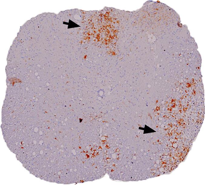
1. Replacement for Fig. 1K: The alternative immunostained (Iba1) image for the Iba1 image in question from the same set of mice used for the study. Sections in DM strain infected mice (K) that demonstrate Iba1+ cells present in the demyelinating plaque (Indicated by arrow).
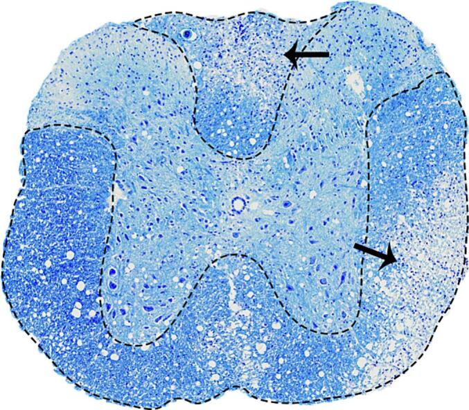
2. Replacement for Fig. 1H: The LFB stained section corresponding to replaced Fig. 1K showing demyelination in regions around the Iba1+ cells (indicated by arrow)