Abstract
Background
Tumor-selective oncolytic viral vectors are promising anticancer therapeutics; however, challenges with dosing and potency in advanced/metastatic cancers have limited efficacy and usage. NG-350A is a next-generation blood-stable adenoviral vector engineered to express an agonist anti-cluster of differentiation (CD)40 antibody without affecting tumor-selectivity and oncolytic potency.
Methods
Intravenous and intratumoral (IT) administration of NG-350A was assessed in a phase Ia/Ib study in patients with metastatic/advanced epithelial tumors (NCT03852511). Dose-escalation was performed separately for intravenous (four dose levels available, each with infusions on Days 1, 3 and 5 of a 57-day treatment period) and IT (single injection on D1 only or injections on Days 1, 8, 15 and 22) administration. The primary objective was safety and tolerability; secondary objectives included determining a recommended dose, pharmacokinetics, and immunogenicity.
Results
In total, 25 heavily pretreated patients received NG-350A (16 with intravenous and 9 with IT administration). Intravenous and IT dosing were both well tolerated, with no evidence of transgene-related or off-target viral toxicity. Intravenous and IT dosing resulted in dose-dependent increases in systemic NG-350A Cmax. Despite both routes of administration inducing anti-virus antibodies, sustained persistence of NG-350A in blood samples was observed up to 7 weeks after the last dose, particularly with higher intravenous dose levels. Delivery of NG-350A to tumors was demonstrated in biopsy samples following both routes of administration; a dose-dependent pattern was seen with intravenous infusion, with four patients remaining positive for vector DNA in biopsies at Day 57. Transgene messenger RNA from replicating NG-350A was detected in 5/12 patients with intravenous treatment and 1/9 patients with IT injection, and sustained increases in inflammatory cytokines were observed following dosing, particularly with higher intravenous dose levels.
Conclusions
This phase 1a study provided initial proof-of-mechanism for NG-350A, with strong evidence of tumor delivery, viral replication and transgene expression—particularly after intravenous dosing. The lack of transgene-related or off-target viral toxicity was consistent with the highly selective delivery and replication of NG-350A, even after systemic delivery. The efficacy of intravenous-dosed NG-350A will now be evaluated in combination with pembrolizumab (NCT05165433), as well as with chemoradiotherapy (NCT06459869).
Trial registration number
Keywords: Oncolytic virus, Immunotherapy, Pharmacokinetics - PK, Pharmacodynamics - PD, Solid tumor
WHAT IS ALREADY KNOWN ON THIS TOPIC
Systemic yet selective delivery of transgene-armed oncolytic viruses has multiple potential advantages, including delivery to all tumor sites, as well as simple and repeatable administration. Some evidence of successful intravenous delivery has been reported, but most therapies in later stage development still use intratumoral (IT) injection or have shown limited evidence of transgene expression.
WHAT THIS STUDY ADDS
The FORTITUDE study supports proof-of-mechanism for NG-350A, a next-generation transgene-armed viral vector, with strong evidence of tumor-selective delivery, replication and transgene expression. Additionally, this study demonstrates that intravenous delivery of NG-350A results in a superior overall pharmacokinetics and pharmacodynamics profile, with no apparent disadvantages versus IT injection.
HOW THIS STUDY MIGHT AFFECT RESEARCH, PRACTICE OR POLICY
The results from this study highlight the importance of designing future tumor-selective vectors for systemic administration. Additionally, this study shows that local expression of immunostimulatory therapies can overcome toxicity that limits non-targeted systemic administration.
Background
Viral vector-based therapies have been explored as anticancer treatments due to their potential for selective oncolytic potency. Historically, development has focused on intratumoral (IT) administration to deliver the virus directly to tumor sites and avoid anti-viral immune responses.1 Although IT administration may increase the therapeutic index of immunotherapies, direct injection of tumors is associated with notable technical and logistical challenges. Additionally, as IT delivery is limited to accessible and injectable tumor lesions, systemic responses are often dependent on abscopal effects which may limit efficacy.1 Among IT administered viral therapies, only talimogene laherparepvec has been approved by the US Food and Drug Administration (for the treatment of unresectable melanoma).2 3
The potential to overcome these limitations has been shown with viral vectors that can be delivered by intravenous administration while retaining tumor specificity.4 This may be a particularly attractive approach if the oncolytic activity of the viral vector can be combined with the selective delivery of immunostimulatory transgenes to all malignant lesions.
Tumor-specific immuno gene (T-SIGn) therapies are next-generation tumor-selective viral vectors derived from enadenotucirev, an “unarmed” blood-stable group B adenovirus 11p/adenovirus three chimeric virus developed by directed evolution to selectively replicate in both primary and metastatic epithelial-derived solid tumors, leading to potent cytotoxicity following intravenous delivery.5 Following multiple phase 1 studies of enadenotucirev that showed promising viral kinetics, tumor delivery and tolerability,4 6 7 the T-SIGn vector NG-350A was developed by incorporating a full-length transgene for an agonist anti-cluster of differentiation (CD)40 antibody downstream of the enadenotucirev major late promoter. This location couples viral replication to anti-CD40 antibody expression (restricting transgene expression to the tumor cells in which NG-350A is actively replicating), in order to combine the tumor-selective oncolytic activity of enadenotucirev with the targeted expression of a potent immunostimulatory monoclonal antibody within the tumor microenvironment. The tumor tissue-targeting nature of NG-350A combined with its CD40 agonist transgene might allow for local delivery of this multimodality approach and a selective transformation of the tumor microenvironment. This approach should achieve a key objective of IT delivery (ie, to achieve higher local bioactive drug concentrations) without the risk of systemic toxicities1 while also targeting all tumor locations to achieve an improved outcome. Additionally, the convenience of intravenous administration would allow for repeat dosing.
Here we present results from a first-in-human clinical trial of NG-350A. Our focus for this study was to determine the safety and tolerability of NG-350A, as well as to assess the impact of IT versus intravenous administration on NG-350A delivery, persistence, and pharmacodynamics. The selection of the dose levels defined in the protocol was guided by studies with the unarmed tumor-selective viral vector enadenotucirev. For enadenotucirev a dose of 3×10¹² viral particles (vp) was initially determined as the maximum tolerated dose.6 The intention to increase the dose intensity per cycle led to the testing of “low-high-high” dose regimens and established the safety of 3×10¹² and 6×10¹² vp when administered on Days 3 and 5 following 1×10¹² on Day 1.7
Methods
FORTITUDE clinical study design
FORTITUDE was a phase Ia/Ib, multicenter, open-label, non-comparative study in patients with metastatic/advanced epithelial tumors (NCT03852511). The study was designed with separate cohorts testing either IT or intravenous administration of NG-350A monotherapy. NG-350A was dose-escalated in parallel in both cohorts using a standard “3+3” design. A Safety Review Committee reviewed outcomes (including dose-limiting toxicities (DLTs)) at each dose level and was responsible for all dose-escalation decisions. After a 57-day study treatment period, patients were followed by imaging until progression of disease (PD); after PD patients were followed for survival and further anticancer therapy.
Dosing regimen
IT dosing
The IT injection dose was dependent on the size of the tumor lesion(s) to be injected. Lesions were injected by repeatedly inserting the needle approximately 0.5–1.0 cm apart and injecting 100–200 µL at each pass. Two dose levels were available: patients in IT Dose Level 1 received IT injections of NG-350A on Day 1 only, whereas patients in IT Dose Level 2 received injections of NG-350A on Days 1, 8, 15 and 22. Each suitable lesion was injected separately, up to a combined total maximum dose (across all injected lesions) of 1×10¹² vp in both IT dose levels.
Intravenous dosing
For the intravenous NG-350A monotherapy dose-escalation cohort, four dose levels (plus an optional de-escalation dose) were planned, each consisting of three intravenous infusions completed within the first 9 days of the study (scheduled on Days 1, 3 and 5). The starting dose level (intravenous Dose Level 1) of 1×10¹¹ vp on Days 1, 3 and 5 was determined based on prior experience with enadenotucirev.6 Three further dose levels, all using a “low-high-high” three administration regimen previously shown to improve tolerability when increasing doses beyond 1×10¹² vp7 were available: 1×10¹² vp on Day 1 followed by 3×10¹² vp on Days 3 and 5 (Dose Level 2; 1-3−3×10¹² vp dose), 1×10¹² vp on Day 1 followed by 6×10¹² vp on Days 3 and 5 (Dose Level 3; 1-6−6×10¹² vp dose) and 1×10¹² vp on Day 1 followed by 10×1012 vp on Days 3 and 5 (Dose Level 4; 1-10−10×1012 vp dose).
Participants
Eligible participants had metastatic or advanced epithelial cancers that had progressed after ≥1 line of systemic therapy and were incurable by local therapy. A full list of the 10 eligible tumor types is provided in the online supplemental appendix. Additional eligibility criteria included age ≥18 years, Eastern Cooperative Oncology Group performance status of 0–1, adequate lung reserve (oxygen saturation on ambient air at sea level ≥95%), and adequate hepatic, bone marrow, and coagulation function. Patients were also required to have adequate renal function, defined as creatinine ≤1.5 mg/dL and estimated glomerular filtration rate ≥60 mL/min/1.73 m2, as well as an absence of proteinuria (spot albumin:creatinine ratio ≤30 mg/g or a 24-hour urinary protein result of <1 g/24 hours). Patients with a history or evidence of significant immunodeficiency; renal, autoimmune or lung disease (including lymphangitic carcinomatosis); or risk factors for bleeding were not eligible.
Objectives, endpoints and assessments
Primary objective
The primary objective was to characterize the safety and tolerability of NG-350A as monotherapy and in combination with pembrolizumab in patients with metastatic or advanced epithelial tumors.
The incidence of adverse events (AEs), serious adverse events (SAEs), DLTs, and AEs leading to discontinuation or death was assessed. AEs were characterized using the National Cancer Institute Common Terminology Criteria for Adverse Events (CTCAE) V.5.0. DLTs were assessed during the first 28 (all intravenous dose levels and IT Dose Level 1) or 35 (IT Dose Level 2) days of treatment.
Secondary objectives
Key secondary objectives included determining a recommended dose of NG-350A for further development, NG-350A pharmacokinetics (PK) and immunogenicity, and the preliminary efficacy of NG-350A.
NG-350A PK were assessed using whole blood samples to measure the concentration of NG-350A on each dosing day (with samples taken pre-dose and immediately post-dose), as well as on Day 15 and at the end of study treatment (EOST) visit scheduled at Day 57. For patients receiving IT Dose Level 2, an additional sample was taken at Day 36. Quantitative PCR (qPCR) was used to detect viral genomic DNA using primers (Eurofins Genomics, Ebersberg, Germany; primer sequences are provided in the online supplemental file 1). Blood samples to measure anti-virus-antibodies in serum were taken pre-dose on Day 1, on Days 8, 15, 22, 29, and at the EOST visit. An electrochemiluminescence ligand binding assay was used as previously described.4
Preliminary antitumor activity (including objective response rate per Response Evaluation Criteria in Solid Tumors (RECIST) V.1.1 and progression-free survival (PFS)) and overall survival (OS) were assessed. Tumor imaging was performed prior to treatment, at the Day 57 EOST visit (or as clinically indicated) and every 8 weeks thereafter during follow-up.
Exploratory objectives
Additional exploratory objectives included cytokine and chemokine responses to NG-350A, NG-350A delivery to the tumor, viral shedding and markers of biological responses to NG-350A, including transgene expression.
Blood samples for cytokine assessments in serum were taken on each NG-350A dosing day (pre-dose and 6–8 hours post-dose), on Days 8, 15, 22, 29 and at the EOST visit. A multiplex Luminex assay was used to analyze the following analytes: IL-2, IL-5, IL-6, IL-10, IL-17A, MCP-1, TNF-α, IFN-γ, IL-13, IL-15, CXCL9 (MIG), CXCL10 (IP-10), CXCL11 (I-TAC), IFN-α2, MIP-1α, IL-8 and IL-12p70. The presence of NG-350A transgene CD40 antibody messenger RNA (mRNA) in the blood was assessed at Days 1, 8, 15, 22, 29, and at the EOST visit in serum by reverse transcription-quantitative polymerase chain reaction (RT-qPCR) analyses (Eurofins Genomics, Ebersberg, Germany; primer sequences provided in the online supplemental file 1).
The presence of NG-350A in tumor tissue was assessed using core needle tumor biopsies collected at screening, on Days 15 and 29, and at the EOST visit. Samples for buccal, rectal and urine shedding were collected pre-dose, on Days 8, 15 and 29, and at the EOST visit. For both biopsy samples and shedding, NG-350A DNA was quantified using the same qPCR as described for the NG-350A PK.
Statistical analyses
Results presented here are descriptive, as formal sample size calculations and statistical hypothesis testing was not performed for this study. The safety analysis set included all patients who received ≥1 dose of study treatment. Efficacy and PK analysis sets included all patients in the safety analysis set who had evaluable baseline and on-treatment assessments. PFS and OS were assessed using the Kaplan-Meier method, with corresponding 95% CIs calculated using the Brookmeyer-Crowley method.
Results
Patient disposition and demographics
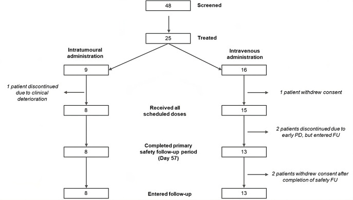
Overall, 48 patients were screened, of whom 25 were treated with NG-350A monotherapy—9 in the IT injection cohort and 16 in the intravenous infusion cohort (figure 1). The most common reasons for screen failure were withdrawal of consent (five patients), inadequate lung reserve (three patients), and inadequate renal function (three patients). The most common tumor types were colorectal cancer (eight patients) and squamous cell carcinoma of the head and neck (four patients). Demographics were generally similar between the IT and intravenous cohorts and were as expected for a mixed advanced/metastatic cancer population (table 1). All patients had received prior therapy, with an overall median of three prior lines of anticancer therapy (range 1–14 in the intravenous cohort and 2–5 in the IT cohort). Additionally, all patients had metastatic disease at baseline, with 60% of patients having ≥3 recorded sites of metastatic disease.
Figure 1. Patient disposition. FU, follow-up; PD, progressive disease.
Table 1. Baseline demographics and disease characteristics.
| Characteristic | IT DL1D1 only(n=6) | IT DL2D1, 8, 15, 22(n=3) | IT overall(n=9) | Intravenous DL11×1011(n=6) | Intravenous DL21-3−3×10¹²(n=4) | Intravenous DL31-6−6×10¹²(n=6) | Intravenous overall(n=16) |
| Median age, years (min, max) | 57.5 (36, 69) | 54 (51, 72) | 54 (36, 72) | 56 (47, 63) | 53 (32, 75) | 55 (50, 78) | 55 (32, 78) |
| Male, n (%) | 3 (50.0) | 3 (100) | 6 (66.7) | 4 (66.7) | 1 (25.0) | 1 (16.7) | 6 (37.5) |
| Race, n (%) | |||||||
| White | 4 (66.7) | 1 (33.3) | 5 (55.6) | 3 (50.0) | 4 (100) | 5 (83.3) | 12 (75.0) |
| Asian | 2 (33.3) | 1 (33.3) | 3 (33.3) | 1 (16.7) | 0 | 1 (16.7) | 2 (12.5) |
| Other | 0 | 1 (33.3) | 1 (11.1) | 2 (33.3) | 0 | 0 | 2 (12.5) |
| Tumor type, n (%) | |||||||
| CRC | 3 (50.0) | 1 (33.3) | 4 (44.4) | 0 | 1 (25.0) | 3 (50.0) | 4 (25.0) |
| SCCHN | 0 | 0 | 0 | 2 (33.3) | 1 (25.0) | 1 (16.7) | 4 (25.0) |
| Pancreatic cancer | 0 | 1 (33.3) | 1 (11.1) | 1 (16.7) | 2 (50.0) | 0 | 3 (18.8) |
| Esophageal/GEJ cancer | 0 | 1 (33.3) | 1 (11.1) | 1 (16.7) | 0 | 1 (16.7) | 2 (12.5) |
| Cholangiocarcinoma | 2 (33.3) | 0 | 2 (22.2) | 1 (16.7) | 0 | 0 | 1 (6.3) |
| Other* | 1 (16.7) | 0 | 1 (11.1) | 1 (16.7) | 0 | 1 (16.7) | 2 (12.5) |
| Median time from diagnosis, months (min, max) | 22 (13, 30) | 37 (16, 46) | 22 (13, 46) | 52 (21, 178) | 35 (23, 47) | 47 (16, 87) | 42 (16, 178) |
| Prior cancer therapies, n (%) | 6 (100) | 3 (100) | 9 (100) | 6 (100) | 4 (100) | 6 (100) | 16 (100) |
| 1–2 prior lines | 2 (33.3) | 1 (33.3) | 3 (33.3) | 3 (50.0) | 2 (50.0) | 2 (33.3) | 7 (43.8) |
| 3+lines | 4 (66.7) | 2 (66.7) | 6 (66.7) | 3 (50.0) | 2 (50.0) | 4 (66.7) | 9 (56.7) |
| Prior antineoplastic agents | 6 (100) | 2 (66.7) | 8 (88.9) | 6 (100) | 4 (100) | 6 (100) | 16 (100) |
| Prior radiotherapy | 1 (16.7) | 2 (66.7) | 3 (33.3) | 6 (100) | 1 (25.0) | 3 (50.0) | 10 (62.5) |
| Prior cancer surgery | 4 (66.7) | 3 (100) | 7 (77.8) | 6 (100) | 4 (100) | 4 (66.7) | 14 (87.5) |
Includes breast cancer, cervical cancer, and prostate cancer.
CRC, colorectal cancer; SCCHN, squamous cell carcinoma of head and neck; GEJ, gastroesophageal junction.
DL1Dose Level 1DL2Dose Level 2DL3Dose Level 3ITintratumoral
Overall, 21/25 patients (84%) completed the study treatment period of 57 days (figure 1). Most patients entered follow-up, although additional imaging follow-up was performed in fewer patients (n=8 entered imaging follow-up and n=5 received follow-up scans).
Dose-escalation
In the IT cohort, six patients were treated at IT Dose Level 1 with no DLTs. Per-protocol, a further three patients were then treated at IT Dose Level 2, with no DLTs observed. Both prespecified IT dose levels were therefore considered tolerable.
In the intravenous cohort, six patients were treated at intravenous Dose Level 1 with a single DLT occurring (blood creatine kinase increased). This dose was considered tolerable, and dosing was escalated to use “low-high-high” dosing regimens designed to increase the total viral dose over the single three-administration cycle. A total of four patients received intravenous Dose Level 2 (1-3−3×10¹² vp dose) with no DLTs occurring. As such, per-protocol, dosing was escalated to intravenous Dose Level 3 (1-6−6×10¹² dose) with no DLTs in the six patients treated at this dose level. Dose Level 3 was therefore considered tolerable; however, no further dose escalation was performed based on experience in trials of other T-SIGn vectors ongoing in parallel. Instead, a further cohort designed to confirm the tolerability of intravenous NG-350A when given in combination with pembrolizumab was added to the study via a protocol amendment. However, after the assessment of one NG-350A plus pembrolizumab dose level, a separate ongoing phase 1a/b study was opened to test the combination in an expanded design. As such, results from this cohort will be reported as part of a subsequent publication on NG-350A plus pembrolizumab.
Patient exposure
Aside from one patient treated at intravenous Dose Level 2 who withdrew consent after their first dose, and one patient in IT Dose Level 2 who missed one dose, all other patients received all scheduled administrations of NG-350A (table 2).
Table 2. Exposure.
| IT DL1D1 only(n=6) | IT DL2D1, 8, 15, 22(n=3) | IT overall(n=9) | Intravenous DL11-1−1×10¹¹(n=6) | Intravenous DL21-3−3×10¹²(n=4) | Intravenous DL31-6−6×10¹²(n=6) | Intravenous overall(n=16) | |
| Patients received planned number of doses (n (%)) | 6 (100) | 2 (66.7) | 8 (88.9) | 6 (100) | 3 (75) | 6 (100) | 15 (93) |
| Number of doses received (n (%)) | NA | NA | |||||
| 1 dose | 6 (100) | 0 | 0 | 1 | 0 | ||
| 2 doses | – | 0 | 0 | 0 | 0 | ||
| 3 doses | – | 1 | 6 | 3 | 6 | ||
| 4 doses | – | 2 | – | – | – | ||
| Cumulative vp administered (mean (min, max); ×1012 vp) | NA | NA | |||||
| Planned | 1.00 | 4.00 | 0.30 | 7.00 | 13.00 | ||
| Actual | 0.47 (0.2, 0.8) | 2.67 (1.0, 4.0) | 0.30 (0.30, 0.30) | 5.50 (1.0, 7.0) | 13.00 (13.0, 13.0) |
DL1Dose Level 1DL2Dose Level 2DL3Dose Level 3ITintratumoralvpviral particles
Due to the technical constraints of IT injection, the maximum combined potential dose of 1×10¹² vp per dosing day, which required at least five separate and distinct injection tracks to achieve a total injection volume of 2 mL within a suitably sized tumor lesion, was not able to be given in the majority of patients. By contrast, all patients in the intravenous cohort received their planned dose.
Safety and tolerability
All patients experienced ≥1 treatment-emergent (TE) AE, and 13 patients (52%) experienced ≥1 Grade 3–4 TEAE (table 3; online supplemental table 1). Overall, the majority of TEAEs were Grade 1–2 and occurred within the initial dosing period (within 8 days). A single AE leading to discontinuation of NG-350A occurred (Clostridium difficile colitis in a patient in intravenous monotherapy cohort 2; unrelated to treatment).
Table 3. Safety summary.
| IT DL1D1 only(n=6) | IT DL2D1, 8, 15, 22(n=3) | IT overall(n=9) | Intravenous DL11-1−1×10¹¹(n=6) | Intravenous DL21-3−3×10¹²(n=4) | Intravenous DL31-6−6×10¹²(n=6) | Intravenous overall(n=16) | |
| Patients with ≥1 TEAE, n (%) | 6 (100) | 3 (100) | 9 (100) | 6 (100) | 4 (100) | 6 (100) | 16(100) |
| Most common TEAEs (>20% of patients) | |||||||
| aPTT prolonged | 4 (67) | 2 (67) | 6 (67) | 3 (50) | 0 | 2 (33) | 5 (31) |
| Fever | 3 (50) | 1 (33) | 4 (44) | 3 (50) | 1 (25) | 2 (33) | 6 (38) |
| Chills | 2 (33) | 0 | 2 (22) | 1 (17) | 3 (75) | 3 (50) | 7 (44) |
| Nausea | 2 (33) | 1 (33) | 3 (33) | 3 (50) | 2 (50) | 1 (17) | 6 (38) |
| Fatigue | 3 (50) | 0 | 3 (33) | 0 | 2 (50) | 2 (33) | 4 (25) |
| Hypokalemia | 1 (17) | 0 | 1 (11) | 3 (50) | 1 (25) | 2 (33) | 6 (38) |
| Weight decreased | 3 (50) | 0 | 3 (33) | 2 (33) | 1 (25) | 0 | 3 (19) |
| Anemia | 3 (50) | 0 | 3 (33) | 1 (17) | 0 | 2 (33) | 3 (19) |
| Patients with ≥1 TE-SAE | 2(33.3) | 1(33.3) | 3(33.3) | 3(50.0) | 2(50.0) | 3(50.0) | 8 (50) |
| Patients with ≥1 TE-SAE related to NG-350A | 1(16.7) | 0 | 1(11.1) | 1(16.7) | 0 | 2(33.3) | 3(18.8) |
| Patients with ≥1 Grade 3–4 TEAE | 4(66.7) | 0 | 4(44.4) | 4(66.7) | 1(25.0) | 4(66.7) | 9(56.3) |
| Patients with ≥1 Grade 3–4 TEAE related to NG-350A | 2(33.3) | 0 | 2(22.2) | 2(33.3) | 0 | 1(50.0) | 3(18.8) |
| Prolonged aPTT | 2 (33.3) | 0 | 2 (22.2) | 1 (16.7) | 0 | 0 | 1 (6.3) |
| Blood creatinine increased | 0 | 0 | 0 | 1 (16.7) | 0 | 0 | 1 (6.3) |
| Decreased appetite | 0 | 0 | 0 | 1 (16.7) | 0 | 0 | 1 (6.3) |
| Acute kidney injury | 0 | 0 | 0 | 0 | 0 | 1 (16.7) | 1 (6.3) |
| Patients with ≥1 TEAE classified as a dose-limiting toxicity | 0 | 0 | 0 | 1(16.7) | 0 | 0 | 1(6.3) |
| Patients with ≥1 TEAE leading to discontinuation of NG-350A | 0 | 0 | 0 | 0 | 1(25.0) | 0 | 1(6.3) |
AEadverse eventaPTTactivated partial thromboplastin timeDL1Dose Level 1DL2Dose Level 2DL3Dose Level 3ITintratumoralSAESerious Adverse EventTEtreatment-emergent
The Grade 3–4 TEAEs reported in the highest proportion of patients were pneumonia (4/25; 16.0%), prolonged activated partial thromboplastin time (aPTT) (3/25; 12%), blood bilirubin increased (2/25; 8%), diarrhea (2/25; 8.0%) and acute kidney injury (2/25; 8%). The only treatment-related Grade 3–4 TEAE that occurred in more than one patient was Grade 3 aPTT prolongation without any clinical bleeding or clotting events (in two patients in the IT arm and one patient in the intravenous arm). A total of 11 of 25 patients experienced ≥1 serious TEAE, including 3 patients (33%) in the IT arm and 8 patients (50%) in the intravenous arm. The only TE-SAE reported in >1 patient was pneumonia (4/25; 16%). All four cases of pneumonia were unrelated to treatment and occurred in patients with lung metastases and/or other predisposing factors. Additional TE-SAEs included an event of Grade 4 serious kidney injury (acute tubular necrosis) that occurred 10 weeks after the last dose of NG-350A and an event of Grade 2 cytokine release syndrome (CRS; both events occurred in patients treated at intravenous Dose Level 3). The kidney injury case was considered related by the investigator, but unrelated by the sponsor due to the delayed timing, the absence of proteinuria during the treatment period, and recent treatment with potentially nephrotoxic antibiotics and/or recent immune checkpoint inhibitor treatment. A kidney biopsy demonstrated acute tubular necrosis, and the kidney injury resolved on short-course prednisone. The event of CRS occurred shortly after dosing in a manner consistent with an acute reaction to infused vp and resolved the following day.
Laboratory abnormalities of proteinuria were commonly seen, with 13/25 (52.0%) patients having ≥1 urine protein dipstick results of 1+ or greater during the study treatment period. This proteinuria was generally transient and mild (maximum 1+: 9/13 patients), with 6/13 (46%) cases resolved to <1+ by the EOST visit. None of the patients with laboratory assessments positive for proteinuria progressed to severe renal AEs. Additionally, 13/25 patients (52.0%; IT cohort 4/9, intravenous cohort 9/16) had a laboratory finding of prolonged aPTT meeting ≥Grade 2 criteria at any point during the treatment period (of whom 3 had had existing aPTT prolongation at baseline). aPTT values improved by at least 1 CTCAE grade in 10/13 (77%) of patients by the EOST visit. No related bleeding or clotting events were observed. Hepatobiliary disorder TEAEs occurred in 2/25 patients (8.0%; Grade 1 hepatitis and jaundice, respectively), who both had liver or pancreatic disease involvement.
NG-350A pharmacokinetics
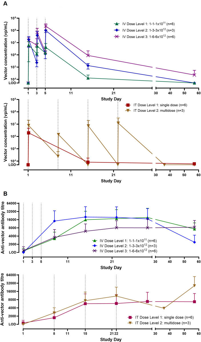
NG-350A Cmax increased with escalating intravenous dosage (figure 2A). Notably, vector DNA was detected in serum for a sustained period, with detection still observed above the limit of detection at the EOST visit (Day 57) for the highest intravenous dose level tested (three out of four patients). NG-350A was also strongly but transiently detected in serum following IT administration (figure 2A). IT dosing on a weekly basis restored circulating virus levels to approximately 1×10⁷ vp after each dose; however, less evidence of sustained presence was observed, with one of three patients having detectable viral presence at Day 57.
Figure 2. NG-350A pharmacokinetics and immunogenicity. (A) Mean NG-350A concentration in blood over time, according to dose level and route of administration. (B) Mean anti-vector antibody titer over time, according to dose level and route of administration. Dashed lines represent scheduled dosing days (first line only for IT Dose Level 1). IT, intratumoral; IV, intravenous; LOD, limit of detection; PK, pharmacokinetics.
Vector immunogenicity
All but four patients had undetectable levels of anti-vector antibodies at baseline (figure 2B). Post-dose antibody titers increased relative to baseline in 88% of patients, with similar rates of seroconversion with IT (7/9 patients) and intravenous (14/15 patients) dosing. No clear impact on PK was observed in those with low levels of anti-vector antibodies at baseline compared with those negative at baseline and no clear dose relationship with antibody titers was observed, with high variability between individual patients. However, overall, there was a trend for titers to increase over time before plateauing at approximately Day 22.
Viral shedding
Limited evidence of viral shedding was observed, with similar profiles observed with IT and intravenous administration. Shedding typically peaked at Day 8 or Day 15, after which little viral DNA could be detected. Among post-treatment samples, only 1 of 75 (1%) urine samples, 7 of 81 (8%) buccal samples and 8 of 79 (10%) rectal samples exceeded the assay limit of quantification (online supplemental table 2).
Detection of NG-350A in tumor biopsies
NG-350A vector DNA was detected by qPCR in tumor biopsies after both IT and intravenous administration (online supplemental table 3). In the intravenous cohort, the proportion of patients positive for vector DNA in tumor increased with dose level, with two out of six evaluable patients in intravenous Dose Level 1 positive (at least one result above the assay limit of quantification) versus six out of seven evaluable patients in intravenous Dose Levels 2 and 3. Notably, four patients in the intravenous arm were positive for vector DNA in biopsies taken at the EOST visit (Day 57).
Anti-CD40 transgene mRNA detection
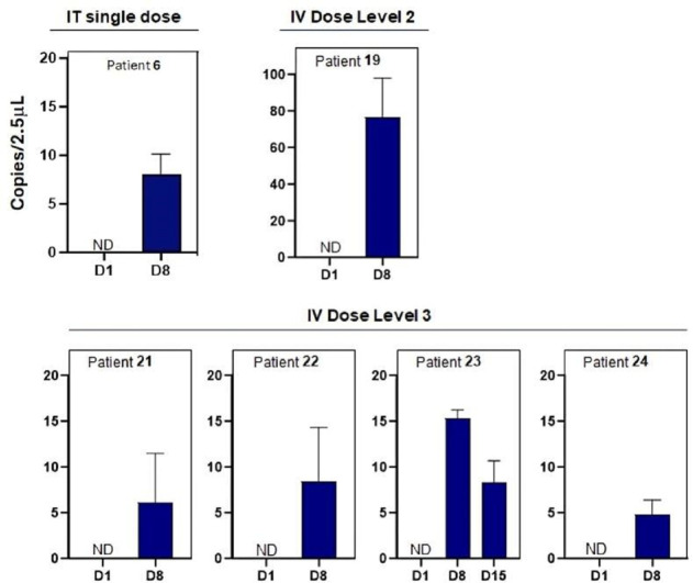
Anti-CD40 transgene was not detectable in core needle biopsies; however, transgene mRNA was detected in serum at D8 or D15 in 1/9 evaluable patients (11%) in the IT cohort (1 patient positive in Dose Level 1) and 5/12 evaluable patients (42%) in the intravenous cohort (all of whom had received higher intravenous dose levels; figure 3).
Figure 3. Detection of CD40 transgene in blood. Copies of CD40 agonist transgene detected in blood for patients positive at one or more time points (not normalized to sample volumes). IT, intratumoral; IV, intravenous; ND, not detected.
Serum cytokines
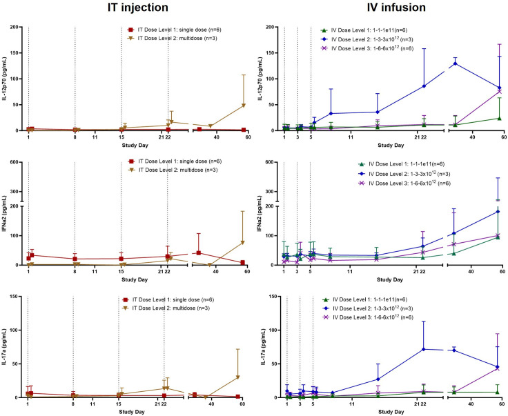
Increases in IL-12p70, IFN-α2, and IL-17a were detected in serum following treatment. These increases were most consistently observed in patients treated with higher intravenous dose levels (intravenous Dose Level 2 and 3; figure 4; online supplemental figure 1). Elevations in IFN-γ and IL-2 were also observed (online supplemental figure 1). These cytokine increases were observed from approximately Day 12 onwards, were sustained, and were restricted to a specific subset of inflammatory cytokines.
Figure 4. NG-350A pharmacodynamics—serum cytokines. Dose-dependent and sustained increases in IL-12p70, IFN-α2, and IL-17a were detected in serum from evaluable patients treated with higher intravenous dose levels. Dashed lines represent scheduled dosing days (first line only for IT Dose Level 1). IT, intratumoral; IV, intravenous.
Efficacy
No objective responses occurred with either IT or intravenous dosing. A best overall response of stable disease was observed for 2/9 (22.2%) patients treated with IT NG-350A, and 7/15 patients (46.7%) treated in the intravenous arm. The target lesion burden over time is shown in online supplemental figure 2. One patient with cholangiocarcinoma (and a Royal Marsden Hospital prognostic score8 of 2) after failure of three prior therapies maintained a stable target lesion burden for over 36 weeks following intravenous NG-350A monotherapy (at Dose Level 1), with no subsequent anticancer therapy until >10 months after beginning NG-350A (despite progression of a non-target lesion, leading to PD per RECIST, after 18 weeks). As shown in online supplemental figure 3, the tumor characterization of this patient changed from “Desert” at screening to “Inflamed” after treatment, with dramatic increases in CD8 (>10-fold) and granzyme B (>8-fold) positive cells in post-treatment biopsies.
Median (95% CI) PFS according to RECIST V.1.1 was 1.8 months (1.7, 3.0 months) with IT monotherapy, and 1.9 months (1.7, 4.1) with intravenous monotherapy. Median (95% CI) OS was 8.2 (4.9, 14.4) months with IT monotherapy, and 6.9 (2.9, 17.1) months with intravenous monotherapy.
Discussion
Here we report the results of the first clinical study of NG-350A, a next-generation tumor-selective T-SIGn therapeutic vector armed with a CD40 agonist monoclonal antibody. Successful delivery of NG-350A to tumor sites was demonstrated with intravenous administration, as well as with IT injections. Intravenous dosing was associated with a dose-dependent systemic PK profile, while IT dosing also resulted in substantial peak vector concentrations in blood. Sustained persistence of NG-350A after dosing was observed in both blood and in tumor biopsies, with NG-350A still detectable at the last on study assessment approximately 7 weeks after the last dose of NG-350A, despite the presence of anti-viral antibodies. Furthermore, data from this study demonstrated that intravenous dosing resulted in mRNA expression of the anti-CD40 antibody payload from the replicating virus.
When comparing intravenous versus IT delivery, it was notable that both routes of administration resulted in relatively similar peak blood concentrations following dosing (given the variability of total IT doses), as well as the induction of anti drug antibodies and known virus-related safety signals in similar proportions of patients (eg, prolonged aPTT and inflammatory reactions to dosing). This suggests that IT dosing did not lead to more tumor-targeted outcomes nor result in less systemic exposure and effects than intravenous dosing. By contrast, low-high to high intravenous dosing resulted in greater evidence of prolonged persistence of NG-350A and a greater proportion of patients positive for replication-dependent transgene expression (56% with low-high to high intravenous doses vs 11% with IT dosing).
PK analyses based on systemic detection require careful interpretation for a replication-competent viral therapy known to be highly tumor-selective (ie, that following initial clearance from blood should be present systemically only through spillover from the tumor). Data from this study are consistent with rapid initial clearance of NG-350A from blood following intravenous and IT delivery, as expected based on the previously characterized half-life of <20 min for the unarmed parent virus enadenotucirev4 6 and the short half-life of cell-free DNA.9 Importantly, however, this study demonstrated the long-term persistence of NG-350A DNA in blood with intravenous but not IT dosing. Given the strong tumor selectivity of the vector, as shown in prior studies of enadenotucirev4 and by the lack of off-tumor replication-associated toxicity in this study, this provides strong evidence of ongoing replication in tumors, resulting in sustained spillover into the blood. The detection of transgene expression—particularly with intravenous delivery—provides additional confirmation of active NG-350A replication, given that transgene expression from T-SIGn vectors is coupled with replication.10 Detection of transgene mRNA in blood is hypothesized to result from transgene expression in tumors cells, resulting in detectable spillover in blood, potentially through tumor-derived exosomes containing transgene mRNA. Further investigation is planned to confirm this hypothesis. Although anti-CD40 protein was not detected, this may be attributable to the challenging nature of detection via small core needle tumor biopsies (given the small amount of available material and assay detection limit), as well as a limited spillover of unbound anti-CD40 from the tumor into the blood to allow systemic detection. Additionally, intravenous, but not IT, NG-350A drove sustained and specific inflammatory cytokine elevations consistent with CD40 agonism that may reflect another spillover effect, for example, systemic release of locally produced inflammatory cytokine consistent with immune activation following NG-350A replication and transgene expression.
NG-350A was well tolerated when given by either intravenous or IT administration with no treatment-related SAEs occurring in more than one patient and no DLTs at the highest dose levels tested. Importantly, the “low-high-high” dosing regimens used resulted in Dose Levels 2 (1-3−3×10¹² vp) and 3 (1-6−6×10¹² vp) being well tolerated, with little evidence of a dose-dependent effect on toxicity. This finding supports previous studies where using an initial low dose (prior to two higher doses) mitigated against inflammatory cytokine reactions that otherwise occurred with doses ≥1×10¹² vp, allowing an overall higher intravenous viral delivery to be achieved than was possible without this initial low dose.6 7 No dose level was considered non-tolerable in this study; however, Dose Level 4 was not assessed based on experience in studies of enadenotucirev and other T-SIGn vectors conducted in parallel.6 11 In these studies, serious reactions (including hypoxia, dyspnea and cytokine release syndrome) occurred following administration of viral doses of 10×1012 vp (equivalent to intravenous Dose Level 4), in some cases even with an initial tolerizing low dose. These reactions appeared to be related to acute innate inflammatory reactions to the infusion of very high numbers of vp (rather than related to transgene expression) and a maximum tolerated intravenous dose of 1-6−6×10¹² vp is thus considered to apply to all T-SIGn vectors, including NG-350A. Intravenous Dose Levels 2 and 3 were identified as having promising PK, pharmacodynamics and tolerability outcomes; however, the small numbers of patients exposed to date mean further testing is required to define a recommended phase 2 dose. Given the tolerability and PK profiles defined in this first-in-human study, future studies will also explore whether multicycle intravenous dosing can further enhance and maintain viral loads within tumors, leading to higher levels of transgene expression and immune activation.
Overall, the safety profile of NG-350A was similar to that of the unarmed vector enadenotucirev,4 6 7 suggesting that the addition of the anti-CD40 transgene in NG-350A did not lead to new safety signals. Most AEs were of low grade and occurred shortly after dosing, reflecting the expected safety profile of an adenoviral vector, and no clear difference in the tolerability of either route of administration (intravenous vs IT) was observed. Inflammatory/cytokine release-related reactions to the injection/infusion of vp were frequently observed with both dosing routes; however, these were typically mild and only one AE requiring discontinuation of therapy occurred (a case of Clostridium difficile colitis considered unrelated to treatment). Consistent with NG-350A-mediated CD40 agonist expression being limited to the tumor, no evidence of anti-CD40 toxicity was seen with intravenous or IT dosing: for example, the liver toxicity and CRS observed with first-generation systemically administered anti-CD40 agonist antibodies112,16 was not seen in this study, where few hepatic AEs and no Grade ≥3 CRS was observed. This finding further supports the rationale for NG-350A, that is, the local and selected release of a CD40 agonist limited to the tumor microenvironment.
Prolonged aPTT was observed with both routes of administration without discernable clinical sequalae; this was previously shown to be related to the transient induction of antiphospholipid antibodies by all enadenotucirev and T-SIGn vectors. Specifically, a lupus anticoagulant (but not the clinically important anticardiolipin or anti-β2-glycoprotein I antibodies associated with antiphospholipid syndrome) is induced, impacting laboratory assessments of aPTT, as has been previously described.17 A signal of acute kidney injury has previously been identified for the parent virus enadenotucirev.7 Patients with impaired renal function or any history of clinically relevant renal toxicity were therefore excluded from this study and renal function was closely monitored in all enrolled patients. Approximately half of all patients treated with NG-350A developed proteinuria; however, this was typically mild and transient, and did not progress to severe renal injury in any case. One event of Grade 4 acute tubular necrosis occurred 10 weeks after the last dose of NG-350A and was considered treatment-related by the treating investigator. This delayed event occurred without proteinuria in a patient who had recently been treated with potentially nephrotoxic antibiotics and an immune checkpoint inhibitor, suggesting possible alternative causalities. Although the potential for adenoviruses, including T-SIGn vectors, to induce infection-associated/post-infectious glomerulonephritis continues to be closely monitored, this study did not reveal clinically significant renal injury to be a common side effect of NG-350A, supporting the revision of inclusion criteria to allow enrollment of a wider population of patients in future studies.
This phase 1a study was not designed to assess efficacy and limited evidence of monotherapy NG-350A clinical activity was observed in this late-stage advanced cancer population. Median PFS was approximately 2 months, a finding that likely reflects the difficult-to-treat population enrolled in this study and is consistent with values reported with checkpoint inhibitors in the often immunotherapy-resistant cancer types enrolled in this study.18,24 Further studies of intravenous-administered NG-350A in combination with additional anticancer modalities will be conducted to determine efficacy.
Conclusions
This first-in-human trial of NG-350A, a next-generation transgene-armed T-SIGn therapeutic vector, demonstrated initial proof-of-mechanism for NG-350A. Delivery via intravenous infusion was successful despite the presence of anti-viral antibodies, resulting in sustained persistence in tumor tissue and the expression of the encoded anti-CD40 monoclonal antibody, as well as the sustained production of inflammatory cytokines. Notably, there were no apparent benefits to IT dosing on delivery, immunogenicity or safety, whereas intravenous delivery resulted in stronger evidence of ongoing virus replication and transgene expression. This suggests that the tumor-selective and blood-stable nature of NG-350A is a significant advantage compared with other oncolytic virus platforms, allowing superior pharmacodynamic activity across both primary and metastatic sites following intravenous administration. Importantly, NG-350A was well-tolerated with either route of administration, with no evidence of the toxicity signals commonly seen with systemic exposure to anti-CD40 agonists. The lack of toxicity signals related to transgene-expression or off-target viral replication suggests that the oncolytic activity, replication and transgene expression from NG-350A remain highly tumor-selective and targeted following intravenous delivery. Given these encouraging results, the intravenous dose regimen of NG-350A evaluated in this study will be assessed in combination with pembrolizumab in the FORTIFY study, as well as in combination with chemoradiotherapy in the FORTRESS study.
supplementary material
Acknowledgements
The authors acknowledge Lyle Walker (Akamis Bio Ltd) for performing the transgene messenger RNA analyses and Dr Korina Pilz of ClinDevConsult GmbH for support with the development of the first draft of the manuscript.
Footnotes
Funding: This study was funded by Akamis Bio Ltd.
Provenance and peer review: Not commissioned; externally peer reviewed.
Patient consent for publication: Not applicable.
Ethics approval: This study involves human participants and was approved by WCG Clinical; IRB20192110. Participants gave informed consent to participate in the study before taking part.
Contributor Information
Aung Naing, Email: anaing@mdanderson.org.
Danny Khalil, Email: khalild@mskcc.org.
Oliver Rosen, Email: oliver.rosen@akamisbio.com.
D Ross Camidge, Email: ROSS.CAMIDGE@CUANSCHUTZ.EDU.
Tom Lillie, Email: tom@thelillies.com.
Rui-Ru Ji, Email: ruiru.ji@akamisbio.com.
Andrea Stacey, Email: andrea.stacey@akamisbio.com.
Matthew Thomas, Email: matthew.thomas@akamisbio.com.
Lee Rosen, Email: lrosen@mednet.ucla.edu.
Data availability statement
Data are available upon reasonable request. All data relevant to the study are included in the article or uploaded as supplementary information.
References
- 1.Melero I, Castanon E, Alvarez M, et al. Intratumoural administration and tumour tissue targeting of cancer immunotherapies. Nat Rev Clin Oncol. 2021;18:558–76. doi: 10.1038/s41571-021-00507-y. [DOI] [PMC free article] [PubMed] [Google Scholar]
- 2.Bezeljak U. Cancer gene therapy goes viral: viral vector platforms come of age. Radiol Oncol. 2022;56:1–13. doi: 10.2478/raon-2022-0002. [DOI] [PMC free article] [PubMed] [Google Scholar]
- 3.Thousand Oaks, CA:: Amgen Inc; 2023. IMLYGIC® (talimogene laherparepvec) prescribing information. [Google Scholar]
- 4.Garcia-Carbonero R, Salazar R, Duran I, et al. Phase 1 study of intravenous administration of the chimeric adenovirus enadenotucirev in patients undergoing primary tumor resection. J Immunother Cancer. 2017;5:71. doi: 10.1186/s40425-017-0277-7. [DOI] [PMC free article] [PubMed] [Google Scholar]
- 5.Kuhn I, Harden P, Bauzon M, et al. Directed evolution generates a novel oncolytic virus for the treatment of colon cancer. PLoS One. 2008;3:e2409. doi: 10.1371/journal.pone.0002409. [DOI] [PMC free article] [PubMed] [Google Scholar]
- 6.Machiels J-P, Salazar R, Rottey S, et al. A phase 1 dose escalation study of the oncolytic adenovirus enadenotucirev, administered intravenously to patients with epithelial solid tumors (EVOLVE) J Immunother Cancer. 2019;7:20. doi: 10.1186/s40425-019-0510-7. [DOI] [PMC free article] [PubMed] [Google Scholar]
- 7.Fakih M, Harb W, Mahadevan D, et al. Safety and efficacy of the tumor-selective adenovirus enadenotucirev, in combination with nivolumab, in patients with advanced/metastatic epithelial cancer: a phase I clinical trial (SPICE) J Immunother Cancer. 2023;11:e006561. doi: 10.1136/jitc-2022-006561. [DOI] [PMC free article] [PubMed] [Google Scholar]
- 8.Arkenau H-T, Barriuso J, Olmos D, et al. Prospective validation of a prognostic score to improve patient selection for oncology phase I trials. J Clin Oncol. 2009;27:2692–6. doi: 10.1200/JCO.2008.19.5081. [DOI] [PubMed] [Google Scholar]
- 9.Khier S, Lohan L. Kinetics of circulating cell-free DNA for biomedical applications: critical appraisal of the literature. Future Sci OA. 2018;4:FSO295. doi: 10.4155/fsoa-2017-0140. [DOI] [PMC free article] [PubMed] [Google Scholar]
- 10.Marino N, Illingworth S, Kodialbail P, et al. Development of a versatile oncolytic virus platform for local intra-tumoural expression of therapeutic transgenes. PLoS One. 2017;12:e0177810. doi: 10.1371/journal.pone.0177810. [DOI] [PMC free article] [PubMed] [Google Scholar]
- 11.Simon G, Subbiah V, Rosen L, et al. 762 First-in-human phase 1a study of NG-641, a tumor-selective vector expressing a FAP-TAc bispecific antibody and immune enhancer module. J Immunother Cancer. 2022;10 doi: 10.1136/jitc-2022-SITC2022.0762. [DOI] [Google Scholar]
- 12.Vonderheide RH. Prospect of targeting the CD40 pathway for cancer therapy. Clin Cancer Res. 2007;13:1083–8. doi: 10.1158/1078-0432.CCR-06-1893. [DOI] [PubMed] [Google Scholar]
- 13.Vonderheide RH, Glennie MJ. Agonistic CD40 antibodies and cancer therapy. Clin Cancer Res. 2013;19:1035–43. doi: 10.1158/1078-0432.CCR-12-2064. [DOI] [PMC free article] [PubMed] [Google Scholar]
- 14.Nowak AK, Cook AM, McDonnell AM, et al. A phase 1b clinical trial of the CD40-activating antibody CP-870,893 in combination with cisplatin and pemetrexed in malignant pleural mesothelioma. Ann Oncol. 2015;26:2483–90. doi: 10.1093/annonc/mdv387. [DOI] [PubMed] [Google Scholar]
- 15.O’Hara MH, O’Reilly EM, Varadhachary G, et al. CD40 agonistic monoclonal antibody APX005M (sotigalimab) and chemotherapy, with or without nivolumab, for the treatment of metastatic pancreatic adenocarcinoma: an open-label, multicentre, phase 1b study. Lancet Oncol. 2021;22:118–31. doi: 10.1016/S1470-2045(20)30532-5. [DOI] [PubMed] [Google Scholar]
- 16.Advani R, Forero-Torres A, Furman RR, et al. Phase I study of the humanized anti-CD40 monoclonal antibody dacetuzumab in refractory or recurrent non-Hodgkin’s lymphoma. J Clin Oncol. 2009;27:4371–7. doi: 10.1200/JCO.2008.21.3017. [DOI] [PubMed] [Google Scholar]
- 17.Khalil DN, Prieto González-Albo I, Rosen L, et al. A tumor-selective adenoviral vector platform induces transient antiphospholipid antibodies, without increased risk of thrombosis, in phase 1 clinical studies. Invest New Drugs . 2023;41:317–23. doi: 10.1007/s10637-023-01345-8. [DOI] [PMC free article] [PubMed] [Google Scholar]
- 18.Ferris RL, Blumenschein G, Jr, Fayette J, et al. Nivolumab for Recurrent Squamous-Cell Carcinoma of the Head and Neck. N Engl J Med. 2016;375:1856–67. doi: 10.1056/NEJMoa1602252. [DOI] [PMC free article] [PubMed] [Google Scholar]
- 19.Bauml J, Seiwert TY, Pfister DG, et al. Pembrolizumab for Platinum- and Cetuximab-Refractory Head and Neck Cancer: Results From a Single-Arm, Phase II Study. J Clin Oncol. 2017;35:1542–9. doi: 10.1200/JCO.2016.70.1524. [DOI] [PMC free article] [PubMed] [Google Scholar]
- 20.Kuang C, Park Y, Augustin RC, et al. Pembrolizumab plus azacitidine in patients with chemotherapy refractory metastatic colorectal cancer: a single-arm phase 2 trial and correlative biomarker analysis. Clin Epigenetics. 2022;14:3. doi: 10.1186/s13148-021-01226-y. [DOI] [PMC free article] [PubMed] [Google Scholar]
- 21.Cohen EEW, Soulières D, Le Tourneau C, et al. Pembrolizumab versus methotrexate, docetaxel, or cetuximab for recurrent or metastatic head-and-neck squamous cell carcinoma (KEYNOTE-040): a randomised, open-label, phase 3 study. Lancet. 2019;393:156–67. doi: 10.1016/S0140-6736(18)31999-8. [DOI] [PubMed] [Google Scholar]
- 22.Eng C, Kim TW, Bendell J, et al. Atezolizumab with or without cobimetinib versus regorafenib in previously treated metastatic colorectal cancer (IMblaze370): a multicentre, open-label, phase 3, randomised, controlled trial. Lancet Oncol. 2019;20:849–61. doi: 10.1016/S1470-2045(19)30027-0. [DOI] [PubMed] [Google Scholar]
- 23.O’Reilly EM, Oh D-Y, Dhani N, et al. Durvalumab With or Without Tremelimumab for Patients With Metastatic Pancreatic Ductal Adenocarcinoma: A Phase 2 Randomized Clinical Trial. JAMA Oncol. 2019;5:1431–8. doi: 10.1001/jamaoncol.2019.1588. [DOI] [PMC free article] [PubMed] [Google Scholar]
- 24.Fountzilas C, Bajor DL, Mukherjee S, et al. Phase Ib/II Study of Cetuximab plus Pembrolizumab in Patients with Advanced RAS Wild-Type Colorectal Cancer. Clin Cancer Res. 2021;27:6726–36. doi: 10.1158/1078-0432.CCR-21-1650. [DOI] [PMC free article] [PubMed] [Google Scholar]
Associated Data
This section collects any data citations, data availability statements, or supplementary materials included in this article.
Supplementary Materials
Data Availability Statement
Data are available upon reasonable request. All data relevant to the study are included in the article or uploaded as supplementary information.


