Abstract
Flaviviruses replicate their genomes in replication organelles (ROs) formed as bud-like invaginations on the endoplasmic reticulum (ER) membrane, which also functions as the site for virion assembly. While this localization is well established, it is not known to what extent viral membrane remodeling, genome replication, virion assembly, and maturation are coordinated. Here, we imaged tick-borne flavivirus replication in human cells using cryo-electron tomography. We find that the RO membrane bud is shaped by a combination of a curvature-establishing coat and the pressure from intraluminal template RNA. A protein complex at the RO base extends to an adjacent membrane, where immature virions bud. Naturally occurring furin site variants determine whether virions mature in the immediate vicinity of ROs. We further visualize replication in mouse brain tissue by cryo-electron tomography. Taken together, these findings reveal a close spatial coupling of flavivirus genome replication, budding, and maturation.
Introduction
Orthoflaviviruses (henceforth flaviviruses) are a large genus of arthropod-borne, positive-sense RNA viruses within the Flaviviridae family. The mosquito-borne Dengue virus alone is estimated to yearly cause hundreds of millions of human infections, some progressing to the severe condition known as dengue shock syndrome1. Human infections with tick-borne flaviviruses are less frequent, but can have severe outcomes. Tick-borne encephalitis virus (TBEV) is the namesake virus of the “TBEV serocomplex” which includes other tick-borne flaviviruses such as Powassan virus and the low-pathogenic Langat virus (LGTV). Pathogenic tick-borne flaviviruses have a strong neurotropism in mammals, and can cause encephalitis with debilitating or deadly outcome in humans2.
After entering the cell through endocytosis, the flavivirus genome is translated as a single, transmembrane polyprotein, which is subsequently cleaved by host and viral proteases into ten individual proteins. Seven of these are the non-structural (NS) proteins, which serve to replicate the viral genome. Of the NS proteins, NS3 and NS5 are cytoplasmic enzymes that serve as protease and helicase (NS3), and RNA-dependent RNA polymerase and methyl transferase (NS5). The remaining NS proteins include the endoplasmic reticulum (ER) lumen-resident peripheral membrane protein NS1, and the integral membrane proteins NS2A, NS2B, NS4A and NS4B. Viral genome replication takes place on a transformed, dilated ER containing multiple bud-like membrane invaginations3,4. These invaginations, referred to as replication organelles (ROs), are the site of viral RNA replication5–9. RO-like membrane rearrangements can be formed by a subset of NS proteins even in the absence of viral RNA replication5,10,11, but require interactions with host ER proteins5–9,12,13. Electron microscopy of resin-embedded, infected cells has shown that the RO is a 80–90 nm, near-spherical bud with a ~10 nm opening towards the cytoplasm14–17. However, due to the destruction of protein structure by resin embedding, the organization of proteins and RNA in the RO is still unknown. Virion assembly also takes place at the ER, when a cytoplasmic complex of viral RNA and C protein interacts with the transmembrane envelope proteins prM and E, followed by budding into the ER lumen. NS2A has been suggested as a key viral protein coupling replication and assembly18–20, and resin-embedding electron microscopy has visualized putative virions in the immediate vicinity of ROs14,16,21. Newly formed, immature virions have a spiky surface covered with extended prM-E trimers22–24. The cleavage of prM by the host-cell protease furin, which is thought to occur in the trans-Golgi, leads to a structurally rearranged, infectious, mature virion with smoother appearance25,26. If virion assembly and maturation are directly linked to ROs is currently unknown.
To shed light on the interactions between flavivirus replication, assembly and maturation, we performed in situ cryo-electron tomography27–31 on human cells and mouse brain tissue infected with LGTV, and a novel, chimeric LGTV carrying TBEV structural proteins. The data suggest a mechanism for RO membrane remodeling, the presence of a protein complex tethering the RO membrane to an apposed ER membrane, and a close proximity of virion assembly and maturation.
Results
Cryo-electron tomography reveals two states of replication organelles in Langat virus-infected cells
To explore the macromolecular architecture of flavivirus ROs, we grew human A549 cells on EM grids and infected them with LGTV. Cells were plunge-frozen at 24 h post infection (h.p.i) and subjected to focused-ion-beam milling, after which lamellas containing the infected cytoplasm were imaged using cryo-electron tomography (cryo-ET) (Table 1). The tomograms revealed a dilated ER inclusive of clustered ROs (Fig. 1 and Supplementary video 1). ROs were clearly identified as near spherical membrane invaginations into the ER lumen, of a kind not present in uninfected cells (Fig. S1A–B). The vicinity of the remodeled ER contained bona fide ribosomes as well as mitochondria immediately apposed to the ER membrane (Fig. 1A–D). ROs frequently appeared in clusters within the lumen of dilated ER, as in Fig. 1A–D in which a single, dilated ER cisterna contained >10 ROs within the field of view (bearing in mind that the RO cluster probably extended beyond the depth of the lamella). The same ER cisterna additionally contained a virus particle with the characteristic spiky appearance of immature flaviviruses (Fig. 1A–B, orange arrow, and Fig. 1D). The majority of ROs contained filamentous densities, presumably the replicating double-stranded form of the viral RNA, within their lumen (Fig. 1A–D). On the other hand, several ROs were devoid of internal filamentous structures (Fig. 1A–C, white arrows). These two types of ROs will henceforth be denoted as ‘filled’ and ‘empty’, respectively (Fig. 1E). In 23 tomograms, 83±14% of ROs were filled (Fig. 1F). The empty ROs were significantly smaller with an average diameter of 46±8 nm (N=25), compared to the filled ROs at 85±5 nm (N=63) (Fig. 1G). In summary, we established a workflow to image flavivirus replication by cryo-ET, revealing that ROs exist in two forms: with and without luminal filamentous densities.
Table 1. Number of tilt series recorded and tomograms analyzed.
“Number of tilt series” refers to the total number of tilt series recorded on a given sample. All of these tilt series were used to reconstruct tomograms, and the “number of tomograms with events” refers to the number of tomograms with virus replication-related events.
| sample | number of tilt series | number of tomograms with events |
|---|---|---|
| LGTV WT | 51 | 23 |
| rLGTVT:prME R86 | 12 | 7 |
| rLGTVT:prME Q86 | 18 | 7 |
| LGTV in ex vivo choroid plexus | 45 | 8 |
Figure 1: In situ cryo-ET uncovers two states of Langat virus replication organelles.
(A) Slice through a tomogram of FIB milled LGTV-infected cell showing viral ROs enclosed within the ER with an immature virion (orange arrow). (B-C) Close up views of the outlined framed region in (A) at their respective Z heights in the tomogram. (A-C) White arrows indicate empty ROs. (D) Segmentation of the tomogram in (A), with color schemes defined for each structure. The immature virion is represented by a subtomogram average. (E) Representative examples of empty and filled viral RO observed in cryo-tomograms of milled LGTV-infected cells. (F) Percentage of filled ROs observed in 23 tomograms of LGTV-infected cells. (G) Size distribution of empty (n=25) and filled (n=63) RO observed in tomograms. The inset represents the different sizes of viral spherules observed in the tomograms. (F-G) Red lines represent the average, each dot corresponds to one analyzed tomogram (see also Table 1). Statistical significance by unpaired two-tailed Student’s t test: ****p < 0.0001. Scale bars 100 nm.
A combination of membrane coat and RNA-induced pressure determines RO morphology
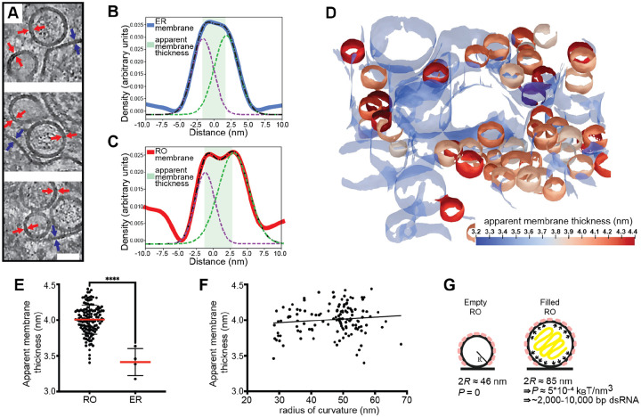
ROs of other viruses depend on the pressure from intraluminal double-stranded RNA (dsRNA) to inflate and stabilize the curved RO membrane32. The presence of empty LGTV ROs speaks against this mechanism for flaviviruses, and we thus reasoned that a coat might confer a spontaneous curvature to the RO membrane. To investigate whether a curvature-inducing coat is present on ROs, we assessed if RO membrane thickness is in line with a layer of surface-bound protein layer. Indeed, by visual inspection of tomograms, the membranes of both empty and filled ROs appeared thicker than the surrounding ER membrane (Fig. 2A). No distinct, repeating macromolecules were visible on the RO membranes. While this does not exclude the presence of a protein coat composed of smaller or membrane-integral proteins, it does make its characterization by subtomogram averaging less likely to be successful. Instead, we extended our previously developed surface morphometrics toolbox to allow for local estimation of membrane thickness33. This software allowes calculation of the average membrane thickness within a user-selected area, based on average density profiles normal to the membrane. Both for single ROs and their surrounding ER membrane, this yielded reliably interpretable density profiles (Fig. 2B–C). Color coding membranes by thickness indicated that RO membranes are consistently thicker than the surrounding ER membrane (Fig. 2D). Indeed, in four tomograms we measured a significant difference in membrane thickness with the ER membrane being 3.4 ± 0.2 nm (N= 4), and RO membranes 4.0 ± 0.2 nm (N =132) (Fig. 2E). On the other hand, the RO membrane thickness appeared largely independent of RO size, as estimated from their radii of curvature (Fig. 2F).
Figure 2: The influence of a membrane coat and viral RNA in shaping the replication organelles.
(A) Slices through tomograms of empty and filled ROs in LGTV-infected cells. Blue and red arrows indicate the thickness of the ER and RO membranes, respectively. Scale bar, 50 nm. (B-C) Membrane thickness estimation by dual Gaussian fitting to radial density plots through a representative ER membrane (B) and RO membrane (C). Solid lines, membrane density profile; dashed lines, fitted composite and dual Gaussian; shaded area, estimated thickness. (D) ER and ROs membranes from a representative tomogram of an LGTV-infected cell, color coded by apparent membrane thickness. ER membrane is partially transparent. (E) Apparent membrane thickness quantification in four tomograms of LGTV-infected cells, comparing individual ROs (n=132) and the surrounding ER (n=4). Red lines, average. Statistical significance by unpaired two-tailed Student’s t test: ****p < 0.0001. (F) Relationship between radius of curvature and apparent membrane thickness for individual ROs (n=132 from six tomograms). (G) Model of the mechanisms determining viral RO size. Two RO states exist in infected cells: empty ROs with a baseline size set independent of luminal RNA, and filled ROs, whose larger size is due to intraluminal pressure from ~2,000–10,000 bp dsRNA.
The observation that flavivirus ROs can form without detectable luminal dsRNA, and the consistent presence of a membrane coat, distinguish them from alphavirus ROs that have a near-identical membrane shape. Based on cryo-ET data, we recently published a mathematical model of alphavirus RO membrane budding, which showed that the pressure from intraluminal dsRNA, together with constraint of the membrane neck, is sufficient for the creation of the RO membrane bud32. We next adapted this mathematical model to explain flavivirus RO membrane remodeling. We assume that the membrane coat generates a spontaneous curvature H0 of the RO membrane. Such a spontaneous curvature can be generated by the protein structure as well as by crowding34. Furthermore, we consider that in ROs that enclose dsRNA, the RNA exerts a pressure P on the membrane. The total energy E of the RO membrane is then composed of an integral over the membrane surface A and the contribution of the pressure, which scales with the volume V,
| (1) |
where the first term describes the bending energy according to the Helfrich model, with κ the bending stiffness and H the mean curvature35. The second term in Eq. 1 contains the membrane tension σ. Motivated by the experimentally observed shapes, we describe the ROs as spheres with a radius R, as schematically depicted in Fig. 2G, simplifying Eq. (1) to
| (2) |
However, there are two unknown factors in Equation (2), the spontaneous curvature H0 and the pressure P in the RO. Based on our observations above, the membrane coat can be assumed to be comparable for empty and filled ROs. Thus, we can take advantage of the imaging of empty ROs to obtain H0 at vanishing pressure, P = 0. Minimizing Eq. (2) with respect to R, we obtain
| (3) |
From the experiments we have obtained an average diameter of the empty ROs to be 2R = 46 nm. By using previously estimated parameters32 for the membrane properties, i.e., σ = 10−5N/m and κ = 10kBT, we predict the spontaneous curvature to be H0 = 0.04nm−1, which corresponds to a radius of curvature 1/H0 = 25nm. Next, we want to predict the influence of the pressure generated by the RNA on the spherule size. Since the RNA does not affect the spontaneous curvature H0, we keep the above prediction and minimize Eq. 2 with respect to R, giving
| (4) |
Now including the predicted H0 = 0.04nm−1 with the measured average diameter of RNA-filled ROs 2R = 85 nm, we obtain the pressure P = 5·10−4 kBTnm−3. To interpret this value, we compare it with our previous study of alphavirus ROs, which shows that a dsRNA with a length of 2,000–10,000 base pairs generates an internal pressure of 10−4 − 10−3 kBTnm−3 (see Materials and Methods, section Estimating RO intraluminal pressure).
Taken together, flavivirus ROs have a consistently thicker membrane than the surrounding ER, consistent with the presence of small, curvature-stabilizing proteins that set a baseline RO size in the absence of luminal RNA. The size increase from empty to filled ROs is consistent with a single copy of the genome in dsRNA form.
Virions form and undergo maturation in the immediate vicinity of replication organelles
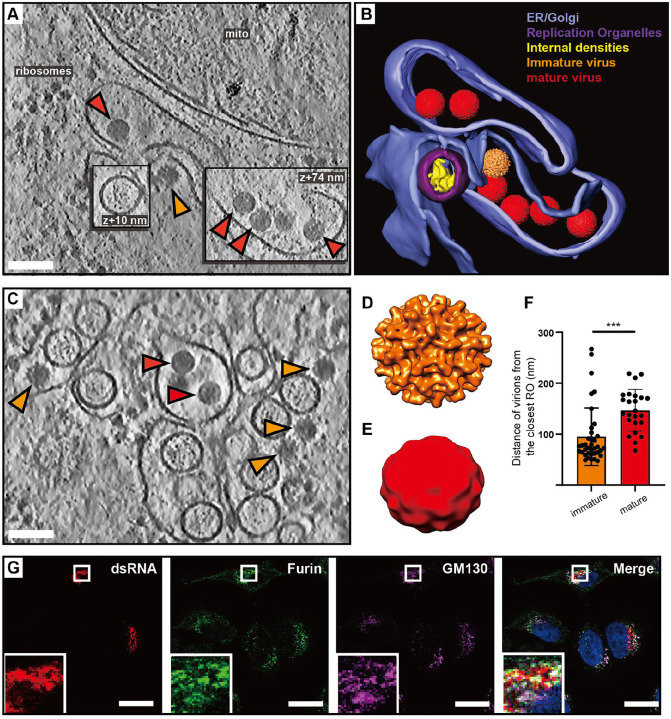
In our cryo-electron tomograms we consistently observed virions in the vicinity of ROs, underscoring the strong association between replication and virion assembly (Fig. 3A–C). We next wished to use the structure preservation in cryo-ET to study the spatial relation between virion budding and maturation. In the tomograms, virus particles at different stages of maturation were distinguishable: spiky particles corresponding to immature virions as well as smooth, mature virions (Fig. 3A–C). Subtomogram averaging on a small number of virions confirmed the distinct morphology of the immature and mature virions (Fig. 3D–E), and a good match between the in situ averages and structures of purified flaviviruses (Fig. S3). The tomograms also included examples of what seemed to be nearly or recently completed virion budding (Fig. 3A–B). In such events, immature virus particles could be observed right at the membrane, across from ROs (Fig. 3A, orange arrow). Both immature and mature virions were consistently found close to ROs, in separate but intertwined membrane compartments (Fig. 3A–C). While immature and mature particles were not observed in the same membrane compartment, seemingly discrete compartments may have been connected beyond the limited thickness of the lamella. To quantitate the relation of immature and mature particles to ROs, we measured the center-center distance of virions to ROs in 5 tomograms. Immature virions were 95±56 nm (N=37), and mature virions 147±41 nm (N=24) from the closest RO (Fig. 3F). The small but significant distance difference (p=0.0003, unpaired t test), together with the observation that immature and mature virions are present in separate compartments, indicates that virion maturation is coupled to a slight spatial separation from ROs but does not necessitate longer-range trafficking. To further investigate the spatial relationship between genome replication, virion assembly and the Golgi apparatus, we performed immunofluorescence light microscopy on fixed, LGTV-infected cells. Furin exhibited co-localization with the Golgi marker GM130 in both uninfected (Fig. S4A–D) and LGTV-infected cells (Fig. 3G–J). Meanwhile, the dsRNA signal was detected in close proximity to and partially overlapping with clusters enriched in furin and GM130 (Fig. 3J). These results corroborate the cryo-ET, reinforcing the close proximity between viral RNA replication, LGTV assembly and maturation within the infected cell. In summary, both virion assembly and maturation can occur in the immediate proximity of ROs, in distinct but intertwined compartments.
Figure 3: Virions form and mature in the immediate vicinity of replication organelles.
(A) Slice through tomogram of LGTV-infected cell showing an immature virion budding across from a RO (orange arrow), and mature virions (red arrows) in adjacent membranes. (B) Segmentation of the tomogram in (A), with color labels defined for each structure. (C) Slice through tomogram of LGTV-infected showing mature (red arrows) and immature (orange arrows) LGTV virions observed near the viral RO. (D-E) Subtomogram averages of immature (D) and mature (E) LGTV from cellular tomograms. (F) The distance from immature (n=37) and mature (n=24) virions to the closest RO, from five tomograms. Statistical significance by unpaired two-tailed Student’s t test: ****p < 0.005. (G) Representative immunofluorescence micrograph of dsRNA, furin and Golgi marker GM130 in LGTV-infected cells at 24 h p.i. Rightmost panel: merge including DAPI-staining of nuclei (blue). Scale bars 100 nm (A,C), 5 μm (G).
The TBEV furin site variants R86 and Q86 differ in replication organelle-proximal virion maturation
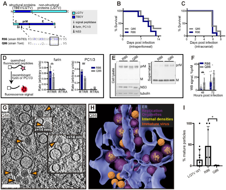
In purified virions, the transition from immature to smooth conformation can be brought about by pH change even in the absence of furin cleavage. Thus, we wanted to test if the observed RO-proximal virion maturation was dependent on the furin site in prM. To do so, we took advantage of our recently characterized chimeric LGTV, which carries the structural proteins prM and E(ectodomain) from TBEV (Fig. 4A). This virus, rLGTVT:prME, is genetically stable and has a low pathogenicity similar to that of wildtype LGTV (Rosendal et al, manuscript in preparation). The rLGTVT:prME prM and E come from the human TBEV isolate 93/783 of the European subtype, whose prM has an unusual arginine (R) in position 86 where most other strains have a glutamine (Q) (Fig. 4A). This residue is located at the P8 position of the furin cleavage site in prM, and previous studies of flaviviruses has shown that modifications here might influence prM cleavage and virus export36. We thus reasoned that R86 may affect furin cleavage efficiency, and provide a naturally occurring tool to study maturation. Hence, we also produced a version of rLGTVT:prME with the more common glutamine 86. These chimeric viruses, henceforth referred to as R86 and Q86, only differ in this amino acid residue. Both viruses replicated with similar kinetics in A549 cells (Fig. S5A), but the R86 virus had a slightly higher lethality in immunocompromised Ips1−/− (IFN-β promoter stimulator 1) mice (5 of 5 mice died with R86, 7 of 10 died with Q86, Fig. 4B). No difference in neurovirulence was detected (Fig. 4C). We next wanted to investigate whether the pathogenicity of R86 as compared to Q86 correlated with different prM cleavage kinetics. In a cleavage assay with a peptide corresponding to residues 81–94 of prM, the R86 sequence was cleaved faster by furin than Q86 (Fig. 4D). We noted that the R86 sequence generates a putative second, minimal recognition site (KR) for other proprotein convertases such as PC1/3 and PC237. In a peptide cleavage assay, the R86 sequence was also cleaved faster than Q86 by PC1/3. However, the cleavage was still completely dependent on the furin recognition site (RTRR), i.e. the putative second PC1/3 cleavage site K85-R86 was not sufficient for cleavage by PC1/3 (Fig. 4D). Next, we looked for the presence of unprocessed prM protein in cell supernatant, which would indicate release of immature virus particles. For both viruses, the bulk of cell-bound M was in the form of uncleaved prM, whereas most M in the supernatant was cleaved (Fig. 4E). Interestingly, the R86 chimeric virus had a significantly lower percentage of uncleaved prM in supernatant at 48 h post infection (Fig. 4F), suggesting that R86 confers a more efficient particle maturation. The biochemical and cell assays thus converge on the interpretation that the R86 sequence variant confers faster furin cleavage.
Figure 4: The TBEV furin site variants R86 and Q86 differ in cleavage efficiency and replication organelle-proximal virion maturation.
(A) The polyprotein of a chimeric LGTV with prM and ecto-E from TBEV strain 93/783, as color coded. Recognition sites for viral and cellular proteases are shown within the structural protein region, and the furin site sequences from TBEV strains 93/783 and Torö are shown highlighting the difference at position 86 (., identical sequence). (B) Percentage survival of Ips1−/− mice infected intraperitoneally with 104 FFU with chimeric LGTV R86 (n=5) or Q86 (n=10). (C) As (B), for but Ips1−/− mice infected intracranially with 102 FFU (n=9, R86 and n=10, Q86). (D) Enzymatic cleavage using recombinant furin or PC1/3 with peptides corresponding to furin site sequences in (A) (RTRR), or peptides with impaired furin sites (RTRA). Data from four independent experiments performed in duplicates are shown, with mean values and standard deviation. (E) prM and M protein levels in cell lysates and supernatant 48 h.p.i. visualized by immunoblotting. Viral NS3 and cellular tubulin included as infection and loading control. Representative blots are shown. (F) Ratio of prM/M intensity quantified in supernatants at 48 and 72 h.p.i. Data from four independent experiments performed in duplicates are shown, with mean values and SD. (G) Slice through tomogram of chimeric LGTV Q86-infected cell showing a predominance of immature virions (orange arrows) within the cytoplasm. Scale bar 100 nm. (H) segmentation of the tomogram in (A), with color labels defined for each structure. (I) Percentage of mature virions in the tomograms of LGTV WT (n=19), chimeric LGTV R86 (n=6), LGTV Q86- infected cells (n=4). (D,F,I) Statistical significance by unpaired two-tailed Student’s t test: *p<0.05, **p<0.01, ***p < 0.001.
Having characterized the different rates of furin cleavage, we then returned to the question of individual virion conformation inside infected cells. We infected cells with R86 and Q86 chimeric viruses and recorded cryo-electron tomograms of the infected cytoplasm as for wildtype LGTV. Both for R86 and Q86, the tomograms showed a similar overall appearance as for wildtype LGTV, including an abundance of filled and empty replication organelles as well as new virions inside dilated ER compartments (Fig. 4G–H, Fig. S5B). In the R86 tomograms, both immature and mature virions were seen, whereas immature virions appeared to predominate in the Q86 tomograms. We calculated the percentage mature virions in a set of tomograms for wildtype LGTV and the chimeric viruses. Whereas the number of virions in individual tomograms was sometimes small, the average fractions mature particles over several tomograms showed a clear trend (Fig. 4I). For Q86, 2.5±5.9% of virions were mature (N=7 tomograms), whereas 46±46% of R86 virions were mature (N=7 tomograms), a significantly higher percentage (p=0.02, unpaired t test). Wildtype LGTV was intermediate to the two recombinant viruses, with 14±25% mature virions. Taken together, a single residue in the distal part of the TBEV furin site affects prM cleavage rates by furin and PC1/3, mean survival in immunocompromised mice, and particle maturation in areas near ROs.
A protein complex connects the replication organelle to an apposed ER membrane
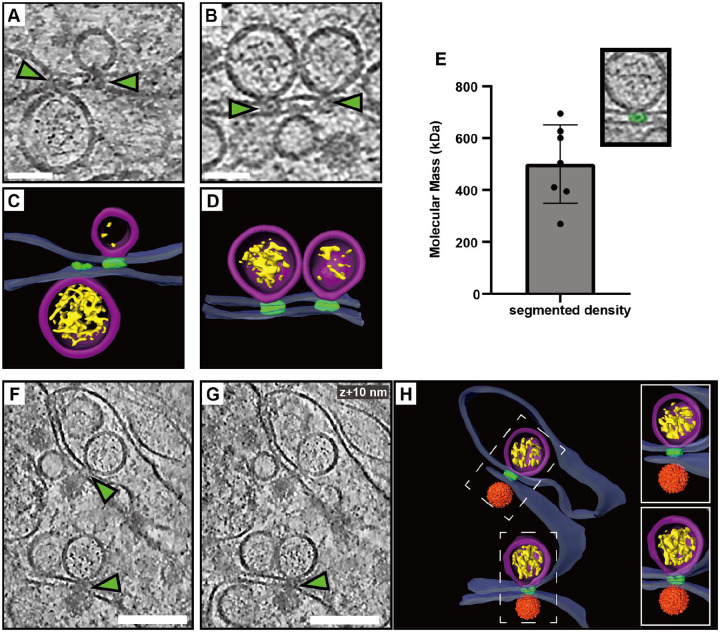
A recurring feature in the cryo-electron tomograms was the close proximity of a second ER membrane to the ER membrane containing the ROs (Figs. 1–2). We noticed that this was consistently mediated by a protein complex present at the membrane neck of the replication organelle, connecting this membrane to the adjacent, second ER membrane (Fig. 5A–D). While limited occurrences of these complexes hindered structural analysis, they consistently appeared at the necks of both filled and empty ROs (Fig. 5A–D). From volume estimates of individual complexes, we estimate their molecular masses to 500±151 kDa (Fig. 5E). Interestingly, we observed similar-looking complexes connecting the replication organelle to the site of immature particle budding in the neighboring ER cisterna (Fig. 5F–H). This observation suggests that this protein complex might play a role in coordinating the packaging of newly synthesized viral RNA into nascent immature virions.
Figure 5: A protein complex zippers replication organelles to an apposed ER membrane.
(A-B) Slices through tomograms of LGTV-infected cells showing complexes (green arrows) located at the neck of the ROs, connecting them to the adjacent ER membrane. (C-D) Segmentation of the tomograms in (A-B). (E) Estimated molecular masses of the complex (n=7). (F-G) Slices through the same tomogram at two different Z heights, showing complexes linking the RO to the site of virus assembly (green arrows). (H) Segmentation of the tomogram shown in (F-G). (C-D,H) Blue, ER membrane; purple, RO; yellow, luminal densities; green, neck complex; orange, immature virions. Scale bars, 100 nm.
Cryo-ET reveals structural signatures of LGTV replication in ex vivo mouse brain
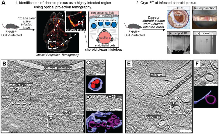
We next wished to explore if the structural features of LGTV replication that we observed in cell lines can also be identified directly in a complex, infected tissue. To do so, we proceeded to set up a workflow for cryo-ET of LGTV-infected mouse brain tissue. We based our approach on our recent publication, in which we imaged entire, ex vivo, LGTV-infected brains from type I interferon receptor knockout (Ifnar−/−) mice using fluorescence optical projection tomography38. In the 3D volumes of infected brains, immunofluorescence staining against LGTV NS5 protein was particularly strong in the choroid plexus (ChP) regions of the brain (Fig. 6A). ChP is an anatomical substructure responsible for secreting cerebrospinal fluid (CSF) into the ventricles, and as such it interfaces both with the blood and the CSF-filled ventricles (Fig. 6A). The ventricular side of the CSF-producing ependymal cells is covered with cilia and microvilli (Fig. 6A). Based on the consistently strong infection of the ChP, we developed a cryo-ET workflow for imaging ChP that was surgically removed from infected brains post mortem. Due to the thickness of this sample, we opted for vitrification through high-pressure freezing. The vitrified tissue was trimmed using a cryo-ultramicrotome after which lamellas were milled in place and subjected to cryo-ET (Fig. 6A). The tomograms of ChP revealed a multitude of subcellular structures such as mitochondria, nuclear pore complexes and a centriole (Fig. S6A–B). In addition, some tomograms contained virus-related features consistent with the observations in infected cell lines. This included mature virus particles encapsulated in vesicles close to membranes containing several ROs (Fig. 6B–D). Another tomogram contained a bona fide empty RO with seemingly thicker membrane than its limiting ER membrane, mirroring the features seen in cellular tomograms (Fig. 6E–F). Taken together, we present the first cryo-ET data on virus replication in brain tissue. The data support our cell line-based findings of RO-proximal virion maturation, and the presence of empty ROs, and provide a proof of principle that neurotropic virus replication can be studied by cryo-ET directly in infected brain samples.
Figure 6: LGTV replication visualized in ex vivo mouse brain using cryo-ET.
(A) Workflow for cryo-ET of ex vivo mouse brain. LGTV-infected brains from Ifnar−/− mice were processed differently based on the imaging method. 1. Fixed and cleared brains were immunostained for the viral protein NS5 (orange/red) and imaged using optical projection tomography. Zoomed insets show infected regions of the choroid plexus, and its anatomy. 2. For cryo-ET, the choroid plexus from unfixed and unstained LGTV-infected brains was (i) high-pressure frozen, (ii) trimmed using a cryo-ultramicrotome, (iii) FIB milled, and (iv) transferred to a cryo-TEM for cryo-ET. (B) Slice through a tomogram of LGTV-infected choroid plexus showing viral replication organelles and smooth virions in proximity, enclosed within membrane vesicles. (C-D) Close-up of the areas indicated in (B) along with their corresponding segmentations. (E) Slice through a tomogram of LGTV-infected choroid plexus showing a bona fide RO with a thicker membrane than the ER, consistent with observations from Fig. 2. (F) Close-up of the area indicated in (E) along with its corresponding segmentation. (B-F) Colors as in Fig. 3. Scale bars, 100 nm.
Discussion
Here, we present an in situ structural study of tick-borne flavivirus replication, using cryo-ET of infected cells and mouse brains. Flaviviruses are part of the vast phylum Kitrinoviricota, which is characterized by ROs housed in membrane buds39. These viruses thus need to encode mechanisms for remodeling host-cell membranes into a high-curvature bud, which is a high-energy and normally transient membrane shape. However, the viruses need to stabilize this bud-shaped membrane throughout hours of viral RNA replication. We recently showed that another genus of Kitrinoviricota, alphaviruses, stabilize their RO membrane through a coat-free mechanism that involves bud neck constraint by a viral protein complex, and inflation of the membrane bud by the intraluminal pressure from the viral RNA32. With this mechanism, the size of the replication organelle is determined by the amount of encapsulated RNA, and there are no membrane buds in the absence of intraluminal RNA. Here, we show that flaviviruses employ a different mechanism to shape the RO membrane. A membrane coat establishes a baseline RO size, allowing for the existence of ROs without intraluminal RNA. The tomograms do not indicate an ordered protein coat lattice on the RO membrane, nor clear individual protein densities, but it is possible that the curvature-generating proteins are too small and/or irregularly distributed to be detected by cryo-ET. We suggest that a strong candidate for this RO curvature generator is the 39 kDa, ER lumen-resident, non-structural protein NS1. NS1 is a peripheral membrane protein that has been reported to remodel liposomes40, and reshape the ER membrane when overexpressed on its own41. The increase in RO size due to intraluminal RNA was back-calculated to stem from the pressure of a single viral genomic RNA copy in dsRNA form (Fig. 2G, Fig. S2). Alphavirus ROs also contain a single genome copy, indicating that this may be a conserved feature across Kitrinoviricota32. Whether the smaller, empty ROs represent assembly intermediates, disassembly intermediates, or dead-end, failed RO assembly events remains to be determined.
A close coupling of flavivirus genome replication and particle budding has been suggested by several lines of evidence14,18–20. Our cryo-electron tomograms clearly resolved the maturation state of individual virions, as supported by the good agreement between the cellular subtomogram averages and structures of purified virions23,25 (Fig. 3D–E, Fig. S4). The tomograms frequently revealed immature virions in the immediate vicinity of ROs, sometimes in membrane compartments opposite to ROs (Figs. 3,6). The observation of a ~500 kDa protein complex, connecting the membranes from which ROs and virions form (Fig. 6), shows that flavivirus ROs have a “crown” or “neck complex” akin to those identified for e.g. coronaviruses, nodaviruses and alphaviruses30,32,42–49. Indeed, of the seven flavivirus non-structural proteins, four are integral membrane proteins of unknown structure and no known enzymatic function. Thus, it is possible that these proteins server a structural role in organizing a membrane-connecting neck complex that coordinates replication and assembly, but larger tomographic data sets would be required to get a decisive subtomogram average from infected cells. Recent publications have highlighted the potential in small-molecule antivirals that target non-enzymatic functions of flavivirus NS proteins50–52. These antivirals were discovered without structural insights into their target proteins, but it is possible that the neck complex we identify here is their target. Either way, a more detailed understanding of the neck complex structure and function may aid the design of improved antiviral strategies.
Contrary to the prevailing model, we observed that furin-dependent virus maturation takes place in the immediate vicinity of ROs (Fig. 3). Thus, the entire sequence replication-assembly-maturation is more closely colocalized than previously thought. The maturation compartments are distinct but intertwined with ROs (Fig. 3), suggesting a revised model of flavivirus maturation, in which a virus-induced reorganization of the secretory pathway places Golgi-like maturation compartments in the immediate proximity of ROs. Studying naturally occurring furin site variants, we could show that a single residue in the distal cleavage site affects the RO-proximal virion maturation, while having no effect on virus release and only a minor effect on lethality in an immunocompromised mouse model (Fig. 4). This suggests that the replication and infection of TBEV is robust to variations in its maturation pathway. A further step towards bridging structural and organismal studies of flavivirus replication is taken by the workflow we present for cryo-ET of infected ex vivo mouse brain tissue. Tomograms of infected choroid plexus revealed clear structural signatures of ROs and clustering mature virions, similar to those in cell lines (Fig. 7). While cryo-ET has recently been used to study Alzheimer’s disease in human brain53, our data are, to the best of our knowledge, the first cryo-ET visualization of infection processes in the brain. Future incorporation of novel lift-out and serial milling techniques into this workflow will allow for faster acquisition of larger cryo-ET data sets on infected brains54,55. This may e.g. enable structural analysis of virion maturation in brain samples, and comparison of replication features between different knock-out mice. In conclusion, our study identifies several novel structural features of tick-borne flavivirus replication, and places them in a cellular context that reveals a high degree of spatial coordination of genome replication, virion assembly and virion maturation.
Materials and Methods
Cell line and culturing
The human A549 lung epithelial cell line was grown in DMEM medium supplemented with 10% fetal bovine serum (FBS) and Penicillin Streptomycin GlutaMAX Supplement (Gibco) at 37°C in a 5% CO2 environment.
Sample preparation for cryo-electron tomography of cells
Ultrafoil Au R2/2 200 mesh grids (200 mesh, Quantifoil Micro Tools GmbH) were glow discharged. Under laminar flow the grids were dipped in ethanol before being placed in μ-Slide 8 Well Chamber (IBIDI) wells. DMEM medium with 10% FBS was added to each well and incubated while cells were being prepared. Fresh medium was added to wells and cells were seeded out at 1.5×10⁴ cells/well. The seeded cells were then placed in a 37°C, 5% CO2 incubator for 24 h. The medium in the wells was then replaced with serum-free DMEM and either Langat virus wildtype (LGTV), recombinant chimeric R86, or recombinant chimeric Q86 added at an MOI of 20. The cells were incubated for one hour before replacing the medium with 2% FBS in DMEM and left to incubate for 24 h. The medium was replaced with fresh DMEM including 2% FBS before being taken for freezing. Plunge freezing into an ethane/propane mix was performed with a Vitrobot (ThermoFisher Scientific) at 22°C, 100% humidity, blotting time of 5 s, and blot force of −5.
Preparation of cryo-lamellas of cells
Lamellas were milled from plunge-frozen cells using the Scios dual beam FIB/SEM microscope (ThermoFisher Scientific). Samples were first coated with a platinum layer using the gas injection system (GIS, ThermoFisher Scientific) operated at 26°C and 12 mm working distance for 10 seconds per grid. Lamella were milled at an angle range of 16°−20°. The cells were milled stepwise using a gallium beam at 30kV with decreasing current starting at 0.5 nA for rough milling and ending at 0.03 nA for final polishing of the lamella. Lamellas were milled to a nominal 200 nm thickness and stored in liquid nitrogen for less than a week before being loaded into a Titan Krios (ThermoFisher Scientific) for data collection.
Cryo-ET data collection on cells
Data were collected using a Titan Krios (ThermoFisher Scientific) at 300 kV in parallel illumination mode. Tilt series acquisition was done using SerialEM56 on a K2 Summit detector (Gatan, Pleasanton, CA) in super-resolution mode. The K2 Summit detector was fitted with a BioQuantum energy filter (Gatan, Pleasanton, CA) operated with a 20eV width silt. Areas to be imaged were selected from low-magnification overview images based on the presence of convoluted cytoplasmic membranes. Tilt series were collected using a 100 μm objective aperture and a 70 μm condenser 2 aperture, after coma-free alignment done using Sherpa (ThermoFisher Scientific). Tilt series were collected using the dose-symmetric scheme with a starting angle of −13° to account for lamella pre tilt. The parameters used for acquisition were: 33,000x nominal magnification with a corresponding object pixel size of 2.145 Å in super-resolution mode, a tilt range of typically −50° to +50°, defocus between −3 μm and −5 μm, tilt increment of 2°, and a total electron dose of 110 e/ Å2.
Image Processing
Motion correction, tilt series alignment, CTF estimation and correction, and tomogram reconstruction was performed as described previously57, using MotionCor258 with Fourier binning of 2, IMOD59,60, and CTFFIND461. For visualization and segmentation, tomograms were 3 times binned using IMOD, resulting in a pixel size of 12.87 Å. Tomograms were denoised using cryoCARE62 or IsoNet63, occasionally combined with a non-local means filter as implemented in Amira (Thermo Fisher Scientific). Segmentation of tomograms was performed in Amira, with initial membrane tracing and segmentation done using MemBrain V264. Subtomogram averages of mature and immature virus particles were incorporated into segmentation using UCSF Chimera65. Amira was used for counting of visually recognizable features (empty and filled ROs, immature and mature particles), measurements of the distances between them, and the volume of the neck complex densities. The neck complex volumes were converted to estimated molecular masses assuming assuming 825 Da/nm3 66.
Subtomogram averaging of virions
From tomograms generated with WARP67 at 10Å/px object pixel size, immature and mature particles were manually picked based on their clearly distinguishable appearance. 84 immature and 51 mature particles were extracted from 11 and 3 tomograms, respectively, with a box size of 80*80*80 voxels. Subtomogram averaging was done in Dynamo68,69, following the same procedure for both the immature and mature data sets. Initially, all particles were translationally and rotationally aligned to a single, high-contrast particle from the respective data sets, without symmetrization. These C1 averages were manually rotated and saved in UCSF Chimera65 to approximately fit Dynamo’s icosahedral convention, after which they were used as a template for a Gold-standard alignment with imposed icosahedral symmetry, using Dynamo’s Adaptive bandpass Filtering function. Gold-standard Fourier shell correlation curves estimated the resolution at a cutoff of 0.143 to 33 Å for the immature particles, and 80Å for the mature particles, respectively. The final averages were filtered to this resolution and masked using the spherical alignment mask.
Estimating RO intraluminal pressure
In recent work32, we demonstrated the relation between the length of an RNA and the volume of the surrounding spherule. Spherules with a volume of V = 103 − 2·103nm3 contain 4·103 − 104 base pairs (Fig. S2A), where the number of base pairs N is well described by
| (S1) |
with L0 = 333nm, , RN = 9.6nm and the length per base pair lbp = 0.256nm. Furthermore, a theoretical model was used to determine the relationship between the scaled pressure and the scaled volume (Fig. S2B). Combining both results, we obtain a relation between the pressure P and the number of base pairs N, which shows that an RNA with a length of 2·103 − 104base pairs corresponds to a pressure of 10−3 − 10−4kBTnm−3 (Fig. S2C).
Immunofluorescence staining
Cells were grown on cover glasses and infected with LGTV as for cryo-ET. At 24 h p.i., cells were fixed with 4% formaldehyde for 20 min at room temperature and then rinsed with PBS. The fixed cells were permeabilized with 0.1% Triton X-100 in PBS for 10 min at room temperature and then rinsed with PBS. The cells were then blocked with 2% BSA in PBS containing 0.05% Tween-20 (PBS-T) for 1 hour at RT. The cells were then stained with primary antibodies against furin (goat polyclonal, dilution 1:50, Invitrogen), GM130 (mouse monoclonal clone 35/GM130, dilution 1:300, BD Transduction) or dsRNA (mouse monoclonal clone J2, dilution 1:1000, Scicons, Nordic MUbio; conjugated to allophycocyanin, Abcam) and secondary fluorescent antibodies (donkey anti-goat Alexa Fluor 488, dilution 1:1000, Invitrogen; goat anti-mouse Alexa Fluor 568, dilution 1:1000, Invitrogen) and DAPI diluted in blocking buffer for 1 hour each. Fluorescence images were acquired using a Leica SP8 confocal microscope with a HC PL APO 63x/1.4 oil CS2 objective (Leica). Confocal fluorescence images were analyzed using ImageJ Fiji software70.
Chimeric viruses
A detailed description of chimeric LGTV (rLGTVT:prME) generation, rescue, and characterization can be found in (Rosendal et al, manuscript in preparation). In brief, the infectious clone of LGTV, strain TP21 (kind gift of Prof. Andres Merits) was used as genetic background into which the prM and ecto-E from TBEV strain 93/783 (GenBank: MT581212.1) was inserted. Point mutation resulting in the R86Q amino acid substitution in the prM protein were introduced by overlapping PCR with primers; For: 5’ GGACGCTGTGGGAAACAGGAAGGCTCACGGACA ‘3, Rev: 5’ TGTCCGTGAGCCTTCCTGTTTCCCACAGCGTCC 3’ (Sigma-Aldrich). RNA was generated from linearized DNA by in vitro transcription and transfected into BHK21 cells using Lipofectamine 2000 (Invitrogen). Supernatant from transfected cells was passaged once in A549 mitochondrial antiviral signaling protein (MAVS−/−) cells, confirmed by sequencing and used for all downstream experiments without further passaging.
RNA isolation and qPCR
RNA was extracted from cell supernatant using Viral RNA kit (Qiagen) according to manufacturer’s instructions. The elution volume was kept constant and cDNA was subsequently synthetized from 10 μl of eluted RNA using high-capacity cDNA Reverse Transcription kit (Thermo Fisher). LGTV RNA was quantified with qPCRBIO probe mix Hi-ROX (PCR Biosystems) and primers recognizing NS371 on a StepOnePlus real-time PCR system (Applied Biosystems).
Western blot
At indicated time points, supernatant was collected and A549 cells infected with rLGTVT:prME R86 or Q86 were lysed in 350 μl of lysis buffer (50 mM Tris-HCl pH7.5 + 150 mM NaCl + 0.1% Triton X-100) complemented with 1x protease inhibitor (cOmplete™ ULTRA, Roche, Basel, Switzerland) on ice for 20 min. Following lysis, cellular debris was removed by centrifugation at 14,000 g for 10 min at 4°C. Supernatant or pre-cleared cell lysate was mixed with Laemmli buffer to final concentration 1x and boiled at 95°C for 5 min. Proteins were separated by standard SDS-PAGE and transferred to an Immobilon®-P PVDF Membrane (GE Healthcare, Chicago, IL, USA). Blots were blocked overnight at 4°C in blocking buffer (PBS + 0.05 % Tween 20 + 2% Amersham ECL Prime Blocking Reagent; Cytiva), stained with primary antibodies against NS372 (chicken polyclonal, diluted 1:1500), tubulin (rabbit polyclonal, diluted 1:4000, Abcam-ab6046) or M73 (in-house rabbit polyclonal serum, diluted 1:500) overnight at 4°C followed by secondary antibodies (goat anti-chicken Alexa-555, donkey anti-rabbit Alexa-647 (dilution 1:2500, Invitrogen, Waltham, MA, USA) for 1 h at room temperature. Blots were visualized on Amersham™ Imager 680 (GE Healthcare).
Enzymatic assays
Synthetic peptides (Biomatik) corresponding to the P13 to P’1 residues of prM from TBEV strain Torö (Dabcyl-GRCGKQEGSRTRRG-E(EDANS)) and 93/783 (Dabcyl-GRCGKREGSRTRRG-E(EDANS)) or corresponding peptides with an impaired furin recognition site (Dabcyl-GRCGKREGSRTRAG-E(EDANS), Dabcyl-GRCGKQEGSRTRAG-E(EDANS)) was used as substrate. Cleavage efficiency was assayed using an adapted fluorogenic peptide assay74. For furin, 3 U furin (Thermo Fisher, Waltham, MA, USA) was mixed with 100 μM of substrate in a total volume of 100 μl reaction buffer (100 mM HEPES pH7.5 + 1 mM CaCl2 + 0.5% Triton X-100) at 30°C for 3h. For proprotein convertase 1/3 (PC1/3), 1 μg recombinant human PC1/3 (R&D Systems, Minneapolis, MN, USA) was mixed with 100 μM of substrate in a total volume of 100 μl reaction buffer (25 mM MES pH 6.0 + 5 mM CaCl2 + 1% (w/v) Brij-35) at 37°C for 1h. The emission at 490 nm measured every 3 min and the average rate calculated by linear regression.
Virus infection of mice
All animal experiments were conducted at the Umeå Centre for Comparative Biology (UCCB), under approval from the regional Animal Research Ethics Committee of Northern Norrland and the Swedish Board of Agriculture, ethical permit A9–2018, A41–2019, and conducted as described previously38,71. Briefly, Mavs−/− mice in C57BL/6 background (kind gift of Nelson O Gekara, Umeå University) were infected by intraperitoneal injection of 104 focus-forming units (FFU) or intracranial injection of 102 FFU of rLGTVT:prME R86 or Q86 diluted in PBS. Ifnar−/− mice were intracranially inoculated with 104 FFU of LGTV and sacrificed when they developed either one pre-defined severe sign or at least three milder signs. Mice were monitored for symptoms of disease and euthanized as previously described criteria for humane endpoint38.
Cryo-ET of LGTV-infected mouse brain tissue
Based on optical projection tomography that visualized the infection distribution in entire, ex vivo brains38, choroid plexuses were surgically removed from brains of LGTV-infected Ifnar−/− mice post mortem. The choroid plexuses were perfused with ice-cold phosphate-buffered saline (PBS) and rapidly transferred to ice-cold artificial CSF75. Just prior to high-pressure freezing, the tissue was placed in a 3 mm copper high-pressure freezing carrier (Wohlwend) which can be clipped into an Autogrid. The sample was covered with a 20% dextran solution in PBS as cryoprotectant and covered with a sapphire disk. The assembled carrier was rapidly vitrified using a Leica EM HPM100 high-pressure freezer. The frozen carrier was trimmed at cryogenic temperatures using a Leica EM FC7 cryo-ultramicrotome with a diamond knife. The copper carrier was trimmed to leave a flat tissue sample on the carrier, measuring 100 μm in width, 20 μm in thickness, and 30 μm in depth76. Frozen carriers were clipped into Autogrids (ThermoFisher) prior to cryo-FIB milling with a Scios dual-beam FIB/SEM microscope (ThermoFisher Scientific).
The sample was coated with a protective platinum layer using a gas injection system for 15 seconds at a working distance of 7 mm. The cryostage was tilted at an angle of approximately 10° for milling. A rough milling was initially performed with an ion beam accelerating voltage of 30 kV and a current ranging from 0.79 to 2.5 nA to reach a thickness of 1 μm. Additionally, the two sides of the lamella were milled above and below to allow cryo-ET data collection by preventing the thick edges of the tissue from obstructing transmission EM imaging76. After rough milling, one edge of the lamella was detached from the main platform to relieve stress. When the lamella reached a thickness of approximately 1 μm, the ion beam current was lowered to 80 to 230 pA for fine milling, resulting in a final lamella thickness of around 200 nm. SerialEM was used to collect tilt series data with tilt angles ranging from 40° to −40° in 2° increments. The total electron dose for a single tilt series was approximately 100 e-/Å2, with defocus between −5 and −10 μm. Tomograms were generated as described above for cells.
Statistics and reproducibility
Data and statistical analysis were performed using Prism (GraphPad Software Inc., USA). Details about replicates, statistical test used, exact values of n, what n represents, and dispersion and precision measures used can be found in figures and corresponding figure legends. Values of p < 0.05 were considered significant. All tomograms shown are representative of larger data sets as indicated in Table 1.
Supplementary Material
Acknowledgements
This project was funded by a Human Frontier Science Program Career Development Award (CDA00047/2017-C to LAC), the Swedish research council (grants 2021-01145 and 2023-02664 to LAC, 2018-05851 and 2020-06224 to AÖW), a Umeå University Medical Faculty strategic grant (LAC), the Knut and Alice Wallenberg Foundation through the Wallenberg Centre for Molecular Medicine Umeå (LAC), Nadia’s Gift Foundation Innovator Award of the Damon Runyon Cancer Foundation (DRR-65-21 to DAG) and the National Institutes of Health (RF1NS125674 to DAG). SD received postdoctoral funding from the European Union under the Marie Skłodowska-Curie grant agreement No 795892. ES, NC and JZ received postdoctoral funding from the Kempe Foundation SMK-1532 (ES and JZ), and Knut and Alice Wallenberg Foundation KAW2015.0284 (NC), through the MIMS Excellence by Choice Postdoctoral Program under the patronage of Emmanuelle Charpentier. BKS received postdoctoral funding from the Wenner-Gren foundation. Cryo-EM was performed at the Umeå Center for Electron Microscopy (UCEM) a SciLifeLab National Cryo-EM facility. Fluorescence microscopy was performed at the Biochemical Imaging Center (BICU) at Umeå University. Both UCEM and BICU received funding from the National Microscopy Infrastructure, NMI (VR-RFI 2019-00217). We are thankful to all members of the VR-TBEV network, and Max Renner for valuable comments and suggestions.
Data availability
The cellular subtomogram averages of immature and mature Langat virus are deposited at the Electron Microscopy Data Bank with accession codes EMD-51640 and EMD-51642, respectively.
References
- 1.Bhatt S., Gething P.W., Brady O.J., Messina J.P., Farlow A.W., Moyes C.L., Drake J.M., Brownstein J.S., Hoen A.G., Sankoh O., et al. (2013). The global distribution and burden of dengue. Nature 496, 504–507. 10.1038/nature12060. [DOI] [PMC free article] [PubMed] [Google Scholar]
- 2.Chambers T.J., and Diamond M.S. (2003). Pathogenesis of flavivirus encephalitis. Adv Virus Res 60, 273–342. 10.1016/s0065-3527(03)60008-4. [DOI] [PMC free article] [PubMed] [Google Scholar]
- 3.Ng M.L. (1987). Ultrastructural studies of Kunjin virus-infected Aedes albopictus cells. J Gen Virol 68 (Pt 2), 577–582. 10.1099/0022-1317-68-2-577. [DOI] [PubMed] [Google Scholar]
- 4.Ko K.K., Igarashi A., and Fukai K. (1979). Electron microscopic observations on Aedes albopictus cells infected with dengue viruses. Arch Virol 62, 41–52. 10.1007/BF01314902. [DOI] [PubMed] [Google Scholar]
- 5.Neufeldt C.J., Cortese M., Acosta E.G., and Bartenschlager R. (2018). Rewiring cellular networks by members of the Flaviviridae family. Nat Rev Microbiol 16, 125–142. 10.1038/nrmicro.2017.170. [DOI] [PMC free article] [PubMed] [Google Scholar]
- 6.Aktepe T.E., and Mackenzie J.M. (2018). Shaping the flavivirus replication complex: It is curvaceous! Cell Microbiol 20, e12884. 10.1111/cmi.12884. [DOI] [PMC free article] [PubMed] [Google Scholar]
- 7.Arakawa M., and Morita E. (2019). Flavivirus Replication Organelle Biogenesis in the Endoplasmic Reticulum: Comparison with Other Single-Stranded Positive-Sense RNA Viruses. Int J Mol Sci 20. 10.3390/ijms20092336. [DOI] [PMC free article] [PubMed] [Google Scholar]
- 8.Ci Y., and Shi L. (2021). Compartmentalized replication organelle of flavivirus at the ER and the factors involved. Cell Mol Life Sci 78, 4939–4954. 10.1007/s00018-021-03834-6. [DOI] [PMC free article] [PubMed] [Google Scholar]
- 9.van den Elsen K., Quek J.P., and Luo D. (2021). Molecular Insights into the Flavivirus Replication Complex. Viruses 13. 10.3390/v13060956. [DOI] [PMC free article] [PubMed] [Google Scholar]
- 10.Romero-Brey I., Merz A., Chiramel A., Lee J.Y., Chlanda P., Haselman U., Santarella-Mellwig R., Habermann A., Hoppe S., Kallis S., et al. (2012). Three-dimensional architecture and biogenesis of membrane structures associated with hepatitis C virus replication. PLoS Pathog 8, e1003056. 10.1371/journal.ppat.1003056. [DOI] [PMC free article] [PubMed] [Google Scholar]
- 11.Yau W.L., Nguyen-Dinh V., Larsson E., Lindqvist R., Overby A.K., and Lundmark R. (2019). Model System for the Formation of Tick-Borne Encephalitis Virus Replication Compartments without Viral RNA Replication. J Virol 93. 10.1128/JVI.00292-19. [DOI] [PMC free article] [PubMed] [Google Scholar]
- 12.Aktepe T.E., Liebscher S., Prier J.E., Simmons C.P., and Mackenzie J.M. (2017). The Host Protein Reticulon 3.1A Is Utilized by Flaviviruses to Facilitate Membrane Remodelling. Cell Rep 21, 1639–1654. 10.1016/j.celrep.2017.10.055. [DOI] [PubMed] [Google Scholar]
- 13.Hoffmann H.H., Schneider W.M., Rozen-Gagnon K., Miles L.A., Schuster F., Razooky B., Jacobson E., Wu X., Yi S., Rudin C.M., et al. (2021). TMEM41B Is a Pan-flavivirus Host Factor. Cell 184, 133–148 e120. 10.1016/j.cell.2020.12.005. [DOI] [PMC free article] [PubMed] [Google Scholar]
- 14.Welsch S., Miller S., Romero-Brey I., Merz A., Bleck C.K., Walther P., Fuller S.D., Antony C., Krijnse-Locker J., and Bartenschlager R. (2009). Composition and three-dimensional architecture of the dengue virus replication and assembly sites. Cell Host Microbe 5, 365–375. 10.1016/j.chom.2009.03.007. [DOI] [PMC free article] [PubMed] [Google Scholar]
- 15.Junjhon J., Pennington J.G., Edwards T.J., Perera R., Lanman J., and Kuhn R.J. (2014). Ultrastructural characterization and three-dimensional architecture of replication sites in dengue virus-infected mosquito cells. J Virol 88, 4687–4697. 10.1128/JVI.00118-14. [DOI] [PMC free article] [PubMed] [Google Scholar]
- 16.Offerdahl D.K., Dorward D.W., Hansen B.T., and Bloom M.E. (2012). A three-dimensional comparison of tick-borne flavivirus infection in mammalian and tick cell lines. PLoS One 7, e47912. 10.1371/journal.pone.0047912. [DOI] [PMC free article] [PubMed] [Google Scholar]
- 17.Gillespie L.K., Hoenen A., Morgan G., and Mackenzie J.M. (2010). The endoplasmic reticulum provides the membrane platform for biogenesis of the flavivirus replication complex. J Virol 84, 10438–10447. 10.1128/JVI.00986-10. [DOI] [PMC free article] [PubMed] [Google Scholar]
- 18.Apte-Sengupta S., Sirohi D., and Kuhn R.J. (2014). Coupling of replication and assembly in flaviviruses. Curr Opin Virol 9, 134–142. 10.1016/j.coviro.2014.09.020. [DOI] [PMC free article] [PubMed] [Google Scholar]
- 19.Kummerer B.M., and Rice C.M. (2002). Mutations in the yellow fever virus nonstructural protein NS2A selectively block production of infectious particles. J Virol 76, 4773–4784. 10.1128/jvi.76.10.4773-4784.2002. [DOI] [PMC free article] [PubMed] [Google Scholar]
- 20.Xie X., Zou J., Zhang X., Zhou Y., Routh A.L., Kang C., Popov V.L., Chen X., Wang Q.Y., Dong H., and Shi P.Y. (2019). Dengue NS2A Protein Orchestrates Virus Assembly. Cell Host Microbe 26, 606–622 e608. 10.1016/j.chom.2019.09.015. [DOI] [PubMed] [Google Scholar]
- 21.Överby A.K., Popov V.L., Niedrig M., and Weber F. (2010). Tick-borne encephalitis virus delays interferon induction and hides its double-stranded RNA in intracellular membrane vesicles. J Virol 84, 8470–8483. 10.1128/JVI.00176-10. [DOI] [PMC free article] [PubMed] [Google Scholar]
- 22.Renner M., Dejnirattisai W., Carrique L., Martin I.S., Karia D., Ilca S.L., Ho S.F., Kotecha A., Keown J.R., Mongkolsapaya J., et al. (2021). Flavivirus maturation leads to the formation of an occupied lipid pocket in the surface glycoproteins. Nat Commun 12, 1238. 10.1038/s41467-021-21505-9. [DOI] [PMC free article] [PubMed] [Google Scholar]
- 23.Anastasina M., Fuzik T., Domanska A., Pulkkinen L.I.A., Smerdova L., Formanova P.P., Strakova P., Novacek J., Ruzek D., Plevka P., and Butcher S.J. (2024). The structure of immature tick-borne encephalitis virus supports the collapse model of flavivirus maturation. Sci Adv 10, eadl1888. 10.1126/sciadv.adl1888. [DOI] [PMC free article] [PubMed] [Google Scholar]
- 24.Newton N.D., Hardy J.M., Modhiran N., Hugo L.E., Amarilla A.A., Bibby S., Venugopal H., Harrison J.J., Traves R.J., Hall R.A., et al. (2021). The structure of an infectious immature flavivirus redefines viral architecture and maturation. Sci Adv 7. 10.1126/sciadv.abe4507. [DOI] [PMC free article] [PubMed] [Google Scholar]
- 25.Fuzik T., Formanova P., Ruzek D., Yoshii K., Niedrig M., and Plevka P. (2018). Structure of tick-borne encephalitis virus and its neutralization by a monoclonal antibody. Nat Commun 9, 436. 10.1038/s41467-018-02882-0. [DOI] [PMC free article] [PubMed] [Google Scholar]
- 26.Kuhn R.J., Zhang W., Rossmann M.G., Pletnev S.V., Corver J., Lenches E., Jones C.T., Mukhopadhyay S., Chipman P.R., Strauss E.G., et al. (2002). Structure of dengue virus: implications for flavivirus organization, maturation, and fusion. Cell 108, 717–725. 10.1016/s0092-8674(02)00660-8. [DOI] [PMC free article] [PubMed] [Google Scholar]
- 27.Quemin E.R.J., Machala E.A., Vollmer B., Prazak V., Vasishtan D., Rosch R., Grange M., Franken L.E., Baker L.A., and Grunewald K. (2020). Cellular Electron Cryo-Tomography to Study Virus-Host Interactions. Annu Rev Virol 7, 239–262. 10.1146/annurev-virology-021920-115935. [DOI] [PubMed] [Google Scholar]
- 28.Nishikiori M., den Boon J.A., Unchwaniwala N., and Ahlquist P. (2022). Crowning Touches in Positive-Strand RNA Virus Genome Replication Complex Structure and Function. Annu Rev Virol 9, 193–212. 10.1146/annurev-virology-092920-021307. [DOI] [PubMed] [Google Scholar]
- 29.Hong Y., Song Y., Zhang Z., and Li S. (2023). Cryo-Electron Tomography: The Resolution Revolution and a Surge of In Situ Virological Discoveries. Annu Rev Biophys 52, 339–360. 10.1146/annurev-biophys-092022-100958. [DOI] [PubMed] [Google Scholar]
- 30.Zimmermann L., Zhao X., Makroczyova J., Wachsmuth-Melm M., Prasad V., Hensel Z., Bartenschlager R., and Chlanda P. (2023). SARS-CoV-2 nsp3 and nsp4 are minimal constituents of a pore spanning replication organelle. Nat Commun 14, 7894. 10.1038/s41467-023-43666-5. [DOI] [PMC free article] [PubMed] [Google Scholar]
- 31.Huang Y., Zhang Y., and Ni T. (2023). Towards in situ high-resolution imaging of viruses and macromolecular complexes using cryo-electron tomography. J Struct Biol 215, 108000. 10.1016/j.jsb.2023.108000. [DOI] [PubMed] [Google Scholar]
- 32.Laurent T., Kumar P., Liese S., Zare F., Jonasson M., Carlson A., and Carlson L.A. (2022). Architecture of the chikungunya virus replication organelle. Elife 11. 10.7554/eLife.83042. [DOI] [PMC free article] [PubMed] [Google Scholar]
- 33.Barad B.A., Medina M., Fuentes D., Wiseman R.L., and Grotjahn D.A. (2023). Quantifying organellar ultrastructure in cryo-electron tomography using a surface morphometrics pipeline. J Cell Biol 222. 10.1083/jcb.202204093. [DOI] [PMC free article] [PubMed] [Google Scholar]
- 34.Alimohamadi H., and Rangamani P. (2018). Modeling Membrane Curvature Generation due to Membrane(−)Protein Interactions. Biomolecules 8. 10.3390/biom8040120. [DOI] [PMC free article] [PubMed] [Google Scholar]
- 35.Helfrich W. (1973). Elastic properties of lipid bilayers: theory and possible experiments. Z Naturforsch C 28, 693–703. [DOI] [PubMed] [Google Scholar]
- 36.Keelapang P., Sriburi R., Supasa S., Panyadee N., Songjaeng A., Jairungsri A., Puttikhunt C., Kasinrerk W., Malasit P., and Sittisombut N. (2004). Alterations of pr-M cleavage and virus export in pr-M junction chimeric dengue viruses. J Virol 78, 2367–2381. 10.1128/jvi.78.5.2367-2381.2004. [DOI] [PMC free article] [PubMed] [Google Scholar]
- 37.Garten W. (2018). Characterization of Proprotein Convertases and Their Involvement in Virus Propagation. In Activation of Viruses by Host Proteases, Böttcher-Friebertshäuser E., Garten W., and Klenk H.D., eds. (Springer Nature; ). 10.1007/978-3-319-75474-1_9. [DOI] [Google Scholar]
- 38.Chotiwan N., Rosendal E., Willekens S.M.A., Schexnaydre E., Nilsson E., Lindqvist R., Hahn M., Mihai I.S., Morini F., Zhang J., et al. (2023). Type I interferon shapes brain distribution and tropism of tick-borne flavivirus. Nat Commun 14, 2007. 10.1038/s41467-023-37698-0. [DOI] [PMC free article] [PubMed] [Google Scholar]
- 39.Ahola T. (2019). New Phylogenetic Grouping of Positive-Sense RNA Viruses Is Concordant with Replication Complex Morphology. mBio 10. 10.1128/mBio.01402-19. [DOI] [PMC free article] [PubMed] [Google Scholar]
- 40.Akey D.L., Brown W.C., Dutta S., Konwerski J., Jose J., Jurkiw T.J., DelProposto J., Ogata C.M., Skiniotis G., Kuhn R.J., and Smith J.L. (2014). Flavivirus NS1 structures reveal surfaces for associations with membranes and the immune system. Science 343, 881–885. 10.1126/science.1247749. [DOI] [PMC free article] [PubMed] [Google Scholar]
- 41.Ci Y., Liu Z.Y., Zhang N.N., Niu Y., Yang Y., Xu C., Yang W., Qin C.F., and Shi L. (2020). Zika NS1-induced ER remodeling is essential for viral replication. J Cell Biol 219. 10.1083/jcb.201903062. [DOI] [PMC free article] [PubMed] [Google Scholar]
- 42.Huang Y., Wang T., Zhong L., Zhang W., Zhang Y., Yu X., Yuan S., and Ni T. (2024). Molecular architecture of coronavirus double-membrane vesicle pore complex. Nature 633, 224–231. 10.1038/s41586-024-07817-y. [DOI] [PMC free article] [PubMed] [Google Scholar]
- 43.Wolff G., Limpens R., Zevenhoven-Dobbe J.C., Laugks U., Zheng S., de Jong A.W.M., Koning R.I., Agard D.A., Grünewald K., Koster A.J., et al. (2020). A molecular pore spans the double membrane of the coronavirus replication organelle. Science 369, 1395–1398. 10.1126/science.abd3629. [DOI] [PMC free article] [PubMed] [Google Scholar]
- 44.Unchwaniwala N., Zhan H., Pennington J., Horswill M., den Boon J.A., and Ahlquist P. (2020). Subdomain cryo-EM structure of nodaviral replication protein A crown complex provides mechanistic insights into RNA genome replication. Proc Natl Acad Sci U S A 117, 18680–18691. 10.1073/pnas.2006165117. [DOI] [PMC free article] [PubMed] [Google Scholar]
- 45.Ertel K.J., Benefield D., Castano-Diez D., Pennington J.G., Horswill M., den Boon J.A., Otegui M.S., and Ahlquist P. (2017). Cryo-electron tomography reveals novel features of a viral RNA replication compartment. Elife 6. 10.7554/eLife.25940. [DOI] [PMC free article] [PubMed] [Google Scholar]
- 46.Tan Y.B., Chmielewski D., Law M.C.Y., Zhang K., He Y., Chen M., Jin J., and Luo D. (2022). Molecular architecture of the Chikungunya virus replication complex. Sci Adv 8, eadd2536. 10.1126/sciadv.add2536. [DOI] [PMC free article] [PubMed] [Google Scholar]
- 47.Laurent T., and Carlson L.A. (2023). The organization of double-stranded RNA in the chikungunya virus replication organelle. PLoS Negl Trop Dis 17, e0011404. 10.1371/journal.pntd.0011404. [DOI] [PMC free article] [PubMed] [Google Scholar]
- 48.Kumar M., and Altan-Bonnet N. (2021). Viral pores are everywhere. Mol Cell 81, 2061–2063. 10.1016/j.molcel.2021.04.025. [DOI] [PubMed] [Google Scholar]
- 49.de Beijer N.L., Snijder E.J., and Barcena M. (2024). A Cool Look at Positive-Strand RNA Virus Replication Organelles: New Insights from Cryo-Electron Microscopy. Annu Rev Biochem 93, 163–187. 10.1146/annurev-biochem-052521-115736. [DOI] [PubMed] [Google Scholar]
- 50.Goethals O., Kaptein S.J.F., Kesteleyn B., Bonfanti J.F., Van Wesenbeeck L., Bardiot D., Verschoor E.J., Verstrepen B.E., Fagrouch Z., Putnak J.R., et al. (2023). Blocking NS3-NS4B interaction inhibits dengue virus in non-human primates. Nature 615, 678–686. 10.1038/s41586-023-05790-6. [DOI] [PMC free article] [PubMed] [Google Scholar]
- 51.Kaptein S.J.F., Goethals O., Kiemel D., Marchand A., Kesteleyn B., Bonfanti J.F., Bardiot D., Stoops B., Jonckers T.H.M., Dallmeier K., et al. (2021). A pan-serotype dengue virus inhibitor targeting the NS3-NS4B interaction. Nature 598, 504–509. 10.1038/s41586-021-03990-6. [DOI] [PubMed] [Google Scholar]
- 52.Kiemel D., Kroell A.H., Denolly S., Haselmann U., Bonfanti J.F., Andres J.I., Ghosh B., Geluykens P., Kaptein S.J.F., Wilken L., et al. (2024). Pan-serotype dengue virus inhibitor JNJ-A07 targets NS4A-2K-NS4B interaction with NS2B/NS3 and blocks replication organelle formation. Nat Commun 15, 6080. 10.1038/s41467-024-50437-3. [DOI] [PMC free article] [PubMed] [Google Scholar]
- 53.Gilbert M.A.G., Fatima N., Jenkins J., O’Sullivan T.J., Schertel A., Halfon Y., Wilkinson M., Morrema T.H.J., Geibel M., Read R.J., et al. (2024). CryoET of beta-amyloid and tau within postmortem Alzheimer’s disease brain. Nature 631, 913–919. 10.1038/s41586-024-07680-x. [DOI] [PMC free article] [PubMed] [Google Scholar]
- 54.Schiotz O.H., Kaiser C.J.O., Klumpe S., Morado D.R., Poege M., Schneider J., Beck F., Klebl D.P., Thompson C., and Plitzko J.M. (2024). Serial Lift-Out: sampling the molecular anatomy of whole organisms. Nat Methods 21, 1684–1692. 10.1038/s41592-023-02113-5. [DOI] [PMC free article] [PubMed] [Google Scholar]
- 55.Nguyen H.T.D., Perone G., Klena N., Vazzana R., Kaluthantrige Don F., Silva M., Sorrentino S., Swuec P., Leroux F., Kalebic N., et al. (2024). Serialized on-grid lift-in sectioning for tomography (SOLIST) enables a biopsy at the nanoscale. Nat Methods 21, 1693–1701. 10.1038/s41592-024-02384-6. [DOI] [PMC free article] [PubMed] [Google Scholar]
- 56.Mastronarde D.N. (2005). Automated electron microscope tomography using robust prediction of specimen movements. J Struct Biol 152, 36–51. S1047–8477(05)00152–8 [pii] 10.1016/j.jsb.2005.07.007. [DOI] [PubMed] [Google Scholar]
- 57.Dahmane S., Kerviel A., Morado D.R., Shankar K., Ahlman B., Lazarou M., Altan-Bonnet N., and Carlson L.A. (2022). Membrane-assisted assembly and selective secretory autophagy of enteroviruses. Nat Commun 13, 5986. 10.1038/s41467-022-33483-7. [DOI] [PMC free article] [PubMed] [Google Scholar]
- 58.Zheng S.Q., Palovcak E., Armache J.P., Verba K.A., Cheng Y., and Agard D.A. (2017). MotionCor2: anisotropic correction of beam-induced motion for improved cryo-electron microscopy. Nat Methods 14, 331–332. 10.1038/nmeth.4193. [DOI] [PMC free article] [PubMed] [Google Scholar]
- 59.Kremer J.R., Mastronarde D.N., and McIntosh J.R. (1996). Computer visualization of three-dimensional image data using IMOD. J. Struct. Biol. 116, 71–76. [DOI] [PubMed] [Google Scholar]
- 60.Xiong Q., Morphew M.K., Schwartz C.L., Hoenger A.H., and Mastronarde D.N. (2009). CTF determination and correction for low dose tomographic tilt series. J Struct Biol 168, 378–387. 10.1016/j.jsb.2009.08.016. [DOI] [PMC free article] [PubMed] [Google Scholar]
- 61.Rohou A., and Grigorieff N. (2015). CTFFIND4: Fast and accurate defocus estimation from electron micrographs. J Struct Biol 192, 216–221. 10.1016/j.jsb.2015.08.008. [DOI] [PMC free article] [PubMed] [Google Scholar]
- 62.Buchholz T.O., Jordan M., Pigino G., and Jug F. (2019). Cryo-CARE: Content-Aware Image Restoration for Cryo-Transmission Electron Microscopy Data. I S Biomed Imaging, 502–506. 10.1109/isbi.2019.8759519. [DOI] [Google Scholar]
- 63.Liu Y.T., Zhang H., Wang H., Tao C.L., Bi G.Q., and Zhou Z.H. (2022). Isotropic reconstruction for electron tomography with deep learning. Nat Commun 13, 6482. 10.1038/s41467-022-33957-8. [DOI] [PMC free article] [PubMed] [Google Scholar]
- 64.Lamm L., Zufferey S., Righetto R.D., Wietrzynski W., Yamauchi K.A., Burt A., Liu Y., Zhang H., Martinez-Sanchez A., Ziegler S., et al. (2024). MemBrain v2: an end-to-end tool for the analysis of membranes in cryo-electron tomography. biorxiv. 10.1101/2024.01.05.574336 [DOI] [Google Scholar]
- 65.Pettersen E.F., Goddard T.D., Huang C.C., Couch G.S., Greenblatt D.M., Meng E.C., and Ferrin T.E. (2004). UCSF Chimera--a visualization system for exploratory research and analysis. Journal of computational chemistry 25, 1605–1612. 10.1002/jcc.20084. [DOI] [PubMed] [Google Scholar]
- 66.Erickson H.P. (2009). Size and shape of protein molecules at the nanometer level determined by sedimentation, gel filtration, and electron microscopy. Biol Proced Online 11, 32–51. 10.1007/s12575-009-9008-x. [DOI] [PMC free article] [PubMed] [Google Scholar]
- 67.Tegunov D., and Cramer P. (2019). Real-time cryo-electron microscopy data preprocessing with Warp. Nat Methods 16, 1146–1152. 10.1038/s41592-019-0580-y. [DOI] [PMC free article] [PubMed] [Google Scholar]
- 68.Castano-Diez D., Kudryashev M., Arheit M., and Stahlberg H. (2012). Dynamo: a flexible, user-friendly development tool for subtomogram averaging of cryo-EM data in high-performance computing environments. J Struct Biol 178, 139–151. 10.1016/j.jsb.2011.12.017. [DOI] [PubMed] [Google Scholar]
- 69.Castano-Diez D., Kudryashev M., and Stahlberg H. (2017). Dynamo Catalogue: Geometrical tools and data management for particle picking in subtomogram averaging of cryo-electron tomograms. J Struct Biol 197, 135–144. 10.1016/j.jsb.2016.06.005. [DOI] [PubMed] [Google Scholar]
- 70.Schindelin J., Arganda-Carreras I., Frise E., Kaynig V., Longair M., Pietzsch T., Preibisch S., Rueden C., Saalfeld S., Schmid B., et al. (2012). Fiji: an open-source platform for biological-image analysis. Nat Methods 9, 676–682. 10.1038/nmeth.2019. [DOI] [PMC free article] [PubMed] [Google Scholar]
- 71.Kurhade C., Zegenhagen L., Weber E., Nair S., Michaelsen-Preusse K., Spanier J., Gekara N.O., Kroger A., and Overby A.K. (2016). Type I Interferon response in olfactory bulb, the site of tick-borne flavivirus accumulation, is primarily regulated by IPS-1. J Neuroinflammation 13, 22. 10.1186/s12974-016-0487-9. [DOI] [PMC free article] [PubMed] [Google Scholar]
- 72.Panayiotou C., Lindqvist R., Kurhade C., Vonderstein K., Pasto J., Edlund K., Upadhyay A.S., and Overby A.K. (2018). Viperin Restricts Zika Virus and Tick-Borne Encephalitis Virus Replication by Targeting NS3 for Proteasomal Degradation. J Virol 92. 10.1128/JVI.02054-17. [DOI] [PMC free article] [PubMed] [Google Scholar]
- 73.Pulkkinen L.I.A., Barrass S.V., Domanska A., Overby A.K., Anastasina M., and Butcher S.J. (2022). Molecular Organisation of Tick-Borne Encephalitis Virus. Viruses 14. 10.3390/v14040792. [DOI] [PMC free article] [PubMed] [Google Scholar]
- 74.Jaimes J.A., Millet J.K., Goldstein M.E., Whittaker G.R., and Straus M.R. (2019). A Fluorogenic Peptide Cleavage Assay to Screen for Proteolytic Activity: Applications for coronavirus spike protein activation. J Vis Exp. 10.3791/58892. [DOI] [PubMed] [Google Scholar]
- 75.Artificial Cerebrospinal Fluid (ACSF) (10x). (2017). Cold Spring Harb Protoc 2017. 10.1101/pdb.rec094342. [DOI] [PubMed] [Google Scholar]
- 76.Zhang J., Zhang D., Sun L., Ji G., Huang X., Niu T., Xu J., Ma C., Zhu Y., Gao N., et al. (2021). VHUT-cryo-FIB, a method to fabricate frozen hydrated lamellae from tissue specimens for in situ cryo-electron tomography. J Struct Biol 213, 107763. 10.1016/j.jsb.2021.107763. [DOI] [PubMed] [Google Scholar]
Associated Data
This section collects any data citations, data availability statements, or supplementary materials included in this article.
Supplementary Materials
Data Availability Statement
The cellular subtomogram averages of immature and mature Langat virus are deposited at the Electron Microscopy Data Bank with accession codes EMD-51640 and EMD-51642, respectively.


