Self-expandable metal stents (SEMSs) are commonly used as a bridge-to-surgery or palliative treatment for obstructive colorectal cancer 1 . Although technical and clinical success rates are high, adverse events such as perforation, migration, and sepsis 2 3 4 may occur owing to the poor visual field due to stool and failure to identify the luminal opening of the tumor, air over-insufflation, and unreasonable guidewire manipulation. Gel immersion can be used to improve the endoscopic view 5 . Herein, we describe a SEMS insertion with a clear view and lower intraluminal pressure using water and gel immersion ( Video 1 ).
Low-pressure insertion of a self-expandable metal stent for obstructive colon cancer using water and gel immersion.
Video 1
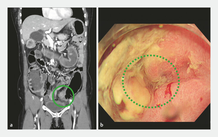
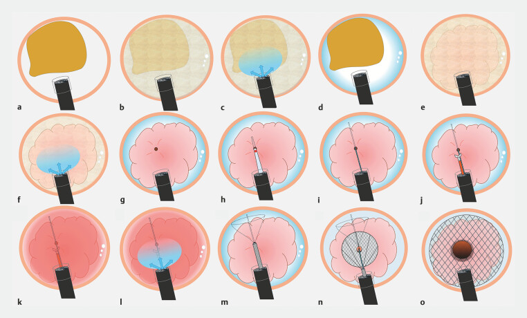
A 55-year-old woman presented with abdominal pain and nausea. She was diagnosed with bowel obstruction to sigmoid colon cancer ( Fig. 1 a ), and a SEMS was inserted as a bridge-to-surgery treatment. First, we removed the gas from the lumen and filled it with water to create underwater conditions ( Fig. 2 a, b ). Because the visual field was poor due to stool and residue, gel was injected (VISCOCLEAR; Otsuka Pharmaceutical Factory, Inc., Tokushima, Japan). The visual field was cleared, and the endoscope reached the tumor ( Fig. 2 c, d ). As the tumor was covered with stool and mucus, it was gently washed with water and gel, and the luminal opening was identified ( Fig. 1 b , Fig. 2 e–g ). Subsequently, the catheter was inserted into the stricture and the proximal colon was confirmed via contrast ( Fig. 2 h ). A wire-guided biopsy was then performed; however, bleeding occurred. The gel injection reduced the momentum of bleeding and improved the endoscopic view ( Fig. 2 i–l ). Finally, the stent was successfully inserted (22 × 120-mm Niti-S Enteral Colonic Uncovered Stent; Taewoong Medical Co., Ltd., Seoul, Korea) ( Fig. 2 m–o ).
Fig. 1.
Computed tomography (CT) and endoscopic image of sigmoid colon cancer. a The CT image shows wall thickening of the sigmoid colon (green circle) and dilation of the proximal colon. b The luminal opening of the tumor (green dotted circle).
Fig. 2.
Schema of the low-pressure insertion of a self-expandable metal stent using water and gel immersion. a View under gas. b Removal of the gas from the lumen and filling it with water. c Injecting the gel. d The endoscopic view clearly changes. e The tumor is covered with stool and mucus. f The tumor is gently washed with water and gel. g The luminal opening is identified. h The catheter is inserted into the stricture. i A guidewire is placed. j Biopsy of the tumor. k Bleeding occurs and negatively impacts the endoscopic view. l Injecting the gel. m A colonic stent is deployed. n Careful deployment of the stent continued. o The stent is inserted successfully.
In conclusion, low-pressure insertion of a SEMS with water and gel immersion may prevent air over-insufflation and ensure a good endoscopic field view. This method may reduce patient discomfort and enable safe stent insertion.
Endoscopy_UCTN_Code_TTT_1AQ_2AF
Acknowledgement
We would like to thank Editage (www.editage.jp) for English language editing.
Footnotes
Conflict of Interest The authors declare that they have no conflict of interest.
Endoscopy E-Videos https://eref.thieme.de/e-videos .
E-Videos is an open access online section of the journal Endoscopy , reporting on interesting cases and new techniques in gastroenterological endoscopy. All papers include a high-quality video and are published with a Creative Commons CC-BY license. Endoscopy E-Videos qualify for HINARI discounts and waivers and eligibility is automatically checked during the submission process. We grant 100% waivers to articles whose corresponding authors are based in Group A countries and 50% waivers to those who are based in Group B countries as classified by Research4Life (see: https://www.research4life.org/access/eligibility/ ). This section has its own submission website at https://mc.manuscriptcentral.com/e-videos .
References
- 1.van Hooft JE, Veld JV, Arnold D et al. Self-expandable metal stents for obstructing colonic and extracolonic cancer: European Society of Gastrointestinal Endoscopy (ESGE) Guideline-Update2020. Endoscopy. 2020;52:389–407. doi: 10.1055/a-1140-3017. [DOI] [PubMed] [Google Scholar]
- 2.Lee YJ, Yoon JY, Park JJ et al. Clinical outcomes and factors related to colonic perforations in patients receiving self-expandable metal stent insertion for malignant colorectal obstruction. Gastrointest Endosc. 2018;87:1548–1557. doi: 10.1016/j.gie.2018.02.006. [DOI] [PubMed] [Google Scholar]
- 3.Sasaki T, Yoshida S, Isayama H et al. Short-term outcomes of colorectal stenting using a low axial force self-expandable metal stent for malignant colorectal obstruction: a Japanese multicenter prospective study. J Clin Med. 2021;10:4936. doi: 10.3390/jcm10214936. [DOI] [PMC free article] [PubMed] [Google Scholar]
- 4.Tomita M, Saito S, Makimoto S et al. Self-expandable metallic stenting as a bridge to surgery for malignant colorectal obstruction: pooled analysis of 426 patients from two prospective multicenter series. Surg Endosc. 2019;33:499–509. doi: 10.1007/s00464-018-6324-8. [DOI] [PMC free article] [PubMed] [Google Scholar]
- 5.Yano T, Takezawa T, Hashimoto K et al. Gel immersion endoscopy: innovation in securing the visual field - Clinical experience with 265 consecutive procedures. Endosc Int Open. 2021;9:E1123–E1127. doi: 10.1055/a-1400-8289. [DOI] [PMC free article] [PubMed] [Google Scholar]


