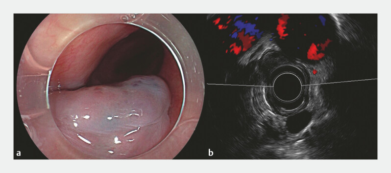
A 44-year-old man presented with symptoms of gastroesophageal reflux disease and dysphagia. Gastroscopy revealed a 3-cm, half-circumferential, bluish-purple esophageal mass located in the mid-esophageal region ( Fig. 1 a ). Computed tomography revealed a soft tissue nodule causing significant stenosis of the esophageal lumen. Endoscopic ultrasound confirmed a well-demarcated, moderately hyperechoic submucosal lesion, characteristic of an esophageal cavernous hemangioma ( Fig. 1 b ). Subsequent to a detailed consultation, endoscopic submucosal tunnel dissection (ESTD) was undertaken ( Video 1 ).
Fig. 1.
Colonoscopy and endoscopic ultrasound. a Gastroscopy revealed a 3-cm, half-circumferential, bluish-purple esophageal mass located in the mid-esophageal region. b Endoscopic ultrasound confirmed a well-demarcated, moderately hyperechoic lesion within the submucosal layer.
Efficacy of endoscopic submucosal tunnel dissection for the management of a large esophageal cavernous hemangioma.
Video 1
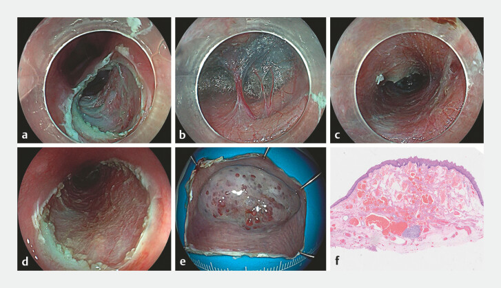
Using a hybrid knife (Erbe Elektromedizin GmbH, Tübingen, Germany), saline mixed with indigo carmine was injected 0.5 cm proximal to the lesion, followed by a 1.5-cm transverse incision to create a submucosal tunnel extending 1 cm distally ( Fig. 2 a ). A significant presence of perforating vessels was observed in the submucosal layer, prompting the use of soft electrocoagulation for meticulous hemostasis ( Fig. 2 b ). An additional 1.5-cm incision was made distally. Incremental dissection along both tunnel margins was performed, achieving complete en bloc resection with a 0.5-cm margin from the tumorʼs edge. Electrocoagulation was applied to exposed vessels to control bleeding, with no damage to the muscular layer ( Fig. 2 c ). The procedure was completed in 30 minutes without complications, including perforation, hemorrhage, or fever. Histopathological analysis confirmed esophageal cavernous hemangioma ( Fig. 2 d ). The patient was discharged on postoperative day four and remained symptom-free during 12 months of follow-up.
Fig. 2.
Endoscopic submucosal tunnel dissection. a A transverse incision was made on the oral side of the lesion to establish the tunnel entry point. b The submucosal layer revealed a notable abundance of perforating vessels. c A submucosal tunnel was meticulously fashioned, extending 1 cm distally from the incision. d Postoperative wound. e The tumor was successfully resected in its entirety. f Histopathological examination confirmed the diagnosis of esophageal cavernous hemangioma.
Esophageal cavernous hemangioma is a rare benign tumor 1 , with management options for asymptomatic cases typically involving observation, whereas symptomatic cases may necessitate intervention. Treatment approaches include esophageal resection, tumor dissection, endoscopic sclerotherapy, and laser therapy 2 . Endoscopic submucosal dissection has been utilized for esophageal hemangiomas 3 4 , and our case illustrates that ESTD enhances submucosal visualization and expedites dissection. This represents the first successful en bloc resection of a symptomatic esophageal cavernous hemangioma via ESTD.
Endoscopy_UCTN_Code_TTT_1AO_2AC
Footnotes
Conflict of Interest The authors declare that they have no conflict of interest.
Endoscopy E-Videos https://eref.thieme.de/e-videos .
E-Videos is an open access online section of the journal Endoscopy , reporting on interesting cases and new techniques in gastroenterological endoscopy. All papers include a high-quality video and are published with a Creative Commons CC-BY license. Endoscopy E-Videos qualify for HINARI discounts and waivers and eligibility is automatically checked during the submission process. We grant 100% waivers to articles whose corresponding authors are based in Group A countries and 50% waivers to those who are based in Group B countries as classified by Research4Life (see: https://www.research4life.org/access/eligibility/ ). This section has its own submission website at https://mc.manuscriptcentral.com/e-videos .
References
- 1.Araki K, Obno S, Egashira A et al. Esophageal hemangiona; a case report and review of the literature. Hepatogastroenterology. 1999;46:3148–3154. [PubMed] [Google Scholar]
- 2.Rodrigues-Pinto E, Pereira P, Macedo G. Bluish discoloration of the esophagus: cavernous hemangioma of the pharynx and larynx with esophageal involvement. Endoscopy. 2015;47:E213–E214. doi: 10.1055/s-0034-1391823. [DOI] [PubMed] [Google Scholar]
- 3.Zhu Z, Wang L, Yin J et al. Endoscopic submucosal dissection for a symptomatic cervical esophageal cavernous hemangioma. Endoscopy. 2022;54:E604–E605. doi: 10.1055/a-1694-3217. [DOI] [PubMed] [Google Scholar]
- 4.Chedgy FJ, Bhattacharyya R, Bhandari P. Endoscopic submucosal dissection for symptomatic esophageal cavernous hemangioma. Gastrointest Endosc. 2015;81:998. doi: 10.1016/j.gie.2014.10.023. [DOI] [PubMed] [Google Scholar]


