Summary
Parsonage-Turner syndrome following monkeypox infection is a rare form of peripheral neuropathy seen in orthopaedic practice and described only once in the literature. We present the case of a man in his 30s with severe shoulder pain and subsequent amyotrophy 2 weeks after monkeypox infection. Our report encompasses the initial findings, radiographic examinations and follow-up over a 6-month period. To confirm the diagnosis, MRI and electrostimulation conduction studies were conducted, highlighting their importance as valuable diagnostic tools in conjunction with a thorough physical examination. Supportive treatment, including physical therapy and pain management, forms the cornerstone of management, while surgical intervention is reserved for refractory cases or when mechanical complications arise. Prognosis varies among individuals. This case report expands the understanding of neurological complications of monkeypox infection. Clinicians should include Parsonage-Turner syndrome in their differential diagnosis for patients presenting with symptoms of peripheral brachial plexus neuropathy following viral infections, including monkeypox.
Keywords: Peripheral nerve disease, Stiff shoulder, Orthopaedic and trauma surgery
Background
Monkeypox is a rare zoonotic disease caused by the monkeypox virus (MPXV), a member of the Orthopoxvirus genus.1 MPXV typically presents with a febrile rash illness, resembling a milder form of smallpox.2 The recent outbreak of May 2022 led to a global public health emergency in non-endemic countries such as Europe and the USA. In Switzerland, reported cases prior to the outbreak are predominantly associated with travel to West and Central African countries. During the recent health crisis, a total of 551 cases were reported in Switzerland.3 4 Neurological complications following MPXV infection are rare, particularly involving the peripheral nervous system.5 6
Parsonage-Turner syndrome (PTS), also known as neuralgic amyotrophy, is a rare peripheral neuropathy with acute-onset severe shoulder pain, followed by weakness and atrophy of the shoulder girdle and upper arm muscles, occasionally seen in orthopaedic practice.7 8 While the exact aetiology and pathophysiology of PTS is not fully understood, it has been associated with various triggers, including viral infection in 20%–52% and vaccination in 15% of cases.9 10 Notably, several case reports have documented PTS following SARS-CoV-2 vaccination and infection.11,13 The triggers along with other genetic, mechanical and autoimmune factors have been described to play a role in the pathophysiology of PTS.7
Diagnosing PTS typically relies on clinical and radiologic findings, although its presentation can be heterogeneous.7 Radiologic signs may present in a progressive sequence, beginning with muscle oedema, followed by atrophy, fatty infiltration and denervation changes.14 The later visualised as increased T2 signal intensity on MRI.14 Nevertheless, these imaging findings may be non-specific and may not always manifest in the early stages of the disease.15
Here, we report the case of a man in his 30s who developed PTS following MPXV infection. Through a review of English literature, we found only one previous case report on PTS following MPXV infection.6 Our clinical and radiological findings are exposed.
Case presentation
Our case involves a left-handed man in his 30s with a medical history of hepatitis C infection, epilepsy resulting from benzodiazepine withdrawal and occasional drug use.
Two weeks after the MPXV exposure and infection, the patient initially presented with debilitating left shoulder pain, loss of force, inability to flex and abduct the arm beyond 90° and a sensation of left upper extremity paralysis. The primary MPXV infection was managed expectantly, with opiate therapy for painful skin lesions.
The patient was referred to our outpatient orthopaedic clinic at the university hospital by his primary care physician (PCP) due to persistent non-traumatic left shoulder pain and dysfunction persisting for several months after MPXV infection. The PCP had already arranged a shoulder MRI.
At the time of consultation (5 months postinfection), the patient had discontinued opioid use, and his left shoulder pain had significantly subsided. However, weakness persisted, limiting his daily activities and professional capabilities.
On clinical examination, we observed amyotrophy of the deltoid, triceps and biceps muscles on the left shoulder and arm. No scapular winging was present, and active range of motion was preserved when compared with the contralateral side. The clinical integrity of the rotator cuff was confirmed. A neurological examination of the left upper limb revealed estimated muscle force at M4+in the deltoid muscle, the rotator cuff, pectoralis major, biceps and triceps. All of which were painless on testing. Reflexes were hypoactive and symmetrical, and sensibility was intact. The patient’s subjective shoulder value (SSV) was 65%, and the Constant Score (CS) was 64 points at 5 months postinfection.16
Investigations
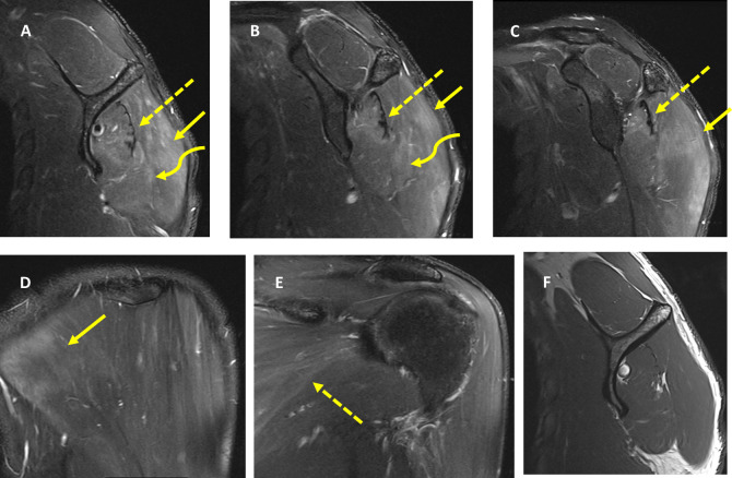
The initial left shoulder MRI (non-contrast enhanced) revealed oedema in the posterior belly of the deltoid muscle and posterior muscles of the rotator cuff (figure 1). Laboratory tests were not conducted. Given our clinical suspicion of PTS, the patient was referred to our hospital’s neurologists. The subsequent workup included an MRI of the brachial plexus and upper left limb electroneuromyography (ENMG) and electromyography (EMG). Gadolinium contrast-enhanced magnetic resonance neurography (MRN) of the left brachial plexus did not indicate any anomalies. The ENMG assessment unveiled a reduction in the amplitude of the left ulnar sensory potential and a latency in F waves which was at the upper limit of the norm. The EMG demonstrated resting activity irregularities within the proximal muscles of the left upper limb, substantiating the presence of a neurogenic sequela. Notably, resting activity exhibited aberrations in the deltoid, biceps brachii and triceps, accompanied by discernible fasciculations and fibrillations. Motor unit recruitment demonstrated normalcy.
Figure 1. MRI at the left shoulder level including the periscapular muscles. (A–C) Sagittal T2-weighted STIR showing denervation oedema of the posterior belly of the deltoid muscle (plain arrow), infraspinatus muscle (dashed arrow) and teres minor muscle (curved arrow). (D) Coronal T2-weighted STIR showing denervation oedema of the posterior belly of the deltoid muscle (plain arrow). (E) Coronal T2-weighted STIR showing denervation oedema of the infraspinatus muscle (dashed arrow). (F) Sagittal T1-weighted showing no muscle atrophy nor fatty infiltration. Moreover, there is no mass or neoplasia along the axillary or suprascapular nerve.
Differential diagnosis
The literature extensively describes the differential diagnosis for PTS, which should be considered by orthopaedic surgeons when encountering non-traumatic shoulder pain (box 1).
Box 1. Differential diagnosis of PTS (adapted from Sathasivam et al26, Traverso et al31, Mareddu et al32).
Differential diagnosis for PTS
Musculoskeletal disorders
Degenerative rotator cuff disease.
Calcific tendinitis.
Pathologies of the biceps tendon.
Cervical spine degenerative and traumatic pathologies.
Subacromial bursitis and impingement.
Adhesive capsulitis.
Shoulder injury related to vaccine administration.
Neurological disorders
Thoracic outlet syndrome.
Cervical radiculopathy/nerve entrapment.
Motor neuron disease.
Herpes zoster.
Mononeuritis multiplex.
-
Brachial plexopathy (trauma, traction, infiltration).
PTS, Parsonage-Turner syndrome.
A thorough history and clinical examination can guide physicians in the diagnostic workup. The majority of musculoskeletal disorders can be largely excluded based on the abrupt onset of pain in PTS.7 Conditions such as cervical radiculopathy, thoracic outlet syndrome and mononeuritis multiplex present with distal sensory abnormalities and less severe pain.17 18 Nerve conduction studies play a crucial role in differentiating between various neurological disorders that may present with similar manifestations, particularly when performed after 3 weeks of symptoms as early testing may be inconclusive.19
Treatment
Based on the clinical and radiologic findings, we confirmed the diagnosis of PTS following MPXV infection. The patient was referred for physical therapy to maintain range of motion and strengthen the affected muscles. Residual pain was managed with over-the-counter analgesics when needed.
Outcome and follow-up
Over several weeks, the patient reported gradual improvement in symptoms, with a near-complete resolution of pain and improved strength and function of the left shoulder girdle muscles. During a follow-up visit 3 months after the initial consultation (8 months postinfection), the patient demonstrated symmetrical force and function, was asymptomatic and could return to work. The SSV increased to 90%, and the CS reached 85 points.
Discussion
Diagnosing PTS in orthopaedic practice, particularly following MPXV infection or other viral infections, can be challenging. To the best of our knowledge, only one other case of PTS has been described related to MPXV infection/vaccination by Nimura et al.6 They published the case of a man in his 40s who experienced severe bilateral shoulder pain 5 days after the initial symptoms following MPXV exposure and vaccination in the USA. Nimura et al6 confirmed PTS in the left shoulder using MRN and ENMG. The MPXV infection was managed with antiviral medication, while PTS was treated with pain management. Notably, our MPXV treatment protocols differ from those in the USA, as we primarily focus on symptomatic management, reserving antiretroviral treatment for severe cases needing hospital admission.3 Unlike Nimura et al,6 in our case, we can reasonably infer that PTS was directly triggered by MPXV infection. In their case, the temporal relationship between infection and vaccination precluded identifying the specific triggering event.6
The exact mechanisms by which viral infections can lead to PTS remain incompletely understood and are believed to be multifactorial, involving environmental, individual, genetic and mechanical factors.20 It has been proposed that viral infections can trigger an immune-mediated response, resulting in inflammation and damage to the brachial plexus.21 In the case of MPXV, it is possible that the virus may directly infect the nerve tissue, initiating neural damage and subsequent development of PTS.
The radiologic findings in our case align with previously described cases of PTS involving the shoulder girdle.14 A retrospective study of 26 PTS patients found that MRI detected abnormalities in all cases, with denervation oedema being the most frequently observed finding, typically located in the supra and infraspinatus muscles.22 While MRI is valuable for excluding alternative diagnoses and complements ENMG, the diagnosis of PTS should not rely solely on radiographic analysis.23 24 Interestingly, a new potentially pathognomonic sign seen early on MRN being described in the literature involves intrinsic, hourglass-like constrictions within the affected nerves.25 However, this was not visualised in our case.
Management options for PTS primarily involve supportive measures, such as physical therapy, pain management and anti-inflammatory medications.20 26 Unless contraindicated, some authors advocate for a 2-week tapered treatment of oral corticosteroids during the acute phase.21 However, no study has definitively demonstrated superior outcomes with corticosteroid use.19 Surgical intervention is indicated in severe cases refractory to conservative treatment (>3 months) and for mechanical complications resulting from muscle atrophy weakness, such as chronic subluxation or dislocation.7 27 Various surgical techniques, including nerve decompression, nerve transfers and tendon transfers, have been described, with varying reported success rates.28 29 The prognosis in PTS varies and largely depends on the extent of neural damage and the severity of symptoms.19 While most patients achieve complete recovery (80%–90%), some may experience chronic pain and disability.8 30
In conclusion, this case report presents a unique manifestation of PTS following MPXV infection in orthopaedic practice. The mechanisms by which viral infections can lead to PTS remain incompletely understood, necessitating further research in this field. Clinicians should be mindful of the potential for PTS following viral infections, including MPXV, and ought to consider this diagnosis in patients presenting with symptoms indicative of peripheral brachial plexus neuropathy.
Learning points.
Orthopaedic surgeons should be aware of the rare presentation of Parsonage-Turner syndrome (PTS) following Monkeypox infection, as it may mimic other shoulder pathologies.
To diagnose PTS in such cases, a comprehensive evaluation combining clinical assessment, radiology and electrostimulation studies is crucial for accurate identification.
The cornerstone of managing PTS in orthopaedics is a supportive approach, including physical therapy and pain management. Surgery might be considered in refractory or complicated cases.
Footnotes
Funding: The authors have not declared a specific grant for this research from any funding agency in the public, commercial or not-for-profit sectors.
Case reports provide a valuable learning resource for the scientific community and can indicate areas of interest for future research. They should not be used in isolation to guide treatment choices or public health policy.
Provenance and peer review: Not commissioned; externally peer reviewed.
Patient consent for publication: Consent obtained directly from patient(s).
Contributor Information
Filippo Gerber, Email: gerber.filippo@gmail.com.
Salim Zenkhri, Email: Salim.Zenkhri@chuv.ch.
Alain Farron, Email: alain.farron@chuv.ch.
Aurélien Traverso, Email: auremt@gmail.com.
References
- 1.Chen N, Li G, Liszewski MK, et al. Virulence differences between monkeypox virus isolates from West Africa and the Congo basin. Virology (Auckl) 2005;340:46–63. doi: 10.1016/j.virol.2005.05.030. [DOI] [PMC free article] [PubMed] [Google Scholar]
- 2.Ali E, Sheikh A, Owais R, et al. Comprehensive overview of human monkeypox: epidemiology, clinical features, pathogenesis, diagnosis and prevention. Ann Med Surg (Lond) 2023;85:2767–73. doi: 10.1097/MS9.0000000000000763. [DOI] [PMC free article] [PubMed] [Google Scholar]
- 3.Marchal O, Sottas C, Pastor D, et al. Mpox (variole du singe): l’œil du dermatologue. Rev Méd Suisse. 2023;19:614–7. doi: 10.53738/REVMED.2023.19.820.614. [DOI] [PubMed] [Google Scholar]
- 4.Mpox (variole du singe) - république et canton de neuchâtel. 2023. https://www.ne.ch/autorites/DFS/SCSP/medecin-cantonal/maladies-vaccinations/Pages/VarioleSinge.aspx Available.
- 5.Money KM, Barnett TA, Rapaka S, et al. Monkeypox-Associated Central Nervous System Disease: A Case Series and Review. Ann Neurol. 2023;93:893–905. doi: 10.1002/ana.26597. [DOI] [PubMed] [Google Scholar]
- 6.Nimura CA, Milani C, Tan ET, et al. Parsonage-turner syndrome following monkeypox infection and vaccination. 2023. https://link.springer.com/10.1007/s00256-023-04298-4 Available. [DOI] [PMC free article] [PubMed]
- 7.Smith CC, Bevelaqua AC. Challenging Pain Syndromes. Phys Med Rehabil Clin N Am. 2014;25:265–77. doi: 10.1016/j.pmr.2014.01.001. [DOI] [PubMed] [Google Scholar]
- 8.van Alfen N, van Engelen BGM. The clinical spectrum of neuralgic amyotrophy in 246 cases. Brain (Bacau) 2006;129:438–50. doi: 10.1093/brain/awh722. [DOI] [PubMed] [Google Scholar]
- 9.Parsonage MJ, Aldren Turner JW. NEURALGIC AMYOTROPHY THE SHOULDER-GIRDLE SYNDROME. The Lancet. 1948;251:973–8. doi: 10.1016/S0140-6736(48)90611-4. [DOI] [PubMed] [Google Scholar]
- 10.Tsairis P. Natural History of Brachial Plexus Neuropathy. Arch Neurol. 1972;27:109. doi: 10.1001/archneur.1972.00490140013004. [DOI] [PubMed] [Google Scholar]
- 11.Queler SC, Towbin AJ, Milani C, et al. Parsonage-Turner Syndrome Following COVID-19 Vaccination: MR Neurography. Radiology. 2022;302:84–7. doi: 10.1148/radiol.2021211374. [DOI] [PMC free article] [PubMed] [Google Scholar]
- 12.Cornea A, Lata I, Simu M, et al. Parsonage-Turner Syndrome Following SARS-CoV-2 Infection: A Systematic Review. Biomedicines. 2023;11:837. doi: 10.3390/biomedicines11030837. [DOI] [PMC free article] [PubMed] [Google Scholar]
- 13.Wright JO, Wiggins W, Smith MS, et al. Shoulder Pain and Dysfunction After Vaccination: A Systematic Review. JBJS Rev. 2023;11 doi: 10.2106/JBJS.RVW.22.00134. [DOI] [PubMed] [Google Scholar]
- 14.Gaskin CM, Helms CA. Parsonage-Turner syndrome: MR imaging findings and clinical information of 27 patients. Radiology. 2006;240:501–7. doi: 10.1148/radiol.2402050405. [DOI] [PubMed] [Google Scholar]
- 15.Kumar I, Verma A, Srivastava A, et al. Utility of magnetic resonance imaging in the diagnosis of unsuspected cases of Parsonage-Turner syndrome: two case reports. J Med Case Reports . 2013;7 doi: 10.1186/1752-1947-7-255. [DOI] [PMC free article] [PubMed] [Google Scholar]
- 16.Gilbart MK, Gerber C. Comparison of the subjective shoulder value and the Constant score. J Shoulder Elbow Surg. 2007;16:717–21. doi: 10.1016/j.jse.2007.02.123. [DOI] [PubMed] [Google Scholar]
- 17.Schreiber AL, Abramov R, Fried GW, et al. Expanding the differential of shoulder pain: Parsonage-Turner syndrome. J Am Osteopath Assoc. 2009;109:415–22. [PubMed] [Google Scholar]
- 18.Mamula CJ, Erhard RE, Piva SR. Cervical Radiculopathy or Parsonage-Turner Syndrome: Differential Diagnosis of a Patient With Neck and Upper Extremity Symptoms. J Orthop Sports Phys Ther . 2005;35:659–64. doi: 10.2519/jospt.2005.35.10.659. [DOI] [PubMed] [Google Scholar]
- 19.Rubin DI. Neuralgic amyotrophy: clinical features and diagnostic evaluation. Neurol. 2001;7:350–6. doi: 10.1097/00127893-200111000-00004. [DOI] [PubMed] [Google Scholar]
- 20.Van Eijk JJJ, Groothuis JT, Van Alfen N. Neuralgic amyotrophy: An update on diagnosis, pathophysiology, and treatment. Muscle Nerve. 2016;53:337–50. doi: 10.1002/mus.25008. [DOI] [PubMed] [Google Scholar]
- 21.van Alfen N. Clinical and pathophysiological concepts of neuralgic amyotrophy. Nat Rev Neurol. 2011;7:315–22. doi: 10.1038/nrneurol.2011.62. [DOI] [PubMed] [Google Scholar]
- 22.Scalf RE, Wenger DE, Frick MA, et al. MRI Findings of 26 Patients with Parsonage-Turner Syndrome. Am J Roentgenol. 2007;189:W39–44. doi: 10.2214/AJR.06.1136. [DOI] [PubMed] [Google Scholar]
- 23.Reza Soltani Z, Sajadi S, Tavana B. A comparison of magnetic resonance imaging with electrodiagnostic findings in the evaluation of clinical radiculopathy: a cross-sectional study. Eur Spine J. 2014;23:916–21. doi: 10.1007/s00586-013-3164-z. [DOI] [PMC free article] [PubMed] [Google Scholar]
- 24.Sneag DB, Rancy SK, Wolfe SW, et al. Brachial plexitis or neuritis? MRI features of lesion distribution in Parsonage-Turner syndrome. Muscle Nerve. 2018;58:359–66. doi: 10.1002/mus.26108. [DOI] [PubMed] [Google Scholar]
- 25.Ripellino P, Arányi Z, van Alfen N, et al. Imaging of neuralgic amyotrophy in the acute phase. Muscle Nerve. 2022;66:709–14. doi: 10.1002/mus.27732. [DOI] [PubMed] [Google Scholar]
- 26.Sathasivam S, Lecky B, Manohar R, et al. Neuralgic amyotrophy. J Bone Joint Surg Br. 2008;90-B:550–3. doi: 10.1302/0301-620X.90B5.20411. [DOI] [PubMed] [Google Scholar]
- 27.Gstoettner C, Mayer JA, Rassam S, et al. Neuralgic amyotrophy: a paradigm shift in diagnosis and treatment. J Neurol Neurosurg Psychiatry . 2020;91:879–88. doi: 10.1136/jnnp-2020-323164. [DOI] [PubMed] [Google Scholar]
- 28.Wheelwright JC, Featherall J, Kazmers NH, et al. Revision Reverse Total Shoulder Arthroplasty With Pectoralis Major Transfer for Anterior Deltoid Deficiency: A Case Report. JBJS Case Connect. 2022;12 doi: 10.2106/JBJS.CC.22.00034. [DOI] [PubMed] [Google Scholar]
- 29.Winter J, Karir A, Clark TA, et al. Surgical Treatment of Parsonage Turner Syndrome With Primary Nerve Transfers: A Case Series and Cadaver Dissection. Ann Plast Surg. 2022;89:301–5. doi: 10.1097/SAP.0000000000003265. [DOI] [PubMed] [Google Scholar]
- 30.Tsairis P, Dyck PJ, Mulder DW. Natural history of brachial plexus neuropathy. Report on 99 patients. Arch Neurol. 1972;27:109–17. doi: 10.1001/archneur.1972.00490140013004. [DOI] [PubMed] [Google Scholar]
- 31.Traverso A, Piasecki K, Gallusser N, et al. Agenesis of the long head of the biceps brachii tendon: ignored variations of the anatomy and the next tendon to disappear? BMJ Case Rep. 2020;13:e234962. doi: 10.1136/bcr-2020-234962. [DOI] [PMC free article] [PubMed] [Google Scholar]
- 32.Mareddu E, Traverso A, Laudato P, et al. Bilateral isolated C5 paralysis of the shoulder: Atypical presentation of a transdiscal C4-C5 cervical spine fracture. BMJ Case Rep. 2021;14:e236323. doi: 10.1136/bcr-2020-236323. [DOI] [PMC free article] [PubMed] [Google Scholar]


