



Abstract
Background
Respiratory distress syndrome (RDS) and transient tachypnoea (TTN) are the two commonest neonatal respiratory disorders. The optimal continuous positive airway pressure (CPAP) to treat them is unknown. We aim to clarify the effect of different CPAP levels on lung aeration and gas exchange in patients with RDS and TTN.
Methods
Prospective, observational, pragmatic, physiological cohort study. CPAP was sequentially increased from 4 to 6 and 8 cmH2O and backwards, with interposed wash-out periods. Lung aeration was assessed with a validated neonatal lung ultrasound score. Gas exchange was non-invasively evaluated with transcutaneous monitoring. Ultrasound score and PtcO2/FiO2 ratio were the co-primary outcomes. PtcCO2 and other oxygenation metrics were the secondary outcomes.
Results
30 neonates with RDS and 30 with TTN were studied. Each CPAP increment significantly (overall always p < 0.001) improved both lung aeration and oxygenation, but the increase from 6 to 8 cmH2O achieved a small absolute benefit. In RDS patients, the absolute improvements were small and the diagnosis of TTN was significantly associated with greater improvement of lung aeration (β= -1.4 (95%CI: -2.4; -0.3), p = 0.01) and oxygenation (β = 39.6 (95%CI: 4.1; 75.1), p = 0.029). Aeration improved in 16 (53.3%) and 27 (90%) patients in the RDS and TTN groups, respectively (p = 0.034). Lung aeration showed significant hysteresis in TTN patients. Secondary outcomes gave similar results.
Conclusions
Increasing CPAP from 4 to 8 cmH2O improves ultrasound-assessed lung aeration and oxygenation in RDS and TTN. The absolute improvements are small when CPAP is beyond 6 cmH2O or for RDS patients.
Supplementary Information
The online version contains supplementary material available at 10.1186/s12931-024-03010-x.
Keywords: Respiratory distress syndrome, Transient tachypnoea of the neonate, Surfactant, Infant
Background
Respiratory distress syndrome (RDS) and transient tachypnoea of the neonate (TTN) are the two most common respiratory disorders needing neonatal intensive care unit (NICU) admission [1]. The cornerstone of their current clinical management consists in early continuous positive airway pressure (CPAP), which is increasingly being used [2]. CPAP recruits the alveoli and improves compliance, eventually increasing the functional residual capacity [3]. Furthermore, CPAP promotes endogenous surfactant production in RDS patients, while it overcomes airway resistance and facilitates alveolar fluid reabsorption in neonates with TTN [4, 5]. These mechanisms eventually translate into reduced work of breathing and improved gas exchange.
In neonates, CPAP is usually provided between 4 and 8 cmH2O, although higher pressure levels have also been safely applied [6–8]. Patients may have variable gestational age, weight or clinical severity, and the optimal pressure is still unknown, hence the choice of CPAP level is only based on local practice and CPAP failure may variably occur.
To overcome this lack of knowledge the assessment of the lung aeration induced by CPAP may be useful. In critically ill adults this may be realized with CT-scan, which is unsuitable for neonates [9]. Conversely, point-of-care lung ultrasound is already available at the bedside and is spreading given its favourable features [10]. Alveolar recruitment guided by quantitative lung ultrasound through the use of dedicated scores is not new in adult critical care [11], and recent data showed that the technique can detect inhomogeneity and changes of lung aeration in ventilated newborn lambs [12, 13]. Thus, quantitative lung ultrasound offers the potential to evaluate the changes of lung aeration due to respiratory support in newborn infants too. Nonetheless, to the best of our knowledge, no study has assessed the effect of different CPAP levels on ultrasound-assessed lung aeration and gas exchange during routine neonatal care. We hypothesize that CPAP increment ameliorates lung aeration and gas exchange in a different degree depending on the pressure level and diagnosis (i.e. RDS or TTN). This hypothesis will be evaluated on lung aeration and a common oxygenation metric as co-primary outcomes, and on secondary oxygenation metrics and carbon dioxide as secondary outcomes.
Methods
Study design and setting
This was a prospective, observational, physiological cohort study conducted in a referral academic NICU from April to October 2022. The study was pragmatic as it used only data routinely recorded during clinical care, which was not changed exclusively for study purposes. Lung ultrasound is the first-line imaging technique for all infants with respiratory failure admitted to our NICU [14]. The study was conducted in accordance with the principle of the Helsinki Declaration and was granted local ethical approval (SLRF/n.22/011). Parental informed consent was obtained upon NICU admission. Data were recorded anonymously, and relevant privacy regulations were respected. STROBE guidelines were followed for the manuscript preparation.
Patients
Inborn neonates admitted to the NICU with signs of respiratory failure needing CPAP were screened daily by one investigator (VS) as this was the prerequisite for the recruitment. They were enrolled if RDS or TTN was diagnosed according to pre-specified integrated consensus criteria [15], based on perinatal history, demographics, laboratory findings, clinical course, and imaging data (supplementary Table 1). None of these criteria was predominant on the others and all had to be fulfilled in order to assign the diagnosis. Imaging consisted of qualitative lung ultrasound findings interpreted according to international guidelines [16], based on classical lung ultrasound semiology [10]. The qualitative lung ultrasound did not comprise any score computation. In detail, ultrasound appearance was considered typical for RDS when it consisted of a homogeneously poorly aerated (“white”) lung with small (< 1 cm) subpleural consolidations and absence of “A-lines” [1]. Conversely, it was considered typical for TTN when an interstitial pattern consisting of well-spaced “B-lines” (without consolidations) was alternated with areas of normal lung [1]. Lung ultrasound was always performed by expert neonatologists proficient in lung ultrasonography (i.e. with at least one year of daily lung ultrasound expertise). Exclusion criteria were major congenital malformations or chromosomopathies, airleaks preventing a comprehensive lung ultrasound examination, early onset sepsis [17], and hemodynamic instability (defined as need for any inotrope). As the clinical course was also considered for the diagnosis, all enrolled cases were independently reviewed by a senior neonatologist at 72 h of life to confirm (or disprove) the initial diagnosis. Upon this review, neonates diagnosed with congenital surfactant anomalies, pulmonary hypoplasia or malformations, persistent pulmonary hypertension or any other neonatal respiratory disorder (i.e. meconium aspiration [18], congenital infectious or biliary pneumonia [19, 20], pulmonary haemorrhage or maternal blood aspiration [21], and NARDS of any type [15]) were also excluded.
Respiratory care
In our clinical routine all neonates received variable flow, nasal mask-delivered CPAP since the delivery room which is adjacent to the NICU, as previously described [1]. CPAP was shortly titrated upon NICU admission, as described below, to assess the severity of the respiratory failure. Appropriately sized nasal masks were used following the manufacturers’ recommendations. Supplemental oxygen was added if 6 cmH2O was not enough to have pre-ductal oxygen saturation (SpO2) between 90 and 95% [22]. Infants remained always supine in a flexed neutral position, with soft nesting rolls surrounding them. The rest of respiratory care was provided according to our NICU protocols as previously published [19], and was essentially based on current evidence and international guidelines. These protocols included surfactant replacement performed, if any, as previously described [23, 24], and always after the measurements described below. Clinical protocols were not changed during the study.
Measurements and data collection
The following measurements were performed upon NICU admission and always before surfactant replacement, if any. CPAP was started at 4 and sequentially increased to 6 and 8 cmH2O. For every CPAP increment, 5’ were elapsed as wash-out adaptation time. This period was used to check pressure transmission and reduce leaks, if needed, repositioning the neonate or gently closing the mouth [25]. Then, CPAP remained unchanged for another 5’ per each pressure level and data were recorded at the end of this period. This timing was consistent with previous preliminary experiences of lung ultrasound-guided recruitment in neonates [26], and is similar to the regional respiratory time constant observed during lung recruitement [27]. Collected data consisted of SpO2, inspired oxygen fraction (FiO2), transcutaneous partial pressure of oxygen (PtcO2) and carbon dioxide (PtcCO2), as well as quantitative lung ultrasound data. The study workflow is depicted in Supplementary Fig. 1. Pre-ductal SpO2 was recorded with an artifact filtering monitor (Nihon Kohden, Shinjuku, Japan) when the pulse wave was regular and smooth [28]. PtcO2 and PtcCO2 were measured with transcutaneous devices (TCM5®, Radiometer, Copenhagen, Denmark), calibrated following the manufacturer’s advice and the American Association for Respiratory Care guidelines [29]. These devices are routinely used upon NICU admission (for a maximum of 15’) and then removed.
Lung aeration was assessed by quantitative lung ultrasound, which has been validated against several techniques [11]. In detail, quantitative ultrasound was performed by calculating the ‘extended lung ultrasound score’ (eLUS), which has been specifically validated for neonates [30]. eLUS includes the evaluation of the dependent lung areas obtained by slightly tilting the infant and slipping the probe between the chest and the mattress [30]. eLUS is calculated on 10 chest areas (5 per side [upper and lower anterior, lateral, upper and lower posterior]) assigning to each area a 0 to 3 value, based on classical ultrasound signs (i.e. 0 for normally aerated (A-pattern), 1 for interstitial-alveolar (B-pattern), 2 for severe interstitial-alveolar (severe B-pattern) and 3 for consolidated (C-pattern) areas) [31]. The total score ranges from 0 (best) to 30 (worst aeration) [30]. Machine settings and technical details were as previously published [31]. When the CPAP increment visibly improved lung aeration, the clinician could decrease CPAP and study lung aeration during both lung inflation and deflation. This was done following the same stepwise procedure as described above, that is lowering CPAP from 8 to 6, and then to 4 cmH2O, as is classically done for alveolar recruitment in intubated neonates [32]. The deflation study was not systematically performed in all patients because it is included in our clinical routine protocol only when the burden of care is mild and the CPAP increment improved lung aeration. eLUS was calculated by expert neonatologists proficient in lung ultrasonography (i.e. with at least one year of daily lung ultrasound expertise). Demographics and basic clinical data were recorded in real time from electronic patient files.
Calculations and outcomes
Lung aeration and oxygenation, respectively expressed as eLUS and PtcO2/FiO2 ratio, were considered as physiological co-primary outcomes. They were chosen to comprehensively describe the effect of pressure both in terms of lung imaging and function. Derived oxygenation metrics (Alveolar-arterial (A-a) gradient, arterial/Alveolar (a/A) ratio and SpO2/FiO2 ratio) were calculated as previously described [33], and considered as physiological secondary outcomes together with PtcCO2.
Statistics
Since this was the first study about the effect of CPAP titration on ultrasound-assessed lung aeration, a formal sample size calculation was unfeasible, and we chose a convenience sample size of 30 patients for each separated cohort of patients affected by each condition of interest (i.e. RDS and TTN). This seemed reasonable, as the capability of quantitative lung ultrasound to detect aeration changes was demonstrated on a similar number of animals [12, 13], and studies on the safety of higher CPAP levels have been performed on smaller sample sizes [6–8]. Data were expressed as number (%), mean (standard deviation (s.d.)) or median [25th – 75th percentile], as appropriate.
Co-primary and secondary outcomes were analysed with repeated measures-ANOVA (RM-ANOVA), followed, when appropriate, by the Bonferroni post hoc test (within-patients analysis) and the Student t-test to compare RDS and TTN groups (between-patient analysis) for each CPAP level. Moreover, we calculated the relative change (Δ) of co-primary outcomes due to each CPAP increment (e.g. ΔPtcO2/FiO2 between 4 and 6 cmH2O = (PtcO2/FiO2 at 6 cmH2O) – (PtcO2/FiO2 at 4 cmH2O) / (PtcO2/FiO2 at 4 cmH2O)) as a percentage and compared them between RDS and TTN patients using the Student t-test.
Subsequently, the improvement in co-primary outcomes obtained by increasing CPAP from 4 to 8 cmH2O (e.g. ΔPtcO2/FiO2 (or eLUS) between 4 and 8 cmH2O = (PtcO2/FiO2 (or eLUS) at 8 cmH2O) – (PtcO2/FiO2 (or eLUS) at 4 cmH2O) / (PtcO2/FiO2 (or eLUS) at 4 cmH2O)) was investigated in multivariate linear regressions. In these models, the relative change (Δ) in primary outcomes (i.e. the difference between eLUS or PtcO2/FiO2 values measured at 8 and 4 cmH2O), was the dependent variable. Gestational age, type of delivery (caesarean or vaginal) and respiratory diagnosis (RDS or TTN) were the independent variables, as they are well known to influence lung aeration and oxygenation [1, 34–36]. No covariate was excluded, multicollinearity was detected as previously described [37], and results were expressed as β-coefficient (95% confidence interval (CI)).
The distribution of lung aeration was analysed based on the classical lung ultrasound patterns observed in the ten scanned chest areas. The distribution was expressed as a percentage of chest areas presenting with each pattern, for each CPAP level and compared using the McNemar-Bowker test for pairwise multiclass nominal data, with Bonferroni correction. Finally, in the subset of patients where lung inflation and deflation were performed, eLUS was compared for each CPAP level with the paired Student t-test. Analyses were performed with R4.2.2 (https://www.r-project.org) and p < 0.05 was considered significant.
Results
We enrolled 30 neonates with RDS and TTN, respectively; the diagnosis was confirmed for each of them (Supplementary Fig. 2). Basic population details are summarized in Table 1. No major clinical problem occurred, and all patients were successfully discharged from the NICU. No patient had consolidation of more than 1 cm at the ultrasound.
Table 1.
Basic characteristics of the two studied cohorts. Data are expressed as number (%), mean (standard deviation) or median [25th – 75th percentile], as appropriate. Prenatal steroids are considered as at least one 12 mg-betamethasone dose given at least 24 h before birth. CRIB-II and Apgar scores are dimensionless variables. Abbreviations: CRIB-II: critical risk index for babies-II; FiO2: inspired oxygen fraction; RDS: respiratory distress syndrome; TTN: transient tachypnoea of the neonate
| RDS (n = 30) |
TTN (n = 30) |
|
|---|---|---|
| Gestational age at birth (weeks) | 30.3 (2.9) | 35.6 (3.2) |
| Birth weight (g) | 1372 (608) | 2480 (779) |
| Male sex | 17 (56.7%) | 17 (56.7%) |
| Prenatal steroids | 22 (73.3%) | 10 (33.3%) |
| Caesarean section | 20 (66.7%) | 15 (50%) |
| CRIB-II score | 8 [6–10] | 3 [2–6] |
| 5’ Apgar score | 8 [6–10] | 9 [7–10] |
| Postnatal age at enrolment (hours) | 1.9 (1.7) | 3.7 (3.1) |
| FiO2 at enrolment | 0.28 (0.1) | 0.22 (0.03) |
In the whole population (i.e. RDS and TTN patients together), the mean eLUS was 15.6 (s.d. 3.7) at 4 cmH2O, and decreased by -0.6 (s.d. 0.9) points at 6 cmH2O, and then by -1.2 (s.d. 1.3) points going from 6 to 8 cmH2O. The mean PtcO2/FiO2 ratio at 4 cmH2O was 276.1 (s.d. 96.7) and increased by 27.1 (s.d. 49.2) points at 6 cmH2O, and then by 10.5 (s.d. 31.2) points going from 6 to 8 cmH2O. The analysis of co-primary outcomes shows that: (1) each CPAP increment significantly (overall p always < 0.001, within-patient and between-patient post-hoc comparisons are shown in Fig. 1 and Supplementary Table 2, respectively) improved lung aeration (i.e. decreased eLUS) and oxygenation (i.e. increased PtcO2/FiO2); the increase from 6 to 8 cmH2O achieved a relatively small oxygenation improvement. (2) CPAP increment improved lung aeration significantly more in TTN than in RDS patients, while the effect on oxygenation was generally similar between the two groups (data are visually depicted in Fig. 1 and detailed as Δ-values in Table 2; raw values are in Supplementary Table 2). (3) In RDS patients, the absolute values of lung aeration and oxygenation improvement were small (Fig. 1; Table 2). Multivariate analysis confirmed that the diagnosis of TTN was associated with the improvement of lung aeration (β= -1.4 (95%CI: -2.4; -0.3), p = 0.01) and oxygenation (β = 39.6 (95%CI: 4.1; 75.1), p = 0.029), while other covariates were non-significant.
Fig. 1.

Co-primary outcomes: absolute values of lung aeration (panel A) and oxygenation (panel B). Open circles and T-bars represent means and standard deviations, respectively. p-values in the right upper corner refer to the overall results of RM-ANOVA; dotted horizontal lines indicate pairwise within-patient post-hoc comparisons. Results for the whole population (ALL) and for the RDS and TTN cohorts are shown. eLUS and PtcO2/FiO2 are dimensionless variables. Abbreviations: CPAP: continuous positive airway pressure; eLUS: extended lung ultrasound score; FiO2: inspired oxygen fraction; PtcO2: transcutaneous partial pressure of oxygen; RDS: respiratory distress syndrome; TTN: transient tachypnoea of the neonate
Table 2.
Co-primary outcomes: relative percent change per CPAP increment. Data are expressed as the mean (standard deviation). The relative change (Δ) induced by each CPAP increment is calculated as a percentage of the lung aeration or oxygenation obtained at the previous CPAP level. p-values refer to the comparison between RDS and TTN groups. Results for the whole population and for the RDS and TTN cohorts are shown. eLUS and PtcO2/FiO2 are dimensionless variables. Abbreviations: CPAP: continuous positive airway pressure; eLUS: extended lung ultrasound score; FiO2: inspired oxygen fraction; PtcO2: transcutaneous partial pressure of oxygen; RDS: respiratory distress syndrome; TTN: transient tachypnoea of the neonate
| CPAP increment (cmH2O) | Whole population (n = 60) | RDS (n = 30) |
TTN (n = 30) |
p |
|---|---|---|---|---|
| Δ (%) | Δ (%) | Δ (%) | ||
| eLUS | ||||
| From 4 to 6 | -4.3 (6.5) | -2.2 (4.5) | -6.2 (7.7) | 0.016 |
| From 6 to 8 | -9.7 (11.9) | -3.3 (4.6) | -16.2 (13.4) | < 0.001 |
| From 4 to 8 | -13.3 (13.8) | -5.3 (6.8) | -21.3 (14.4) | < 0.001 |
| PtcO 2 /FiO 2 | ||||
| From 4 to 6 | 12.5 (23.2) | 9.4 (15.6) | 15.5 (28.8) | 0.150 |
| From 6 to 8 | 3.3 (10.1) | 1.7 (10.4) | 4.8 (9.9) | 0.217 |
| From 4 to 8 | 15.8 (25.5) | 10.7 (6) | 21 (31.8) | 0.028 |
The distribution of lung aeration according to ultrasound patterns significantly changed with the CPAP increment, but the changes were different in RDS and TTN patients (Fig. 2A-B). In the former, chest areas with severe B-patterns were significantly decreased by increasing CPAP from 4 to 6, and from 6 to 8 cmH2O, but there were no other modifications. In TTN patients, severe B areas were reduced and, in addition, chest zones with B-patterns were significantly reduced and normally aerated (A-pattern) areas increased. At the end of the CPAP increments (i.e. at 8 cmH2O), eLUS was decreased in 16 (53.3%) and 27 (90%) patients in the RDS and TTN groups, respectively (p = 0.034). No lung aeration worsening and no direct change from severe B- to A-pattern with a single CPAP increment was observed in any patient. Figure 2C-D shows illustrative pictures of lung aeration changes in RDS and TTN patients.
Fig. 2.

Distribution of ultrasound patterns of lung aeration. Panels A and B depict changes of lung aeration patterns with increasing CPAP in patients with RDS and TTN, respectively. Data are expressed as a percentage of scanned chest areas. Green, yellow, and orange columns represent, respectively, the percentage of chest zones with normal (A-pattern), alveolar-interstitial (B-pattern) and severe alveolar-interstitial (severe B-pattern) lung ultrasound appearance, respectively. The width of diagonal bands depicts how many chest zones were changed by a given CPAP increment and p-values of significant changes are indicated within the bands (McNemar-Bowker test for pairwise multi-class nominal data, with Bonferroni correction). Panels C and D show illustrative cases of RDS and TTN, respectively. In the latter, with increasing CPAP level, the shift from a severe B- to a B- and an A-pattern is more evident than in the former. Images were captured on the right upper chest zone after 5’ of (4, 6 and 8 cmH2O) CPAP application, in a transversal view with a high frequency, micro-linear, “hockey stick”-shaped probe placed between the ribs; technical details are available in [31]. Abbreviations: CPAP: continuous positive airway pressure
Lung aeration during both CPAP increase and decrease could have been studied in a subset of TTN patients (n = 9). Significant hysteresis was evident (Fig. 3): at 4 cmH2O, eLUS was 11.2 (s.d. 2.7) and 9.9 (s.d. 3) during inflation and deflation, respectively (p = 0.029); at 6 cmH2O, eLUS was 10.1 (s.d. 2.6) and 9 (s.d. 2.8) during inflation and deflation, respectively (p = 0.015).
Fig. 3.
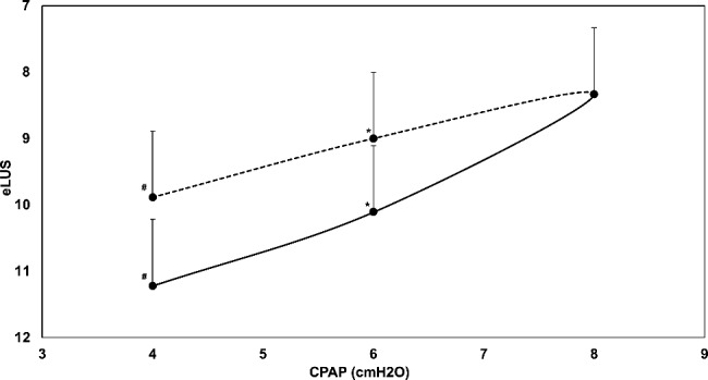
Ultrasound-assessed lung aeration during CPAP increment (i.e. inflation) and decrement (i.e. deflation) in TTN patients. Data were obtained on a subset of neonates with TTN (n = 9). Black and hatched lines represent inflation and deflation, respectively. Black circles and T-bars represent means and standard deviations, respectively. Symbols represents the paired comparison of lung aeration between inflation and deflation at the same CPAP level (#p = 0.029; *p = 0.015). Abbreviations: CPAP: continuous positive airway pressure; eLUS: extended lung ultrasound score; TTN: transient tachypnoea of the neonate
Supplementary Figs. 3–4 shows the analysis of secondary outcomes. The analysis of derived oxygenation metrics gave results similar to those observed in terms of PtcO2/FiO2 ratio (Supplementary Fig. 3). In the whole population, PtcCO2 slightly decreased with increasing CPAP level, though the decrease was small and unlikely to be clinically meaningful (Supplementary Fig. 4).
Discussion
In a “real-world” clinical setting, we investigated the effect of increasing CPAP within the values commonly used in neonatal care and found that: (1) CPAP increment improves ultrasound-assessed lung aeration and oxygenation, although the absolute improvements are small at 8 cmH2O or for RDS patients; (2) eLUS improves significantly more in patients affected by TTN than in those with RDS, and (3) within the tested CPAP range, lung hysteresis is evident in some neonates with TTN. To the best of our knowledge, this is the first clinical study on the topic and clarify the effect of different CPAP levels on ultrasound-assessed lung aeration and gas exchange.
Our data suggest that increasing CPAP from 6 to 8 cmH2O may not have clinically meaningful effects in neonates with RDS. The reduced clinical significance of CPAP increments in RDS is demonstrated by the small absolute changes in PaO2/FiO2, eLUS and lung aeration pattern distribution, despite their statistical significance. For instance, a mean PaO2/FiO2 or eLUS difference of less than 10 and 1 points, respectively, is unlikely to impact on clinical outcomes. Conversely, when the integration of imaging and clinical data indicates a diagnosis of TTN, increasing CPAP may entail a relevant change in eLUS and PaO2/FiO2.
This is coherent with the different pathobiology of RDS and TTN, which are due to primary surfactant deficiency and excessive alveolar fluid accumulation, respectively [38, 39]. Consequently, RDS neonates have lower compliance than patients with TTN [40, 41]. Thus, lungs affected by RDS are less aerated and may require the application of pressure levels beyond the tested range to obtain a relevant ultrasound change. Consistently: (1) lung ultrasound findings changed and showed hysteresis in invasively ventilated preterm lambs with RDS when lungs were inflated at a maximum of 35 cmH2O; [12, 13] (2) increasing CPAP to 10 cmH2O in neonates showed an increased lung volume and hysteresis pattern using electrical impedance tomography [42]. The application of CPAP beyond 8 cmH2O is, however, not considered routine care in every NICU: the safety and comfort of these pressure levels are not entirely ascertained, their use might delay surfactant replacement beyond its optimal time window and eventually reduce its efficacy [34]. Thus, we cannot provide any clue regarding the effect of CPAP at higher levels which need to be investigated in dedicated studies with a different design. Conversely, the excessive fluid accumulation typical of TTN may be overcome by CPAP levels usually provided in neonatal practice, which are also similar to those applied in adult critical care for lung oedema [43].
Our findings on oxygenation are solid since they were similar with all the calculated oxygenation metrics. Furthermore, the general correspondence between eLUS and oxygenation metrics is coherent with data used to validate lung ultrasound scores for clinical use in neonates [30, 33]. Carbon dioxide seems to follow a similar trend since it was not reduced by increasing CPAP beyond 6 cmH2O, its absolute values were within or close to the norm and the magnitude of its change was small. This is consistent with the restrictive pathophysiology of RDS and TTN and the previously reported data on the course of their gas exchange using CPAP early from birth, at a fixed level, during the first 72 h of life [1].
Our study also demonstrates that quantitative lung ultrasound findings can changes following 2–4 cmH2O CPAP increment, despite some degree of leaks that may unavoidably occur in the clinical setting, for instance around the nostrils [44]. This can inform the use of quantitative lung ultrasound at the bedside and is consistent with data reported by a small preliminary study showing the improvement of quantitative lung ultrasound findings 4–6 h after the application of 4–8 cmH2O CPAP [45]. Interestingly, ultrasound was unable to detect small lung volume changes in preterm lambs using different lung ultrasound scores not validated for clinical use; [12] nonetheless, a 4 cmH2O increment represents a relatively wide range in clinical practice and our hysteresis data are consistent with those produced in that experimental setting [12]. Thus, quantitative lung ultrasound may help to provide gentle respiratory support by avoiding pressure increase when this does not provide visible benefits [46]. Conversely, our study was physiological and focused on eLUS and gas exchange, thus we cannot provide data about the effect of different CPAP levels on major clinical outcomes.
Our work has strengths and limitations. The main strength resides in the real-world nature of the study, which has been nested within the framework of routine clinical practice. Thus, the study deems variables that may not be considered in experimental settings or with other designs. Given the ease of use of lung ultrasound and its increasingly widespread application, our results can be easily reproduced in other centres. Conversely, the same study design entails some main limitations. First, there is no randomization or blinding: this was unavoidable since quantitative lung ultrasound is included in our clinical management, and it would have been unethical doing otherwise. Thus, the study was pragmatically performed during routine care; to obtain a blinding a different study design would have been needed. As consequences: (1) we cannot exclude that the eLUS improvement would be due to the total time elapsed on CPAP rather than to the incremental CPAP level, and (2) physicians could see the pressure level and the vital parameters during ultrasound scans since it is impossible to conceal patient conditions. Nonetheless, previous studies have demonstrated equally optimal inter-observer agreement for lung ultrasound interpretation, irrespective of blindness [31, 33, 47, 48]. Second, we recruited a relatively small convenience sample size, as a formal calculation was unfeasible but our choice seems justified in light of previous studies [6–8, 12, 13]. Third, we used a relatively short wash-out as this was embedded in our routine clinical practice, whereas a longer time could have allowed to detect more differences. However, the wash-out time was similar to that used in the unique preliminary study available in this area and findings obtained during alveolar recruitment in intubated neonates [26, 27]. Also, wash-out was equal to the time spent under each CPAP level before performing any measurement. Another limitation is that the hysteresis was demonstrated in a small subgroup of TTN patients. Nonetheless, the procedure was not entirely (i.e. inflation and deflation) realized in patients who did not show a relevant improvement when CPAP was increased and, given the pragmatic nature of the study, when the clinical workload did not allow it. However, the detection of hysteresis is an ancillary finding that is consistent with data obtained in animal models [12, 13]. Gas exchange was monitored with non-invasive devices, which is accepted by international consensus [15], while the use of arterial lines and repeated blood gas analyses would be unethical in these patients. Our data have been produced with variable flow CPAP and cannot be generalised to patients assisted with other types of CPAP. Finally, the RDS cohort consisted of relatively mature neonates and data may not be directly applicable to extremely preterm neonates.
Conclusions
CPAP increments within 4 and 8 cmH2O improve quantitative lung ultrasound findings and oxygenation in term and late preterm infants with TTN, as well as in relatively mature preterm neonates with RDS. However, the absolute improvements is small when CPAP is beyond 6 cmH2O or for RDS patients. Quantitative lung ultrasound findings changed between commonly used CPAP levels and hence the technique can be helpful in guiding respiratory support in these patients.
Electronic supplementary material
Below is the link to the electronic supplementary material.
Acknowledgements
N.A.
Abbreviations
- A-a
Alveolar-arterial gradient
- a/A
Arterial/Alveolar
- CPAP
Continuous positive airway pressure
- CRIB-II
Critical risk index for babies-II
- eLUS
Extended lung ultrasound score
- FiO2
Inspired oxygen fraction
- NICU
Neonatal intensive care unit
- PtcCO2
Partial pressure of carbon dioxide
- PtcO2
Partial pressure of oxygen
- RDS
Respiratory distress syndrome
- SpO2
Peripheral arterial hemoglobin oxygen saturation
- TTN
Transient tachypnoea of the neonate
Author contributions
VS: data collection, interpretation and analysis, manuscript draft preparation; BL, LV, GR, SDLO, LP, MM : data collection and interpretation; SSA and RBA: data and diagnosis interpretation and review; DDL: data interpretation and analysis, study conception and design, general supervision. All authors reviewed the manuscript for important intellectual content, approved the final version to be published and agreed to be accountable for all aspects of the work in ensuring that questions related to the accuracy or integrity of any part of the work are appropriately investigated and resolved.
Funding
None.
Data availability
The deidentified datasets generated during and/or analysed during the current study are available from the corresponding author on reasonable request for research purposes and provided that all relevant regulations are respected.
Declarations
Ethics approval and consent to participate
Approved by the French Critical Care Ethics Commission n.22/011 on March 15, 2022. Parental informed consent was obtained upon NICU admission.
Consent for publication
N.A.
Competing interests
The authors declare no competing interests.
Conflict of interest
none.
Footnotes
The original online version of this article was revised: The error in the author name has been corrected.
Publisher’s note
Springer Nature remains neutral with regard to jurisdictional claims in published maps and institutional affiliations.
Change history
8/2/2025
The original online version of this article was revised: The error in the author name has been corrected
Change history
8/6/2025
A Correction to this paper has been published: 10.1186/s12931-025-03333-3
References
- 1.Pezza L, Sartorius V, Loi B, Regiroli G, Centorrino R, Lanciotti L, et al. Evolution of ultrasound-assessed lung aeration and gas exchange in respiratory distress syndrome and transient tachypnea of the neonate. J Pediatr. 2023;256:44–e522. 10.1016/j.jpeds.2022.11.037. [DOI] [PubMed] [Google Scholar]
- 2.Roberts CL, Badgery-Parker T, Algert CS, Bowen JR, Nassar N. Trends in use of neonatal CPAP: a population-based study. BMC Pediatr. 2011;11:89. 10.1186/1471-2431-11-89. [DOI] [PMC free article] [PubMed] [Google Scholar]
- 3.Diblasi RM. Nasal continuous positive airway pressure (CPAP) for the respiratory care of the newborn infant. Respir Care. 2009;54:1209–35. [PubMed] [Google Scholar]
- 4.Cavicchioli P, Zimmermann LJ, Cogo PE, Badon T, Giordano G, Torresin M, et al. Endogenous surfactant turnover in preterm infants with respiratory distress syndrome studied with stable isotope lipids. Am J Respir Crit Care Med. 2001;163:55–60. [DOI] [PubMed] [Google Scholar]
- 5.Martherus T, Kuypers KLAM, Böhringer S, Dekker J, Witlox RSGM, Hooper SB, et al. Feasibility and effect of physiological-based CPAP in Preterm infants at Birth. Front Pediatr. 2021;9:777614. [DOI] [PMC free article] [PubMed] [Google Scholar]
- 6.Mukerji A, Shah PS, Shivananda S, Yee W, Read B, Minski J, et al. Survey of noninvasive respiratory support practices in Canadian neonatal intensive care units. Acta Paediatr. 2017;106:387–93. [DOI] [PubMed] [Google Scholar]
- 7.Mukerji A, Wahab MGA, Mitra S, Mondal T, Paterson D, et al. High continuous positive airway pressure in neonates: a physiological study. Pediatr Pulmonol. 2019;54:1039–44. [DOI] [PubMed] [Google Scholar]
- 8.Mukerji A, Abdul Wahab MG, Razak A, Rempel E, Patel W, Mondal T, et al. High CPAP vs. NIPPV in preterm neonates — a physiological cross-over study. J Perinatol. 2021;41:1690–6. [DOI] [PMC free article] [PubMed] [Google Scholar]
- 9.Chiumello D, Formenti P, Coppola S. Lung recruitment: what has computed tomography taught us in the last decade? Ann Intensive Care. 2019;9:12. 10.1186/s13613-019-0497-8. [DOI] [PMC free article] [PubMed] [Google Scholar]
- 10.Raimondi F, Yousef N, Migliaro F, Capasso L, De Luca D. Point-of-care lung ultrasound in neonatology: classification into descriptive and functional applications. Pediatr Res. 2021;90:524–31. 10.1038/s41390-018-0114-9. [DOI] [PMC free article] [PubMed] [Google Scholar]
- 11.Mongodi S, De Luca D, Colombo A, Stella A, Santangelo E, Corradi F, et al. Quantitative lung Ultrasound: technical aspects and clinical applications. Anesthesiology. 2021;134:949–65. 10.1097/ALN.0000000000003757. [DOI] [PubMed] [Google Scholar]
- 12.Sett A, Kenna KR, Sutton RJ, Perkins EJ, Sourial M, Chapman JD, et al. Lung ultrasound of the dependent lung detects real-time changes in lung volume in the preterm lamb. Arch Dis Child Fetal Neonatal Ed. 2023;108:51–6. [DOI] [PMC free article] [PubMed] [Google Scholar]
- 13.Sett A, Foo GWC, Kenna KR, Sutton RJ, Perkins EJ, Sourial M, et al. Quantitative lung ultrasound detects dynamic changes in lung recruitment in the preterm lamb. Pediatr Res. 2023;93:1591–8. [DOI] [PMC free article] [PubMed] [Google Scholar]
- 14.Escourrou G, De Luca D. Lung ultrasound decreased radiation exposure in preterm infants in a neonatal intensive care unit. Acta Paediatr. 2016;105:e237–9. 10.1111/apa.13369. [DOI] [PubMed] [Google Scholar]
- 15.De Luca D, van Kaam AH, Tingay DG, Courtney SE, Danhaive O, Carnielli VP, et al. The Montreux definition of neonatal ARDS: biological and clinical background behind the description of a new entity. Lancet Respir Med. 2017;5:657–66. 10.1016/S2213-2600(17)30214-X. [DOI] [PubMed] [Google Scholar]
- 16.Singh Y, Tissot C, Fraga MV, Yousef N, Cortes RG, Lopez J, et al. International evidence-based guidelines on point of Care Ultrasound (POCUS) for critically ill neonates and children issued by the POCUS Working Group of the European Society of Paediatric and neonatal intensive care (ESPNIC). Crit Care. 2020;24(1):65. 10.1186/s13054-020-2787-9. [DOI] [PMC free article] [PubMed] [Google Scholar]
- 17.De Luca D, Baroni S, Vento G, Piastra M, Pietrini D, Romitelli F, et al. Secretory phospholipase A2 and neonatal respiratory distress: pilot study on broncho-alveolar lavage. Intensive Care Med. 2008;34:1858–64. 10.1007/s00134-008-1224-3. [DOI] [PubMed] [Google Scholar]
- 18.Piastra M, Yousef N, Brat R, Manzoni P, Mokhtari M, De Luca D, et al. Lung ultrasound findings in meconium aspiration syndrome. Early Hum Dev. 2014;90:S41–3. [DOI] [PubMed] [Google Scholar]
- 19.Dell’Orto V, Bourgeois-Nicolaos N, Rouard C, Romain O, Shankar-Aguilera S, Doucet-Populaire F, et al. Cell Count Analysis from Nonbronchoscopic Bronchoalveolar Lavage in Preterm infants. J Pediatr. 2018;200:30–e372. 10.1016/j.jpeds.2018.04.074. [DOI] [PubMed] [Google Scholar]
- 20.De Luca D, Alonso A, Autilio C. Bile acid-induced lung injury: update of reverse translational biology. Am J Physiol Lung Cell Mol Physiol. 2022;323:L93–106. [DOI] [PubMed] [Google Scholar]
- 21.De Luca D, Tingay DG, van Kaam AH, Courtney SE, Kneyber MCJ, Tissieres P, et al. Epidemiology of neonatal ARDS: prospective, multicenter, international cohort study. Pediatr Crit Care Med. 2022;23:524–34. 10.1097/PCC.0000000000002961. [DOI] [PubMed] [Google Scholar]
- 22.Gizzi C, Klifa R, Pattumelli MG, Massenzi L, Taveira M, Shankar-Aguilera S, et al. Continuous positive Airway pressure and the Burden of Care for transient tachypnea of the neonate: Retrospective Cohort Study. Am J Perinatol. 2015;32:939–43. 10.1055/s-0034-1543988. [DOI] [PubMed] [Google Scholar]
- 23.Raschetti R, Yousef N, Vigo G, Marseglia G, Centorrino R, Ben-Ammar R, et al. Echography-guided surfactant therapy to improve timeliness of surfactant replacement: a Quality Improvement Project. J Pediatr. 2019;212:137–e1431. 10.1016/j.jpeds.2019.04.020. [DOI] [PubMed] [Google Scholar]
- 24.Fortas F, Loi B, Centorrino R, Regiroli G, Ben-Ammar R, Shankar-Aguilera S, et al. Enhanced INSURE (ENSURE): an updated and standardised reference for surfactant administration. Eur J Pediatr. 2022;181:1269–75. 10.1007/s00431-021-04301-x. [DOI] [PMC free article] [PubMed] [Google Scholar]
- 25.Jourdain G, De Tersant M, Dell’Orto V, Conti G, De Luca D. Continuous positive airway pressure delivery during less invasive surfactant administration: a physiologic study. J Perinatol. 2018;38:271–7. 10.1038/s41372-017-0009-3. [DOI] [PubMed] [Google Scholar]
- 26.Pierro M, Chioma R, Ciarmoli E, Villani P, Storti E, Copetti R. Lung ultrasound guided pulmonary recruitment during mechanical ventilation in neonates: a case series. J Neonatal Perinat Med. 2022;15:357–65. [DOI] [PubMed] [Google Scholar]
- 27.Miedema M, de Jongh FH, Frerichs I, van Veenendaal MB, van Kaam AH. Regional respiratory time constants during lung recruitment in high-frequency oscillatory ventilated preterm infants. Intensive Care Med. 2012;38:294–9. [DOI] [PMC free article] [PubMed] [Google Scholar]
- 28.Loi B, Regiroli G, Foligno S, Centorrino R, Yousef N, Vedovelli L, et al. Respiratory and haemodynamic effects of 6 h-pronation in neonates recovering from respiratory distress syndrome, or affected by acute respiratory distress syndrome or evolving bronchopulmonary dysplasia: a prospective, physiological, crossover, controlled cohort study. EClinicalMedicine. 2022;55:101791. 10.1016/j.eclinm.2022.101791. [DOI] [PMC free article] [PubMed] [Google Scholar]
- 29.Restrepo RD, Hirst KR, Wittnebel L, Wettstein R. AARC Clinical Practice Guideline: Transcutaneous Monitoring of Carbon Dioxide and Oxygen: 2012. Respir Care. 2012;57:1955–62. [DOI] [PubMed] [Google Scholar]
- 30.Loi B, Vigo G, Baraldi E, Raimondi F, Carnielli VP, Mosca F, et al. Lung Ultrasound to monitor extremely Preterm infants and predict BPD: Multicenter Longitudinal Cohort Study. Am J Respir Crit Care Med. 2021;203:1398–409. 10.1164/rccm.202008-3131OC. [DOI] [PubMed] [Google Scholar]
- 31.Gomond-Le Goff C, Vivalda L, Foligno S, Loi B, Yousef N, De Luca D, et al. Effect of different probes and expertise on the interpretation reliability of point-of-care lung Ultrasound. Chest. 2020;157:924–31. 10.1016/j.chest.2019.11.013. [DOI] [PubMed] [Google Scholar]
- 32.De Jaegere A, van Veenendaal MB, Michiels A, van Kaam AH. Lung recruitment using oxygenation during open lung high-frequency ventilation in Preterm infants. Am J Respir Crit Care Med. 2006;174:639–45. [DOI] [PubMed] [Google Scholar]
- 33.Brat R, Yousef N, Klifa R, Reynaud S, Shankar Aguilera S, De Luca D, et al. Lung Ultrasonography score to evaluate oxygenation and surfactant need in neonates treated with continuous positive Airway pressure. JAMA Pediatr. 2015;169:e151797. 10.1001/jamapediatrics.2015.1797. [DOI] [PubMed] [Google Scholar]
- 34.De Luca D, Autilio C, Pezza L, Shankar-Aguilera S, Tingay DG, Carnielli VP. Personalized medicine for the management of RDS in Preterm neonates. Neonatology. 2021;118:127–38. 10.1159/000513783. [DOI] [PubMed] [Google Scholar]
- 35.Martelius L, Janér C, Süvari L, Helve O, Lauerma K, Pitkänen O, et al. Delayed lung liquid absorption after Cesarean Section at Term. Neonatology. 2013;104:133–6. [DOI] [PubMed] [Google Scholar]
- 36.Ramachandrappa A, Jain L. Elective cesarean section: its impact on neonatal respiratory outcome. Clin Perinatol. 2008;35:373–93. [DOI] [PMC free article] [PubMed] [Google Scholar]
- 37.Piastra M, De Luca D, Marzano L, Stival E, Genovese O, Pietrini D, et al. The number of failing organs predicts non-invasive ventilation failure in children with ALI/ARDS. Intensive Care Med. 2011;37:1510–6. 10.1007/s00134-011-2308-z. [DOI] [PubMed] [Google Scholar]
- 38.Avery ME, Mead J. Surface properties in relation to atelectasis and hyaline membrane disease. Am J Dis Child. 1959;97:517–23. [DOI] [PubMed] [Google Scholar]
- 39.McGillick EV, Te Pas AB, van den Akker T, Keus JMH, Thio M, Hooper SB. Evaluating clinical outcomes and physiological perspectives in studies investigating respiratory support for babies born at term with or at risk of transient tachypnea: a narrative review. Front Pediatr. 2022;10:878536. 10.3389/fped.2022.878536. [DOI] [PMC free article] [PubMed] [Google Scholar]
- 40.Martelius L, Süvari L, Janér C, Helve O, Kaskinen A, Kirjavainen T, et al. Lung ultrasound and static lung compliance during postnatal adaptation in healthy term infants. Neonatology. 2015;108:287–92. [DOI] [PubMed] [Google Scholar]
- 41.Biasucci DG, Loi B, Centorrino R, Raschetti R, Piastra M, Pisapia L, et al. Ultrasound-assessed lung aeration correlates with respiratory system compliance in adults and neonates with acute hypoxemic restrictive respiratory failure: an observational prospective study. Respir Res. 2022;23:360. 10.1186/s12931-022-02294-1. [DOI] [PMC free article] [PubMed] [Google Scholar]
- 42.Bhatia R, Davis PG, Tingay DG. Regional volume characteristics of the Preterm Infant receiving first intention continuous positive Airway pressure. J Pediatr. 2017;187:80–e882. 10.1016/j.jpeds.2017.04.046. [DOI] [PubMed] [Google Scholar]
- 43.Bello G, De Santis P, Antonelli M. Non-invasive ventilation in cardiogenic pulmonary edema. Ann Transl Med. 2018;6:355–355. [DOI] [PMC free article] [PubMed] [Google Scholar]
- 44.Centorrino R, Dell’Orto V, Gitto E, Conti G, Luca D. Mechanics of nasal mask-delivered HFOV in neonates: a physiologic study. Pediatr Pulmonol. 2019;54:1304–10. 10.1002/ppul.24358. [DOI] [PubMed] [Google Scholar]
- 45.Perri A, Fattore S, D’Andrea V, Sbordone A, Patti ML, Nobile S, et al. Lowering of the neonatal lung Ultrasonography score after nCPAP Positioning in neonates over 32 weeks of gestational age with neonatal respiratory distress. Diagnostics (Basel). 2022;12(8):1909. [DOI] [PMC free article] [PubMed] [Google Scholar]
- 46.Dargaville PA, Tingay DG. Lung protective ventilation in extremely preterm infants. J Paediatr Child Health. 2012;48:740–6. [DOI] [PubMed] [Google Scholar]
- 47.Brusa G, Savoia M, Vergine M, Bon A, Copetti R, Cattarossi L. Neonatal lung Sonography: Interobserver Agreement between Physician interpreters with varying levels of experience. J Ultrasound Med. 2015;34:1549–54. [DOI] [PubMed] [Google Scholar]
- 48.Sartorius V, Loi B, Vivalda L, Regiroli G, de la Rubia Ortega S, Centorrino R, et al. Ultra-high frequency lung ultrasound in preterm neonates: a test validation study on interpretation agreement and reliability. Arch Dis Child Fetal Neonatal Ed. 2023;108:607–11. 10.1136/archdischild-2023-325300. [DOI] [PubMed] [Google Scholar]
Associated Data
This section collects any data citations, data availability statements, or supplementary materials included in this article.
Supplementary Materials
Data Availability Statement
The deidentified datasets generated during and/or analysed during the current study are available from the corresponding author on reasonable request for research purposes and provided that all relevant regulations are respected.