Abstract
Background/Aims
Despite advances in antiviral therapy for hepatitis C virus (HCV) infection, hepatocellular carcinoma (HCC) still develops even after sustained viral response (SVR) in patients with advanced liver fibrosis or cirrhosis. This meta-analysis investigated the predictive performance of vibration-controlled transient elastography (VCTE) and fibrosis 4-index (FIB-4) for the development of HCC after SVR.
Methods
We searched PubMed, MEDLINE, EMBASE, and the Cochrane Library for studies examining the predictive performance of these tests in adult patients with HCV. Two authors independently screened the studies’ methodological quality and extracted data. Pooled estimates of sensitivity, specificity, and area under the curve (AUC) were calculated for HCC development using random-effects bivariate logit normal and linear-mixed effect models.
Results
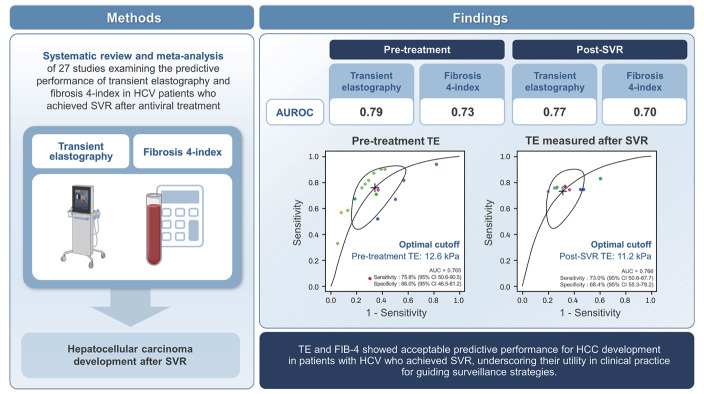
We included 27 studies (169,911 patients). Meta-analysis of HCC after SVR was possible in nine VCTE and 15 FIB-4 studies. Regarding the prediction of HCC development after SVR, the pooled AUCs of pre-treatment VCTE >9.2–13 kPa and FIB-4 >3.25 were 0.79 and 0.73, respectively. VCTE >8.4–11 kPa and FIB-4 >3.25 measured after SVR maintained good predictive performance, albeit slightly reduced (pooled AUCs: 0.77 and 0.70, respectively). The identified optimal cut-off value for HCC development after SVR was 12.6 kPa for pre-treatment VCTE. That of VCTE measured after the SVR was 11.2 kPa.
Conclusions
VCTE and FIB-4 showed acceptable predictive performance for HCC development in patients with HCV who achieved SVR, underscoring their utility in clinical practice for guiding surveillance strategies. Future studies are needed to validate these findings prospectively and validate their clinical impact.
Keywords: Vibration-controlled transient elastography, Fibrosis 4-index, Hepatitis C virus, Hepatocellular carcinoma, Prediction
Graphical Abstract
INTRODUCTION
Hepatitis C virus (HCV) is a significant cause of hepatocellular carcinoma (HCC), accounting for 8.2–12.0% of HCC cases in South Korea and contributing to 34% of HCC cases in the United States [1,2]. The majority of HCV-related HCC cases are preceded by cirrhosis, because HCV is an RNA virus that does not integrate into the host’s genome, making it less likely to be the primary initiator of tumorigenesis [3]. Although cases of HCC have been documented in individuals with minimal or no fibrosis, most HCV-related HCC are observed in patients with advanced fibrosis or cirrhosis, with an annual incidence rate of 2–4% in HCV-related cirrhosis [4,5].
Regarding HCV-related HCC, treating existing HCV infections is the most effective way to prevent the development of HCC. Previously, achievement of sustained virological response (SVR) with interferon (IFN) decreased the risk of HCC [6]. In the last decade, direct-acting antivirals (DAAs) have emerged as the standard treatment for HCV infection due to their remarkable effectiveness [7]. Although SVR achieved with DAA therapy has been found to reduce HCC risk by >70%, HCC still develops in a substantial proportion of patients, especially among those with advanced fibrosis or cirrhosis and patients who have other oncogenic factors in the post-SVR period [8].
Therefore, it is crucial to identify patients at risk of developing HCC, necessitating continuous surveillance while also identifying those who can safely terminate their follow-up monitoring. The recommendations for HCC surveillance vary across guidelines for HCV patients in the post-SVR period with advanced fibrosis (METAVIR score F3) or cirrhosis (METAVIR score F4) [9,10]. Since non-invasive surrogates such as vibration-controlled transient elastography (VCTE) or the Fibrosis-4 index (FIB-4) are currently used to assess the fibrotic burden, it is clinically important to establish the criteria of these non-invasive surrogates for selecting candidates for surveillance of HCC after SVR. Moreover, antiviral therapy can improve liver damage, potentially decreasing the risk of HCV-related HCC development [11,12]. Therefore, dynamic changes in non-invasive surrogates in the post-SVR period should be considered in the risk assessment of HCC development after SVR.
This meta-analysis explored the performance and optimal cut-off values of VCTE and FIB-4, measured before and after SVR, in predicting HCC development among patients with HCV.
MATERIALS AND METHODS
This study followed the preferred reporting items for systematic review and meta-analysis of diagnostic test accuracy guidelines (Supplementary Table 1).
Eligibility
We included studies that examined VCTE-based liver stiffness (LS) or FIB-4 measured before and after achieving SVR with antiviral therapy in patients with HCV. For this analysis, only studies involving patients with HCV who achieved SVR after antiviral therapy were included. Studies published in peer-reviewed journals in any language were considered if they met the following criteria: i) included adults aged ≥18 years with HCV; ii) provided data for at least 10 patients; and iii) reported estimates for sensitivity, specificity, positive predictive value (PPV), negative predictive value (NPV), and area under the curve (AUC) for predicting HCC development after SVR.
Exclusion criteria
Studies were excluded if they met the following criteria: i) included patients with coinfection with hepatitis B virus and/or HIV, ii) included patients who did not receive antiviral therapy for HCV, iii) comprised individuals with a history of HCC or liver transplantation, and iv) lacked sufficient data to calculate predictive performance measures. In cases where the studies had missing data or did not report predictive performance, specifically for patients with HCV within a mixed cohort of patients with liver disease, the corresponding author was contacted via email to request the necessary information or results. Studies were excluded if no responses were received within 28 days.
Search strategy and selection process
A comprehensive web-based literature search of PubMed/MEDLINE, EMBASE, and CENTRAL (Cochrane Library) was systematically performed for articles published until June 30th, 2023 (see Supplementary Table 2 for specific search terms). The search was performed collaboratively by an experienced medical librarian (CHJ) and a hepatologist (HAL). Additionally, the reference lists of the included studies were manually searched to identify further relevant research.
Study selection
The search results were imported into an online platform for systematic review management (Covidence; Veritas Health Innovation, Melbourne, Australia; www.covidence.org), where duplicates were automatically eliminated. Initially, titles and abstracts were screened to identify potentially relevant papers, and a full-text assessment of eligibility was conducted. Two researchers independently screened titles, abstracts, and full papers. Any disagreements were resolved through a consensus among the researchers; if a consensus could not be reached, a senior team member made the final decision. In cases where multiple reports from the same study existed, the most comprehensive or recent publication was selected based on consensus among the reviewers.
Data extraction
Two researchers independently extracted data using a standardized data extraction form. Any disagreements were resolved by consensus or, if necessary, consultation with a senior member of the review team. The data included study characteristics (country, year of publication, and study type), patient characteristics (age, sex, and laboratory findings), viral factors (HCV genotype and HCV RNA levels), number of HCC cases, type of antiviral therapy (IFN or DAAs), timing of non-invasive surrogates, and predictive performance of non-invasive surrogates (including cut-off values, sensitivity, specificity, PPV, NPV, and AUC). Additionally, the necessary data for calculating the true positives, false positives, true negatives, and false negatives were extracted. In cases where this information was not explicitly provided in the study, the values were computed based on the reported diagnostic test sensitivity, specificity, and prevalence. The database search for this study commenced on June 1, 2023, and the article review process was completed on September 26, 2023.
Methodological quality assessment
Two independent reviewers assessed the risk of bias and the relevance of the study findings to the review question using the Quality Assessment of Diagnostic Accuracy Studies-2 (QUADAS-2) tool [13]. Disagreements were resolved through consensus between the reviewers whenever possible. If a consensus could not be reached, a third member of the review team made the final decision.
Evaluation of predictive accuracy
Tables containing performance indices data were extracted and reconstructed to assess the performance of non-invasive surrogates in predicting the development of HCC after SVR. Sensitivity, specificity, PPV, NPV, positive likelihood ratio (PLR), and negative likelihood ratio (NLR), along with their respective 95% confidence intervals (CIs), were calculated based on study-specific estimates. Graphical descriptive analysis of the included studies was conducted using forest plots.
A meta-analysis was conducted when three or more studies with adequate information were available for the same non-invasive surrogate at the same measurement period. In cases where multiple cut-off values were reported, studies were categorized into specific ranges of cut-off values for meta-analysis. A bivariate logit-normal random effects model was employed to estimate mean sensitivity, mean specificity, and their respective variances and covariances. Summary receiver operating characteristic (sROC) curves were created, including 95% CIs, which indicates that the ‘true value’ would typically be within this region 95% of the time, based on the available data. The 95% CIs corresponding to the summary AUC values were estimated using 500 bootstrap iterations.
A linear mixed-effects model was used to analyze data from individual studies reporting more than two cut-offs [14,15]. This model allows for the calculation of summary sensitivities and specificities across different cut-offs as well as the determination of PPV and NPV based on the prevalence of the target condition. Sensitivity and specificity were aggregated at each recommended cut-off to generate a multiple-threshold sROC curve. Furthermore, the PPV and NPV were derived, and the cut-offs necessary to meet the minimum acceptable criteria were identified. The statistical software R, utilizing the mada and diagmeta packages (Version 3.6.1; R Foundation for Statistical Computing, Vienna, Austria), was used for all analyses.
RESULTS
Search results
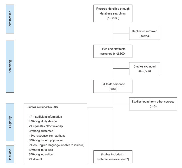
Out of the 3,263 articles initially identified and imported into Covidence from electronic database searches, 2,600 articles were screened after removing duplicates. From these, 64 articles were selected for full-text review based on electronic searches, with an additional three articles identified through manual searching of the reference lists. Ultimately, 27 studies (26 full-text reports and one conference abstract) were included in the meta-analysis, as depicted in Figure 1.
Figure 1.
PRISMA flow diagram of studies included in meta-analysis. PRISMA, Preferred Reporting Items for Systematic Reviews and Meta-analyses.
Study characteristics
The characteristics of the TE [8,16-23] and FIB-4 [15,16,22-40] studies included in the systematic review are summarized in Table 1. Eleven single-center and 16 multicenter studies were included. The meta-analysis included 17 studies from Asia and 10 from non-Asian regions, comprising seven prospective cohort studies and 20 retrospective studies. Three studies evaluated both the VCTE and FIB-4.
Table 1.
A summary of studies included in the systematic literature review and meta-analysis
| Reference | Publication year | Country | Study design | Number of patients | Number of HCCs | Treatment | Test | Time of measurement | Cutoff |
|---|---|---|---|---|---|---|---|---|---|
| Sohn et al. [23] | 2024 | South Korea | Multicenter, retrospective | 1,248 | 34 | DAA | VCTE | Pre-treatment | 14.5 kPa |
| SVR12 | 14.5 kPa | ||||||||
| Nakai et al. [19] | 2022 | Japan | Multicenter, retrospective | 567 | 30 | DAA | VCTE | Pre-treatment | 9.2 kPa |
| VCTE | SVR24 | 8.4 kPa | |||||||
| Pons et al. [8] | 2020 | Spain | Multicenter, prospective | 572 | 25 | DAA | VCTE | Pre-treatment | 20.0 kPa |
| VCTE | SVR48 | 10.0 kPa | |||||||
| Rinaldi et al. [20] | 2019 | Italy | Multicenter, prospective | 258 | 35 | DAA | VCTE | Pre-treatment | 27.8 kPa |
| Wang et al. [21] | 2016 | Taiwan | Single center, retrospective | 376 | 21 | IFN | VCTE | Pre-treatment | 12.0 kPa |
| Liu et al. [17] | 2023 | Taiwan | Single center, retrospective | 466 | 40 | DAA | VCTE | Pre-treatment | 12.0 kPa |
| VCTE | SVR | 10.0 kPa | |||||||
| FIB-4 | Pre-treatment | 4.6 | |||||||
| FIB-4 | SVR | 3.7 | |||||||
| Kuo et al. [16] | 2022 | Taiwan | Single center, retrospective | 697 | 28 | DAA | VCTE | Pre-treatment | 11.0 kPa |
| FIB-4 | SVR | 3.6 | |||||||
| Morisco et al. [18] | 2021 | Italy | Multicenter, prospective | 687 | 26 | DAA | VCTE | Pre-treatment | 20.0 kPa |
| Ciancio et al. [24] | 2023 | Italy | Single center, prospective | 1,000 | 71 | DAA | FIB-4 | Pre-treatment | 3.25 |
| Zou et al. [25] | 2022 | China | Single center, retrospective | 701 | 27 | DAA | FIB-4 | Pre-treatment | 3.25 |
| Ideno et al. [26] | 2023 | Japan | Single center, retrospective | 690 | 71 | DAA | FIB-4 | Pre-treatment | 3.25 |
| Kumada et al. [27] | 2022 | Japan | Single center, retrospective | 1,384 | 51 | DAA, IFN | FIB-4 | Pre-treatment | 3.25 |
| Azzi et al. [28] | 2022 | France | Multicenter, prospective | 3,531 | 153 | DAA | FIB-4 | SVR | 3.25 |
| Caviglia et al. [29] | 2022 | Italy | Single center, retrospective | 575 | 57 | DAA | FIB-4 | SVR | 3.38 |
| Tada et al. [30] | 2022 | Japan | Multicenter, retrospective | 3,058 | 107 | DAA | FIB-4 | SVR | 3.25 |
| Ampuero et al. [31] | 2022 | Spain | Multicenter, prospective | 1,054 | 56 | DAA | FIB-4 | Pre-treatment | 3.25 |
| Tahata et al. [32] | 2021 | Japan | Multicenter, prospective | 1,473 | 52 | DAA | FIB-4 | Pre-treatment | 3.25 |
| Kumada et al. [33] | 2021 | Japan | Single center, retrospective | 1,352 | 55 | DAA, IFN | FIB-4 | Pre-treatment | 1.50 |
| Matsumae et al. [34] | 2023 | Japan | Multicenter, prospective | 524 | 24 | DAA | FIB-4 | Pre-treatment | 2.625 |
| Myojin et al. [35] | 2022 | Japan | Multicenter, retrospective | 964 | 50 | DAA | FIB-4 | Pre-treatment | 3.25 |
| Ide et al. [36] | 2019 | Japan | Multicenter, prospective | 2,552 | 70 | DAA | FIB-4 | Pre-treatment | 4.6 |
| Nagaoki et al. [37] | 2020 | Japan | Single center, retrospective | 298 | 29 | DAA, IFN | FIB-4 | Pre-treatment | 5.0 |
| Hiraoka et al. [38] | 2019 | Japan | Multicenter, retrospective | 1,069 | 22 | DAA | FIB-4 | SVR24 | 3.25 |
| Ioannou et al. [39] | 2019 | US | Multicenter, retrospective | 48,135 | 1,509 | DAA, IFN | FIB-4 | Pre-treatment | 3.25 |
| Kramer et al. [15] | 2022 | US | Multicenter, retrospective | 92,567 | 3,247 | DAA | FIB-4 | Pre-treatment | 3.25/1.45 |
| Ogawa et al. [22] | 2020 | Japan | Single center, retrospective | 290 | 16 | DAA | FIB-4 | SVR12 | 3.25 |
| VCTE | SVR12 | 10.0 kPa | |||||||
| Tamaki et al. [40] | 2021 | Japan | Multicenter, retrospective | 3,823 | 148 | DAA | FIB-4 | SVR24, 48 | 3.25 |
HCC, hepatocellular carcinoma; DAA, direct acting antiviral; VCTE, vibration-controlled transient elastography; IFN, interferon; FIB-4, fibrosis-4 index; SVR, sustained viral response; kPa, kilopascal; SVR12, 12 weeks after SVR; SVR24, 24 weeks after SVR; SVR48, 48 weeks after SVR.
Study quality
The methodological quality of the studies assessed using the QUADAS-2 tool is summarized in Supplementary Figure 1. None of the studies exhibited a high risk of bias.
Patient characteristics
In total, 169,911 patients with HCV infection who achieved SVR with antiviral therapy were included in this meta-analysis. Supplementary Table 3 describes the characteristics of the patients in the studies included in the meta-analysis. The mean and median age ranged from 54.1 to 69.0 years. LS measured using VCTE had a mean or median range of 7.1–25.5 kPa, whereas FIB-4 had a mean or median range of 1.86–3.98.
Predictive performance of VCTE for HCC development after SVR
The predictive performances of VCTE and FIB-4 for HCC development after achieving SVR were evaluated. The pooled predictive AUC of a pre-treatment VCTE (eight studies) was 0.73, with a sensitivity of 65.7%, specificity of 69.5%, PLR of 2.22, and NLR of 0.48 (Table 2, Supplementary Fig. 2A). The pooled risk ratio of pre-treatment VCTE (nine studies) was 3.88 (95% CI 2.21–18.16) for HCC development after SVR (Table 3, Supplementary Fig. 3A). When limited to a pre-treatment VCTE cut-off of 9.2–13 kPa (five studies), the predictive performance improved, with a pooled diagnostic AUC of 0.79, sensitivity of 75.1%, specificity of 71.7%, PLR of 2.67, and NLR of 0.39. The cutoff 9.2–13.0 kPa of pre-treatment VCTE (six studies) had a pooled risk ratio of 4.56 (95% CI 3.05–6.81) for HCC development after SVR (Table 3).
Table 2.
The diagnostic performance of vibration-controlled transient elastography and FIB-4 in chronic hepatitis C patients with sustained virological response
| Test | Time of measurement | Cutoff | Number of studies | Number of patients | Number of HCCs | AUC | Sensitivity (%) (95% CI) | Specificity (%) (95% CI) | Positive likelihood ratio (95% CI) | Negative likelihood ratio (95% CI) |
|---|---|---|---|---|---|---|---|---|---|---|
| VCTE | Pre-treatment | Total (9.2–27.8 kPa) | 8 | 4,871 | 239 | 0.73 | 65.7 (45.5–0.81) | 69.5 (56.8–79.8) | 2.22 (1.65–2.99) | 0.48 (0.28–0.85) |
| 9.2–13 kPa | 5 | 3,354 | 153 | 0.79 | 75.1 (61.9–84.9) | 71.7 (50.3–86.3) | 2.67 (1.85–3.86) | 0.39 (0.29–0.51) | ||
| After SVR | Total (8.4–11 kPa) | 6 | 3,840 | 171 | 0.77 | 76.6 (69.3–82.6) | 63.9 (51.5–74.7) | 2.20 (1.62–3.00) | 0.37 (0.28–0.49) | |
| FIB–4 | Pre-treatment | Total (1.45–5) | 15 | 154,408 | 5,386 | 0.72 | 73.4 (66.1–79.6) | 60.5 (52.7–67.8) | 1.88 (1.67–2.11) | 0.50 (0.45–0.56) |
| 3.25 | 13 | 86,435 | 4,330 | 0.73 | 70.9 (62.4–78.1) | 64.9 (55.5–73.3) | 2.05 (1.73–2.43) | 0.48 (0.42–0.55) | ||
| 3.25–5.0 | 5a | 40,534 | 3,339 | 0.66 | 73.3 (63.2–81.5) | 52.4 (42.3–62.3) | 1.56 (1.34–1.81) | 0.50 (0.40–0.63) | ||
| After SVR | Total (2.73–3.7) | 9 | 14,757 | 605 | 0.71 | 61.6 (55.0–67.8) | 73.7 (66.9–79.6) | 2.34 (1.98–2.77) | 0.55 (0.50–0.60) | |
| 3.25 | 7 | 13,019 | 480 | 0.70 | 57.9 (50.2–65.2) | 75.4 (68.0–81.6) | 2.37 (1.89–2.85) | 0.58 (0.51–0.65) |
HCC, hepatocellular carcinoma; AUC, area under the receiver operating characteristic; CI, confidence interval; FIB-4, fibrosis-4 index; SVR, sustained virological response; VCTE, vibration-controlled transient elastography; kPa, kilopascal.
Studies including only patients with liver cirrhosis.
Table 3.
Risk ratio for predicting the occurrence of hepatocellular carcinoma in chronic hepatitis C patients who achieved sustained viral response
| Test | Time of measurement | Cutoff | Number of studies | Number of patients | Number of HCCs | I2 (%) | P-value | Risk ratio | 95% CI |
|---|---|---|---|---|---|---|---|---|---|
| VCTE | Pre-treatment | Total (8.4–27.8 kPa) | 9 | 6,744 | 324 | 88.0 | <0.01 | 3.88 | 2.21–18.16 |
| 9.2–13.0 kPa | 6 | 4,347 | 188 | 3.1 | 0.39 | 4.56 | 3.05–6.81 | ||
| 17.3–27.8 kPa | 3 | 1,343 | 80 | 0.0 | 0.40 | 4.68 | 2.21–9.90 | ||
| After SVR | Total (8.4–11 kPa) | 7 | 4,238 | 190 | 60.0 | 0.02 | 3.93 | 2.17–7.11 | |
| FIB–4 | Pre-treatment | Total (1.45–5) | 15 | 155,444 | 5,427 | 95.0 | <0.01 | 2.30 | 1.64–3.10 |
| <3.25 | 2 | 64,521 | 898 | 77.2 | 0.03 | 4.14 | 0.94–18.19 | ||
| 3.25 | 14 | 86,614 | 4,355 | 81.6 | <0.01 | 2.45 | 1.68–3.13 | ||
| 3.25–5.0 | 4 | 4,309 | 174 | 38.5 | 0.18 | 1.32 | 0.95–1.82 | ||
| After SVR | Total (2.73–3.7) | 10 | 16,913 | 681 | 89.0 | <0.01 | 2.22 | 1.62–3.03 | |
| 3.25 | 6 | 13,019 | 480 | 0.0 | 0.58 | 3.05 | 2.46–3.80 | ||
| 3.25–3.7 | 3 | 1,738 | 125 | 0.0 | 0.81 | 3.00 | 2.03–4.44 |
HCC, hepatocellular carcinoma; CI, confidence interval; FIB-4, fibrosis-4 index; SVR, sustained virological response; VCTE, vibration-controlled transient elastography; kPa, kilopascal.
For the VCTE measured after SVR (six studies with a cutoff range of 8.4–11 kPa), the pooled predictive AUC was 0.77, with a sensitivity of 76.6%, specificity of 63.9%, PLR of 2.20, and NLR of 0.37 (Table 2, Supplementary Fig. 2B). The pooled risk ratio of VCTE measured after SVR (seven studies) was 3.93 (95% CI: 2.17–7.11) (Table 3, Supplementary Fig. 3B).
Predictive performance of FIB-4 for HCC development after SVR
The pooled predictive AUC of a pre-treatment FIB-4 (15 studies) was 0.72, with a sensitivity of 73.4%, specificity of 60.5%, PLR of 1.88, and NLR of 0.50 (Table 2, Supplementary Fig. 4A). In the risk ratio analysis, a pre-treatment FIB-4 (15 studies) had a pooled risk ratio of 2.30 (95% CI: 1.64–3.10) for HCC development after SVR (Table 3, Supplementary Fig. 5A). When limited to a pre-treatment FIB-4 cut-off of 3.25 (13 studies), the pooled predictive AUC was 0.73, with a sensitivity of 70.9%, specificity of 64.9%, PLR of 2.05, and NLR of 0.48, which was superior compared to those of higher cut-off values. The cut-off of 3.25 of a pre-treatment FIB-4 (14 studies) had a pooled risk ratio of 2.45 (95% CI: 1.68–3.13) for HCC development after SVR (Table 3).
For the FIB-4 measured after SVR (nine studies), the pooled diagnostic AUC was 0.71, with a sensitivity of 61.6%, specificity of 73.7%, PLR of 2.34, and NLR of 0.55 (Table 2, Supplementary Fig. 4B). The FIB-4 measured after the SVR (10 studies) had a pooled risk ratio of 2.22 (95% CI: 1.62–3.03) (Table 3, Supplementary Fig. 5B). When limited to a FIB-4 measured after SVR cut-off of 3.25 in (six studies), the pooled predictive AUC was 0.70, with a sensitivity of 57.9%, specificity of 75.4%, PLR of 2.37, and NLR of 0.58. The FIB-4 measured after the SVR cut-off of 3.25 had a pooled risk ratio of 3.05 (95% CI: 2.46–3.80) (Table 3).
Optimal cut-off of VCTE to predict HCC development after SVR
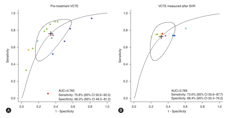
The optimal VCTE cut-off for predicting HCC development after SVR was investigated in studies with multiple cut-off values (Fig. 2, Supplementary Fig. 6). For the pretreatment VCTE (eight studies), the optimal cut-off for HCC was 12.6 kPa with a sensitivity of 75.8% and specificity of 66.0%. The optimal cut-off of VCTE measured after SVR for HCC development after SVR (six studies) was 11.2 kPa with a sensitivity of 73.0% and specificity of 68.4%.
Figure 2.
The performance of VCTE for predicting HCC after the achievement of SVR. Multiple-threshold sROC curve of pre-treatment VCTE (A) and VCTE measured after SVR (B). sSROC, summary receiver operating characteristic curve; VCTE, vibration-controlled transient elastography; SVR, sustained virological response; HCC, hepatocellular carcinoma; CI, confidence interval.
DISCUSSION
The development of HCC after achieving SVR in patients with HCV infection has garnered significant attention owing to the evolving landscape of antiviral therapies, particularly with the advent of DAAs. Despite these advancements, HCC can still develop in a subset of patients after achieving an SVR, particularly in those with advanced liver fibrosis or cirrhosis, underscoring the need for effective predictive tools and surveillance strategies. This meta-analysis of 27 studies, including 169,911 patients, presented acceptable performance of VCTE and FIB-4 measured before and after SVR to predict HCC development after SVR in patients with HCV. These findings highlight the substantial role of non-invasive surrogates in identifying patients at increased risk of HCC development after SVR, emphasizing their utility in clinical practice for guiding surveillance strategies after SVR.
The results of this meta-analysis have profound implications for clinical practice. First, pre-treatment VCTE >9.2–13 kPa and FIB-4 >3.25 had pooled diagnostic AUC of 0.73 and 0.79, respectively, for the prediction of HCC after SVR. This indicates that the integration of VCTE and FIB-4 into the routine assessment of patients with HCV before antiviral therapy is required, which might facilitate the early identification of individuals with a non-invasive surrogate-based high fibrotic burden who require careful surveillance owing to the increased risk of post-SVR HCC development. Recent studies support these results [15-17]; however, the significance lies in the increased reliability of the predictive performance of non-invasive surrogates for HCC after SVR by conducting a systematic review and meta-analysis of the largest number of studies reported to date.
Second, VCTE and FIB-4 measured after SVR showed good performance in predicting HCC development after SVR. Although the predictive performance of VCTE and FIB-4 measured after SVR was slightly lower than that of pre-treatment VCTE and FIB-4, the pooled AUC values for VCTE and FIB-4 at both time points were maintained above 0.7, which underscores the dynamics of non-invasive surrogates and their clinical implications for HCC surveillance strategies during antiviral therapy for HCV. In a recent multicenter retrospective study that examined patients who achieved SVR following DAA therapy, those who experienced an increase in FIB-4 from baseline to 2 years after SVR had elevated HCC risks compared to those whose FIB-4 levels stabilized or declined after treatment [15]. In addition, two studies regarding the VCTE measured after SVR also showed acceptable predictive performance for HCC development after SVR [17,19]. Consequently, VCTE and FIB-4 measured after SVR can serve as important indicators for assessing HCC risk after SVR, emphasizing that dynamic assessment of non-invasive surrogates, rather than a single pre-treatment assessment, is essential for accurate identification of patients at increased risk of HCC after SVR. However, caution is needed when interpreting these non-invasive surrogate measurements after SVR because they do not precisely correlate with the fibrosis stage after SVR [41].
Third, VCTE had higher diagnostic accuracy than FIB-4 before and after SVR, which may be because various systemic conditions, such as inflammation, abnormalities in other organs, or other acute illnesses, can influence FIB-4. In addition, the diagnostic accuracy of FIB-4 in assessing fibrotic burden has been reported to be low in young and older adults [42,43]. However, LS measured using VCTE can also be overestimated by intrahepatic inflammation [44,45]. Considering the high cost and limited accessibility of the test, FIB-4 is useful as a primary screening test for identifying high-risk groups for HCC after SVR in patients with HCV infection.
Fourth, to establish simplified and effective screening criteria for high-risk groups with HCC after SVR, various cutoff values were comprehensively analyzed to determine the optimal cut-off value. In this study, the optimal cut-off values for HCC were found to be 12.6 kPa for pre-treatment VCTE and 11.2 kPa for VCTE measured after SVR. However, the identified cut-off values were derived from the analysis of various studies with diverse clinical characteristics; therefore, further validation through multiple studies, especially prospective studies, is necessary. Currently, there are no established criteria for non-invasive surrogates to select patients in need of HCC surveillance. Moreover, considering that most patients with HCV infection do not undergo liver biopsy for fibrosis assessment, a simplified strategy may be helpful in clinical decision-making. Owing to insufficient data, we could not conduct an sROC analysis for an optimal FIB-4 cut-off. However, a meta-analysis showed that FIB-4 >3.25 measured before and after SVR was considered the most useful cut-off value in predicting HCC development after SVR. The identification of optimal cut-off values for VCTE and FIB-4 enhances the precision of risk stratification, enabling clinicians to tailor surveillance and management strategies more effectively. The study findings support the use of simple, specific cut-off values, thereby enhancing the clinical utility of VCTE and FIB-4 in HCC surveillance after achieving SVR. Furthermore, this approach optimizes patient care and contributes to the efficient allocation of healthcare resources by focusing efforts on those most at risk.
Despite these robust findings, this study has some limitations. First, a substantial number of included studies were retrospective, potentially introducing bias and limiting causal inference. Second, heterogeneity in study design, patient characteristics, and diagnostic thresholds across the studies may have influenced the pooled estimates. Furthermore, the lack of standardized cut-off values for non-invasive surrogates underscores the need for further research to establish widely applicable thresholds. The generalizability of our findings is limited by the heterogeneity in patient characteristics across the included studies. The applicability of these findings to specific subgroups uncertain. Third, the variability in the follow-up duration and time of assessment of VCTE and FIB-4 may have influenced the generalizability of the results. Future research should aim to address these limitations through prospective studies with standardized methodologies and longer follow-up periods to validate the identified cut-off values and further refine the risk stratification models. Fourth, although VCTE and FIB-4 measured before and after SVR showed significant predictive performance, the clinical implications of the dynamic change in these tests on the risk of HCC development after SVR were not assessed. Further prospective studies assessing non-invasive surrogates, pre- and post-SVR, are needed to reveal the changes in the results of non-invasive tests and their clinical relevance to HCC development after SVR. Fifth, the inability to perform detailed sensitivity or subgroup analyses due to insufficient data and variability in the cut-off values used across studies is a notable limitation. Sixth, due to time constraints and limitations in accessible data, we couldn’t perform individual participant data analysis, so there is a limitation in overcoming the diversity of participants included in the study. Seventh, although the cut-off value derived in this study aims to achieve the highest diagnostic accuracy, its application in clinical settings may have limitations, particularly concerning the NPV, which is essential for HCC surveillance. Eighth, although the likelihood of overlapping patients between the studies is small, there is a possibility of some patient overlap. This could potentially inflate the sample size and introduce bias, affecting the accuracy and validity of the meta-analysis results. Finally, considering the modest predictive accuracies of FIB-4 and VCTE in this study, exploring the integration of additional biomarkers with VCTE or FIB-4 could enhance the predictive performance for HCC development after SVR, offering a more comprehensive approach to patient management.
In conclusion, this systematic review and meta-analysis reinforces the importance of VCTE and FIB-4 in predicting the risk of HCC development after SVR in patients with HCV. These tools can guide the implementation of targeted surveillance strategies by identifying patients at an increased risk of HCC after SVR, ultimately facilitating the early detection and management of HCC. Future research for prospective validation of optimal cut-off values and the integration of non-invasive surrogates into clinical practice guidelines for HCC surveillance in patients with HCV who achieved SVR are needed.
Acknowledgments
This study was registered with PROSPERO (registration number: CRD42024567498).
The authors thank the Clinical Practice Guideline Committee for Noninvasive Tests (NIT) to Assess Liver Fibrosis in Chronic Liver Disease of the Korean Association for the Study of the Liver (KASL) for providing the opportunity to conduct this research.
Abbreviations
- AUC
area under the curve
- CI
confidence interval
- DAAs
direct-acting antivirals
- FIB-4
fibrosis 4-index
- HCC
hepatocellular carcinoma
- HCV
hepatitis C virus
- IFN
interferon
- LS
liver stiffness
- NLR
negative likelihood ratio
- NPV
negative predictive value
- PLR
positive likelihood ratio
- PPV
positive predictive value
- QUADAS-2
Quality Assessment of Diagnostic Accuracy Studies-2
- sROC
summary receiver operating characteristic
- SVR
sustained viral response
- VCTE
vibration-controlled transient elastography
Study Highlights
• This systematic review and meta-analysis explores the predictive performance of VCTE and FIB-4 for HCC development after SVR in patients with HCV infection.
• It includes data from 27 studies, encompassing 169,911 patients, revealing that both pre-treatment VCTE and FIB-4 offer acceptive predictive performance for HCC development after SVR, with optimal cut-off values identified for early detection.
• The identified optimal cut-off values for predicting HCC after SVR are 12.6 kPa for pre-treatment VCTE and 11.2 kPa for VCTE measured after the SVR, with FIB-4 >3.25 also showing high predictive accuracy.
• These results affirm the importance of VCTE and FIB-4 in clinical practice, enabling targeted surveillance strategies for HCV patients achieving SVR, thus facilitating early intervention for those at risk of developing HCC.
Footnotes
Authors’ contribution
HAL and YJK conceptualized and designed the study. HAL and HAL performed the statistical analyses. All the authors interpreted the findings. HAL and MNK drafted the manuscript. HAL, MNK, HAL, MC, JWH, and YJK critically revised the manuscript for important intellectual content. All authors provided their final approval for the version to be submitted.
Conflicts of Interest
The authors declare no conflicts of interest relevant to this work.
SUPPLEMENTAL MATERIAL
Supplementary material is available at Clinical and Molecular Hepatology website (http://www.e-cmh.org).
PRISMA-DTA checklist
Electronic search strategy
Patient characteristics of included studies in the systematic literature review and meta-analysis
Methodological quality summary of included studies. Red circles: high risk of bias, Yellow circles: unclear risk of bias, Green circles: low risk of bias.
Forest plots of studies included in the diagnostic accuracy analysis for the prediction of HCC using VCTE measured at (A) pre-treatment and (B) after SVR. HCC, hepatocellular carcinoma; VCTE, vibration-controlled transient elastography; SVR, sustained virological response.
Forest plots of studies included in the risk ratio analysis for the prediction of HCC using VCTE measured at (A) pre-treatment and (B) after SVR. HCC, hepatocellular carcinoma; VCTE, vibration-controlled transient elastography; SVR, sustained virological response.
Forest plots of studies included in the diagnostic accuracy analysis for the prediction of HCC using FIB-4 measured at (A) pre-treatment and (B) after SVR. HCC, hepatocellular carcinoma; FIB-4, fibrosis-4 index; SVR, sustained virological response.
Forest plots of studies included in the risk ratio analysis for the prediction of HCC using FIB-4 measured at (A) pre-treatment and (B) after SVR. HCC, hepatocellular carcinoma; FIB-4, fibrosis-4 index; SVR, sustained virological response.
The performance of VCTE for predicting HCC after the achievement of SVR. Multiple-threshold ROC curves of pre-treatment VCTE (A) and VCTE measured after SVR (B). ROC, receiver operating characteristic curve; VCTE, vibration-controlled transient elastography; SVR, sustained virological response; HCC, hepatocellular carcinoma.
REFERENCES
- 1.Lee JH, Yoon, et al. Hepatocellular Carcinoma in Korea: an Analysis of the 2015 Korean Nationwide Cancer Registry. J Liver Cancer. 2022;22:207. doi: 10.17998/jlc.21.1.58.e1. [DOI] [PMC free article] [PubMed] [Google Scholar]
- 2.Karim MA, Singal AG, Kum HC, Lee YT, Park S, Rich NE, et al. Clinical characteristics and outcomes of nonalcoholic fatty liver disease-associated hepatocellular carcinoma in the United States. Clin Gastroenterol Hepatol. 2023;21:670–680.e18. doi: 10.1016/j.cgh.2022.03.010. [DOI] [PMC free article] [PubMed] [Google Scholar]
- 3.De Mitri MS, Poussin K, Baccarini P, Pontisso P, D’Errico A, Simon N, et al. HCV-associated liver cancer without cirrhosis. Lancet. 1995;345:413–415. doi: 10.1016/s0140-6736(95)90400-x. [DOI] [PubMed] [Google Scholar]
- 4.Hajarizadeh B, Grebely J, Dore GJ. Epidemiology and natural history of HCV infection. Nat Rev Gastroenterol Hepatol. 2013;10:553–562. doi: 10.1038/nrgastro.2013.107. [DOI] [PubMed] [Google Scholar]
- 5.McGlynn KA, Petrick JL, El-Serag HB. Epidemiology of hepatocellular carcinoma. Hepatology. 2021;73 Suppl 1:4–13. doi: 10.1002/hep.31288. [DOI] [PMC free article] [PubMed] [Google Scholar]
- 6.Yu ML, Lin SM, Chuang WL, Dai CY, Wang JH, Lu SN, et al. A sustained virological response to interferon or interferon/ribavirin reduces hepatocellular carcinoma and improves survival in chronic hepatitis C: a nationwide, multicentre study in Taiwan. Antivir Ther. 2006;11:985–994. [PubMed] [Google Scholar]
- 7.Lee HW, Lee H, Kim BK, Chang Y, Jang JY, Kim DY. Cost-effectiveness of chronic hepatitis C screening and treatment. Clin Mol Hepatol. 2022;28:164–173. doi: 10.3350/cmh.2021.0193. [DOI] [PMC free article] [PubMed] [Google Scholar]
- 8.Pons M, Rodríguez-Tajes S, Esteban JI, Mariño Z, Vargas V, Lens S, et al. Non-invasive prediction of liver-related events in patients with HCV-associated compensated advanced chronic liver disease after oral antivirals. J Hepatol. 2020;72:472–480. doi: 10.1016/j.jhep.2019.10.005. [DOI] [PubMed] [Google Scholar]
- 9.European Association for the Study of the Liver EASL recommendations on treatment of hepatitis C: Final update of the series☆. J Hepatol. 2020;73:1170–1218. doi: 10.1016/j.jhep.2022.10.006. [DOI] [PubMed] [Google Scholar]
- 10.Bhattacharya D, Aronsohn A, Price J, Lo Re V, AASLD-IDSA HCV Guidance Panel Hepatitis C guidance 2023 update: AASLD-IDSA recommendations for testing, managing, and treating hepatitis C virus infection. Clin Infect Dis. 2023 May 25; doi: 10.1093/cid/ciad319. doi: [DOI] [PubMed] [Google Scholar]
- 11.Shiratori Y, Ito Y, Yokosuka O, Imazeki F, Nakata R, Tanaka N, et al. Antiviral therapy for cirrhotic hepatitis C: association with reduced hepatocellular carcinoma development and improved survival. Ann Intern Med. 2005;142:105–114. doi: 10.7326/0003-4819-142-2-200501180-00009. [DOI] [PubMed] [Google Scholar]
- 12.D’Ambrosio R, Aghemo A, Fraquelli M, Rumi MG, Donato MF, Paradis V, et al. The diagnostic accuracy of Fibroscan for cirrhosis is influenced by liver morphometry in HCV patients with a sustained virological response. J Hepatol. 2013;59:251–256. doi: 10.1016/j.jhep.2013.03.013. [DOI] [PubMed] [Google Scholar]
- 13.Whiting PF, Rutjes AW, Westwood ME, Mallett S, Deeks JJ, Reitsma JB, et al. QUADAS-2: a revised tool for the quality assessment of diagnostic accuracy studies. Ann Intern Med. 2011;155:529–536. doi: 10.7326/0003-4819-155-8-201110180-00009. [DOI] [PubMed] [Google Scholar]
- 14.Shim SR. Meta-analysis of diagnostic test accuracy studies with multiple thresholds for data integration. Epidemiol Health. 2022;44:e2022083. doi: 10.4178/epih.e2022083. [DOI] [PMC free article] [PubMed] [Google Scholar]
- 15.Kramer JR, Cao Y, Li L, Smith D, Chhatwal J, El-Serag HB, et al. Longitudinal associations of risk factors and hepatocellular carcinoma in patients with cured hepatitis C virus infection. Am J Gastroenterol. 2022;117:1834–1844. doi: 10.14309/ajg.0000000000001968. [DOI] [PMC free article] [PubMed] [Google Scholar]
- 16.Kuo YH, Kee KM, Hung CH, Lu SN, Hu TH, Chen CH, et al. Liver stiffness-based score at sustained virologic response predicts liver-related complications after eradication of hepatitis C virus. Kaohsiung J Med Sci. 2022;38:268–276. doi: 10.1002/kjm2.12465. [DOI] [PMC free article] [PubMed] [Google Scholar]
- 17.Liu YC, Cheng YT, Chen YC, Hsieh YC, Jeng WJ, Lin CY, et al. Comparing predictability of non-invasive tools for hepatocellular carcinoma in treated chronic hepatitis C patients. Dig Dis Sci. 2023;68:323–332. doi: 10.1007/s10620-022-07621-6. [DOI] [PubMed] [Google Scholar]
- 18.Morisco F, Federico A, Marignani M, Cannavò M, Pontillo G, Guarino M, et al. Risk factors for liver decompensation and HCC in HCV-cirrhotic patients after DAAs: a multicenter prospective study. Cancers (Basel) 2021;13:3810. doi: 10.3390/cancers13153810. [DOI] [PMC free article] [PubMed] [Google Scholar]
- 19.Nakai M, Yamamoto Y, Baba M, Suda G, Kubo A, Tokuchi Y, et al. Prediction of hepatocellular carcinoma using age and liver stiffness on transient elastography after hepatitis C virus eradication. Sci Rep. 2022;12:1449. doi: 10.1038/s41598-022-05492-5. [DOI] [PMC free article] [PubMed] [Google Scholar]
- 20.Rinaldi L, Guarino M, Perrella A, Pafundi PC, Valente G, Fontanella L, et al. Role of liver stiffness measurement in predicting HCC occurrence in direct-acting antivirals setting: a reallife experience. Dig Dis Sci. 2019;64:3013–3019. doi: 10.1007/s10620-019-05604-8. [DOI] [PubMed] [Google Scholar]
- 21.Wang JH, Yen YH, Yao CC, Hung CH, Chen CH, Hu TH, et al. Liver stiffness-based score in hepatoma risk assessment for chronic hepatitis C patients after successful antiviral therapy. Liver Int. 2016;36:1793–1799. doi: 10.1111/liv.13179. [DOI] [PubMed] [Google Scholar]
- 22.Ogawa E, Takayama K, Hiramine S, Hayashi T, Toyoda K. Association between steatohepatitis biomarkers and hepatocellular carcinoma after hepatitis C elimination. Aliment Pharmacol Ther. 2020;52:866–876. doi: 10.1111/apt.15976. [DOI] [PubMed] [Google Scholar]
- 23.Sohn W, Park SY, Lee TH, Chon YE, Kim IH, Lee BS, et al. Effect of direct-acting antivirals on disease burden of hepatitis C virus infection in South Korea in 2007-2021: a nationwide, multicentre, retrospective cohort study. EClinicalMedicine. 2024;73:102671. doi: 10.1016/j.eclinm.2024.102671. [DOI] [PMC free article] [PubMed] [Google Scholar]
- 24.Ciancio A, Ribaldone DG, Spertino M, Risso A, Ferrarotti D, Caviglia GP, et al. Who should not be surveilled for HCC development after successful therapy with DAAS in advanced chronic hepatitis C? Results of a long-term prospective study. Biomedicines. 2023;11:166. doi: 10.3390/biomedicines11010166. [DOI] [PMC free article] [PubMed] [Google Scholar]
- 25.Zou Y, Yue M, Jia L, Wang Y, Chen H, Wang Y, et al. Repeated measurement of FIB-4 to predict long-term risk of HCC development up to 10 years after SVR. J Hepatocell Carcinoma. 2022;9:1433–1443. doi: 10.2147/JHC.S389874. [DOI] [PMC free article] [PubMed] [Google Scholar]
- 26.Ideno N, Nozaki A, Chuma M, Ogushi K, Hara K, Moriya S, et al. Fib-4 index predicts prognosis after achievement of sustained virologic response following direct-acting antiviral treatment in patients with hepatitis C virus infection. Eur J Gastroenterol Hepatol. 2023;35:219–226. doi: 10.1097/MEG.0000000000002479. [DOI] [PubMed] [Google Scholar]
- 27.Kumada T, Toyoda H, Yasuda S, Ito T, Tsuji K, Fujioka S, et al. Factors linked to hepatocellular carcinoma development beyond 10years after viral eradication in patients with hepatitis C virus. J Viral Hepat. 2022;29:919–929. doi: 10.1111/jvh.13728. [DOI] [PubMed] [Google Scholar]
- 28.Azzi J, Dorival C, Cagnot C, Fontaine H, Lusivika-Nzinga C, Leroy V, et al. Prediction of hepatocellular carcinoma in hepatitis C patients with advanced fibrosis after sustained virologic response. Clin Res Hepatol Gastroenterol. 2022;46:101923. doi: 10.1016/j.clinre.2022.101923. [DOI] [PubMed] [Google Scholar]
- 29.Caviglia GP, Troshina G, Santaniello U, Rosati G, Bombaci F, Birolo G, et al. Long-term hepatocellular carcinoma development and predictive ability of non-invasive scoring systems in patients with HCV-related cirrhosis treated with direct-acting antivirals. Cancers (Basel) 2022;14:828. doi: 10.3390/cancers14030828. [DOI] [PMC free article] [PubMed] [Google Scholar]
- 30.Tada T, Kurosaki M, Tamaki N, Yasui Y, Mori N, Tsuji K, et al. A validation study of after direct-acting antivirals recommendation for surveillance score for the development of hepatocellular carcinoma in patients with hepatitis C virus infection who had received direct-acting antiviral therapy and achieved sustained virological response. JGH Open. 2022;6:20–28. doi: 10.1002/jgh3.12690. [DOI] [PMC free article] [PubMed] [Google Scholar]
- 31.Ampuero J, Carmona I, Sousa F, Rosales JM, López-Garrido Á, Casado M, et al. A 2-step strategy combining FIB-4 with transient elastography and ultrasound predicted liver cancer after HCV cure. Am J Gastroenterol. 2022;117:138–146. doi: 10.14309/ajg.0000000000001503. [DOI] [PubMed] [Google Scholar]
- 32.Tahata Y, Sakamori R, Yamada R, Kodama T, Hikita H, Hagiwara H, et al. Prediction model for hepatocellular carcinoma occurrence in patients with hepatitis C in the era of direct-acting anti-virals. Aliment Pharmacol Ther. 2021;54:1340–1349. doi: 10.1111/apt.16632. [DOI] [PubMed] [Google Scholar]
- 33.Kumada T, Toyoda H, Yasuda S, Tada T, Tanaka J. Usefulness of serial FIB-4 score measurement for predicting the risk of hepatocarcinogenesis after hepatitis C virus eradication. Eur J Gastroenterol Hepatol. 2021;33(1S Suppl 1):e513–e521. doi: 10.1097/MEG.0000000000002139. [DOI] [PubMed] [Google Scholar]
- 34.Matsumae T, Kodama T, Tahata Y, Myojin Y, Doi A, Nishio A, et al. Thrombospondin-2 as a predictive biomarker for hepatocellular carcinoma after hepatitis C virus elimination by directacting antiviral. Cancers (Basel) 2023;15:463. doi: 10.3390/cancers15020463. [DOI] [PMC free article] [PubMed] [Google Scholar]
- 35.Myojin Y, Hikita H, Tahata Y, Doi A, Kato S, Sasaki Y, et al. Serum growth differentiation factor 15 predicts hepatocellular carcinoma occurrence after hepatitis C virus elimination. Aliment Pharmacol Ther. 2022;55:422–433. doi: 10.1111/apt.16691. [DOI] [PubMed] [Google Scholar]
- 36.Ide T, Koga H, Nakano M, Hashimoto S, Yatsuhashi H, Higuchi N, et al. Direct-acting antiviral agents do not increase the incidence of hepatocellular carcinoma development: a prospective, multicenter study. Hepatol Int. 2019;13:293–301. doi: 10.1007/s12072-019-09939-2. [DOI] [PubMed] [Google Scholar]
- 37.Nagaoki Y, Imamura M, Teraoka Y, Morio K, Fujino H, Ono A, et al. Impact of viral eradication by direct-acting antivirals on the risk of hepatocellular carcinoma development, prognosis, and portal hypertension in hepatitis C virus-related compensated cirrhosis patients. Hepatol Res. 2020;50:1222–1233. doi: 10.1111/hepr.13554. [DOI] [PubMed] [Google Scholar]
- 38.Hiraoka A, Kumada T, Ogawa C, Kariyama K, Morita M, Nouso K, et al. Proposed a simple score for recommendation of scheduled ultrasonography surveillance for hepatocellular carcinoma after direct acting antivirals: multicenter analysis. J Gastroenterol Hepatol. 2019;34:436–441. doi: 10.1111/jgh.14378. [DOI] [PubMed] [Google Scholar]
- 39.Ioannou GN, Beste LA, Green PK, Singal AG, Tapper EB, Waljee AK, et al. Increased risk for hepatocellular carcinoma persists up to 10 years after HCV eradication in patients with baseline cirrhosis or high FIB-4 scores. Gastroenterology. 2019;157:1264–1278.e4. doi: 10.1053/j.gastro.2019.07.033. [DOI] [PMC free article] [PubMed] [Google Scholar]
- 40.Tamaki N, Kurosaki M, Yasui Y, Mori N, Tsuji K, Hasebe C, et al. Change in fibrosis 4 index as predictor of high risk of incident hepatocellular carcinoma after eradication of hepatitis C virus. Clin Infect Dis. 2021;73:e3349–e3354. doi: 10.1093/cid/ciaa1307. [DOI] [PMC free article] [PubMed] [Google Scholar]
- 41.Broquetas T, Herruzo-Pino P, Mariño Z, Naranjo D, Vergara M, Morillas RM, et al. Elastography is unable to exclude cirrhosis after sustained virological response in HCV-infected patients with advanced chronic liver disease. Liver Int. 2021;41:2733–2746. doi: 10.1111/liv.15058. [DOI] [PubMed] [Google Scholar]
- 42.Tatler AL. Recent advances in the non-invasive assessment of fibrosis using biomarkers. Curr Opin Pharmacol. 2019;49:110–115. doi: 10.1016/j.coph.2019.09.010. [DOI] [PubMed] [Google Scholar]
- 43.Graupera I, Thiele M, Serra-Burriel M, Caballeria L, Roulot D, Wong GL, et al. Low accuracy of FIB-4 and NAFLD fibrosis scores for screening for liver fibrosis in the population. Clin Gastroenterol Hepatol. 2022;20:2567–2576.e6. doi: 10.1016/j.cgh.2021.12.034. [DOI] [PubMed] [Google Scholar]
- 44.Sagir A, Erhardt A, Schmitt M, Häussinger D. Transient elastography is unreliable for detection of cirrhosis in patients with acute liver damage. Hepatology. 2008;47:592–595. doi: 10.1002/hep.22056. [DOI] [PubMed] [Google Scholar]
- 45.Arena U, Vizzutti F, Corti G, Ambu S, Stasi C, Bresci S, et al. Acute viral hepatitis increases liver stiffness values measured by transient elastography. Hepatology. 2008;47:380–384. doi: 10.1002/hep.22007. [DOI] [PubMed] [Google Scholar]
Associated Data
This section collects any data citations, data availability statements, or supplementary materials included in this article.
Supplementary Materials
PRISMA-DTA checklist
Electronic search strategy
Patient characteristics of included studies in the systematic literature review and meta-analysis
Methodological quality summary of included studies. Red circles: high risk of bias, Yellow circles: unclear risk of bias, Green circles: low risk of bias.
Forest plots of studies included in the diagnostic accuracy analysis for the prediction of HCC using VCTE measured at (A) pre-treatment and (B) after SVR. HCC, hepatocellular carcinoma; VCTE, vibration-controlled transient elastography; SVR, sustained virological response.
Forest plots of studies included in the risk ratio analysis for the prediction of HCC using VCTE measured at (A) pre-treatment and (B) after SVR. HCC, hepatocellular carcinoma; VCTE, vibration-controlled transient elastography; SVR, sustained virological response.
Forest plots of studies included in the diagnostic accuracy analysis for the prediction of HCC using FIB-4 measured at (A) pre-treatment and (B) after SVR. HCC, hepatocellular carcinoma; FIB-4, fibrosis-4 index; SVR, sustained virological response.
Forest plots of studies included in the risk ratio analysis for the prediction of HCC using FIB-4 measured at (A) pre-treatment and (B) after SVR. HCC, hepatocellular carcinoma; FIB-4, fibrosis-4 index; SVR, sustained virological response.
The performance of VCTE for predicting HCC after the achievement of SVR. Multiple-threshold ROC curves of pre-treatment VCTE (A) and VCTE measured after SVR (B). ROC, receiver operating characteristic curve; VCTE, vibration-controlled transient elastography; SVR, sustained virological response; HCC, hepatocellular carcinoma.


