Abstract
Reverse genetics was used to analyze the host range of two avian influenza viruses which differ in their ability to replicate in mouse and human cells in culture. Engineered viruses carrying sequences encoding amino acids 362 to 581 of PB2 from a host range variant productively infect mouse and human cells.
Influenza A viruses are important pathogens of humans, but their predominant hosts are birds. Since their genomes are comprised of segmented RNA, the viruses have the ability to reassort genes with influenza viruses from other species, resulting in the emergence of new pandemics. Both the 1957 Asian (H2N2) and 1968 Hong Kong (HK/68) (H3N2) pandemics originated by reassortment; in 1957, the pandemic virus acquired three genes (PB1, HA, and NA) from the avian influenza virus gene pool and retained five other genes from a circulating human strain (16), and in 1968 the Hong Kong pandemic virus acquired two genes (PB1 and HA) from the duck reservoir by reassortment and kept six genes from the human virus. In addition, viruses residing in birds may acquire mutations which might allow them to directly infect humans. This may explain how the H5N1 influenza viruses in Hong Kong in 1997 infected 18 humans and caused the deaths of six people (6, 31).
The HA gene is thought to be a determinant of host range because of its role in host cell recognition and attachment. Avian influenza viruses bind preferentially to terminal sialic acid (SA) α2,3-galactosyl sugars, whereas human strains preferentially bind to terminal SA α2,6-galactose sequences (5, 10, 21). However, the Hong Kong-origin H5N1 viruses isolated from humans show receptor-binding properties that are typical of avian but not human viruses (22) and yet they were still able to replicate and cause disease and death in humans, although there was no evidence for transmission from infected individuals. These observations indicate that receptor specificity is not the sole factor determining host range and also that an intermediate host (15, 19, 28) is not necessarily required for the first stage of transmission from birds to humans.
Previous work has established a role for the viral protein PB2 in determining influenza virus host range in tissue culture. PB2 is a component of the RNA polymerase, along with two other viral proteins, PB1 and PA. Within PB2, residue 627 is always glutamate in avian isolates but lysine in viruses from humans. Altering residue 627 alone changed the host range of an avian PB2 single-gene reassortant so that it replicated in mammalian cells (30). Recently, HK/97 H5N1 viruses carried several mutations in internal proteins (including variation at PB2 residue 627) which allowed them to infect humans. Different human isolates produced clear differences in pathogenicity when inoculated into mice (12), but variation at PB2 residue 627 did not correlate with mouse pathogenicity. This suggests that other amino acids within the PB2 protein may be important for determining host range (13). Other H5 isolates (7) also differ in mouse pathogenicity, but the gene(s) responsible for the different phenotypes has not been identified.
Here, two avian H7 influenza viruses, Rostock (H7N1) and Dobson (H7N7), which vary in their replication in mouse and human cells, have been studied as a model for the genetic basis of influenza virus host range. Both of the avian influenza A virus strains Rostock (clone S3) and the parental Dobson clone form clear plaques on chicken embryo fibroblasts (CEF), but it has been reported that only fowl plague virus (FPV) Dobson forms plaques on mammalian BHK cells (1). Following multiple passages of the Dobson virus through mammalian cells, a host range mutant known as Dobson 4H was isolated which forms plaques in mouse L cells. Previous work with reassortant viruses showed that the extended host range was conferred by segment 1 RNA which encodes PB2 (1, 2, 20).
All experiments with influenza virus strains A/FPV/Dobson/27 (H7N7) (Dobson), A/FPV/Dobson-4H/27 (H7N7) (Dobson 4H), and A/FPV/Rostock-S3 (H7N1) (Rostock S3) were done in an approved category 4 high-containment laboratory at the Institute for Animal Health.
To examine differences in the amino acid sequence of PB2 of each virus, viral RNA was isolated from the allantoic fluid from infected eggs (3), and cDNA was prepared by reverse transcription (RT)-PCR, cloned, and sequenced. The PB2 protein of Dobson 4H differs from that of Dobson by 6 amino acids and from that of Rostock S3 by 23 residues (Table 1). Residue 627, previously identified as a determinant of host range (30), was conserved among Rostock, Dobson, and Dobson 4H PB2s; all show the amino acid Glu at this position, typical of avian virus. This observation shows that the host range phenotype of Dobson 4H is conferred by a different locus within the PB2 gene from that previously reported.
TABLE 1.
Amino acid differences in PB2 polypeptides of avian influenza viruses Rostock S3, Dobson, and Dobson 4Ha
| Amino acid no. | Residue
|
||
|---|---|---|---|
| Rostock S3 | Dobson | Dobson 4H | |
| 70 | K | R | R |
| 116 | T | K | K |
| 122 | V | I | I |
| 132 | P | H | P |
| 134 | H | R | R |
| 153 | N | D | D |
| 156 | A | S | S |
| 187 | R | K | K |
| 271 | T | A | A |
| 293 | K | R | R |
| 339 | N | K | K |
| 341 | K | E | E |
| 398 | V | I | I |
| 402 | I | M | M |
| 444 | T | V | V |
| 461 | I | V | I |
| 470 | S | N | N |
| 482 | K | K | R |
| 512 | I | L | L |
| 567 | V | D | D |
| 569 | A | T | T |
| 570 | T | M | M |
| 611 | D | N | D |
| 647 | M | M | I |
| 680 | G | N | N |
| 701 | D | D | N |
Changes unique to Dobson 4H, which can form plaques in mouse L cells, are marked in bold.
We compared our Rostock sequence with the two previously published sequences (23, 27). We found that our stock of Rostock clone S3 differed at 4 nucleotides from that of sequences reported by Roditi and Roberston (27), resulting in two coding changes, E203V and M402I. In addition, our Rostock cDNA differs from that of van der Werf and coworkers (23) at 8 nucleotides, resulting in five coding changes (M64I, T116K, I402M, I512L, and T570I). These differences are likely to reflect the different passage history of the viruses and may explain the lower level of replication of model RNAs by our Rostock PB2 clone in Vero cells (Fig. 1) in comparison with that reported by Naffakh et al. in COS-1 cells (23).
FIG. 1.
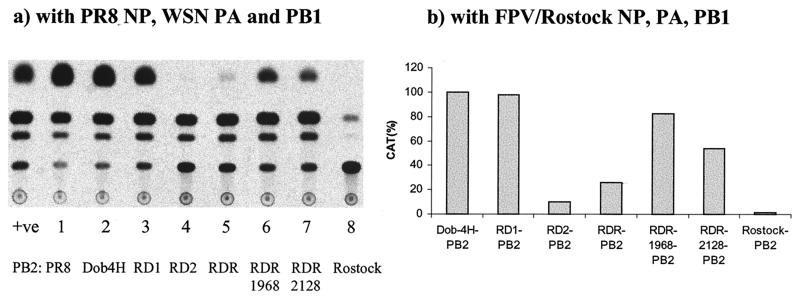
CAT expression levels supported by different PB2 genes measured in a cell-based replication assay. Vero cells were transfected using DOTAP with plasmids directing expression of the PB2 gene along with either pGT-h-NP, derived from A/PR8/34, and pGT-h-PA or pGT-h-PB1, derived from A/WSN/33 (8) (a), or pHMG-FPV-NP, pHMG-FPV-PA, or pHMG-FPV-PB1, derived from A/FPV/Rostock/34 (23) (b), and pPolI-CAT-RT (26). At 48 h posttransfection, cell extracts were prepared and tested for CAT levels using previously described methods (25) (a) or a CAT ELISA kit (b).
The three polymerase proteins (PB1, PB2, and PA) and the nucleoprotein (NP) can transcribe and replicate an influenza virus-like RNA (vRNA) molecule (26). To assess the level of replication supported by different avian PB2 genes in a mammalian cell environment, we inserted the PB2 genes of Rostock and Dobson 4H viruses into the mammalian expression vector pcDNA3. Plasmids pGT-h-NP, pGT-h-PA, and pGT-h-PB1 (2, 1, and 1 μg, respectively), derived from A/PR8/34 and A/WSN/33 (8, 26), and pHMG-FPV-NP, pHMG-FPV-PA, and pHMG-FPV-PB1 (2, 1, and 1 μg, respectively), derived from A/FPV/Rostock/34 (23), were transfected along with 2 μg of pPolI-CAT-RT and 1 μg of either pGT-h-PB2, pcDNA3-Dobson 4H PB2, or pcDNA3-Rostock PB2 into 106 Vero cells in suspension using DOTAP (Boehringer, Mannheim, Germany). Forty-eight hours posttransfection the cells were tested for the expression of chloramphenicol acetyltransferase (CAT) by enzyme-linked immunosorbent assay (ELISA; Roche Molecular Biochemicals) or enzyme assay (25).
Similar levels of CAT were obtained with the Dobson 4H PB2 (Fig. 1a, lane 2) and the human PR8 PB2 (lane 1). This indicates that this avian PB2 protein is compatible with human components of the polymerase complex in Vero cells. On the other hand, substituting Rostock PB2 in this mammalian system gave a very low signal (Fig. 1a, lane 8). This confirms observations made in COS-1 cells that the Rostock PB2 is incompatible with the other human polymerase components (23). In addition, the inability of Rostock PB2 to function well in this system may reflect a defect in the activity of this protein in a mammalian cell environment. Indeed, similar results were obtained when the different PB2s were expressed in combination with avian influenza A virus A/FPV/Rostock/34 PB1 and PA polymerase proteins and NP (Fig. 1b). Dobson 4H PB2 and PR8 PB2 gave rise to high expression of the influenza virus reporter, whereas the expression from our Rostock PB2 was much lower. We also tested the expression of the model RNA driven by the Rostock PB2 gene obtained from van der Werf (23). This PB2 gene gave an intermediate level of replication in Vero cells (data not shown), suggesting that some of the sequence differences between the two Rostock isolates may influence their ability to function in a mammalian cell.
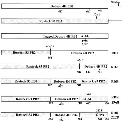
A series of chimeric PB2 cDNAs were constructed in order to map sequence differences between Rostock S3 PB2 and Dobson 4H required for the PB2 protein to complement the other polymerase components and direct replication of a vRNA in Vero cells (Fig. 2). These chimeras had (i) the entire carboxyl region of Rostock S3 from amino acids 362 to 759 exchanged for that of Dobson 4H (RD1), (ii) amino acids 582 to 759 exchanged (RD2); (iii) a central region from amino acids 362 to 581 exchanged (RDR); (iv and v) additional single coding changes resulting in Ile being changed to Met at residue 647 (nucleotide 1968) or Asp changed to Asn at residue 701 (nucleotide 2128) within the RDR chimera (RDR1968 and RDR2128, respectively).
FIG. 2.
PB2 gene constructs to identify the sequences which control host range. A genetic tag was generated by introducing a single silent mutation into the cDNA of Dobson 4H PB2. A single A-to-G substitution at position 1779 resulted in the destruction of a KpnI restriction enzyme site. To construct chimeric RD1 PB2, the 398 residues in the carboxyl region of Dobson 4H PB2 from amino acid 362 (corresponding to nucleotide 1111) were fused to the N terminus of Rostock PB2. In the second chimera, RD2, the exchanged region contains the carboxyl-terminal 178 amino acids of Dobson 4H fused to Rostock sequences. In the third chimera, RDR, only the region encompassing amino acids 362 to 581 (corresponding to nucleotides 1111 to 1772) of Dobson 4H has been exchanged into Rostock. The chimeras RDR1968 and RDR2128 were constructed by introducing single coding changes at residue 647 (nucleotide 1968) or residue 701 (nucleotide 2128) into RDR PB2.
The results showed that insertion of sequence from Dobson 4H PB2 into Rostock PB2 enhanced replication in Vero cells. Thus, when the chimeric PB2s RD1, RDR, RDR1968, and RDR2128 were expressed in the system, significant levels of CAT activity were obtained (Fig. 1a, lanes 3, 5, 6, and 7), although the signal was lower than that for PR8 PB2 or the complete Dobson 4H PB2 (lanes 1 and 2). On the other hand, chimera RD2 gave rise to replication barely higher than Rostock (Fig. 1a, lanes 4 and 8). Similar results were obtained in parallel experiments in which chimeric PB2s were analyzed with A/FPV/Rostock/34 polymerase proteins and NP to replicate the CAT vRNA-like substrate (Fig. 1b). From these results, it would appear that the region between amino acids 362 and 581 contains key amino acid substitutions to promote replication in this system and that single amino acid changes at residues 647 and 701 also contribute to the phenotype.
We next attempted to generate viruses in which RNA segment 1 derives from either the PB2 gene of Dobson 4H or chimeras of sequences from Dobson 4H and Rostock PB2 in order to establish which regions of PB2 might be responsible for productive infection in human and mouse cells. To this end, cDNAs for wild-type Dobson 4H PB2 and the five chimeric PB2s of Rostock and Dobson 4H were cloned into a pPoll-Ribozyme cassette vector (26) by PCR amplification from the pUC19-PB2 plasmids. Additionally, as a genetic marker to monitor rescued transfectant virus, a silent mutation at nucleotide 1779 (A→G), which destroys the KpnI site, was introduced to produce plasmids encoding tagged Dobson 4H PB2.
Vero cells were transfected with pGT-h-NP, pGT-h-PA, pGT-h-PB1, and pGT-h-PB2 (2, 1, 1, and 1 μg, respectively) and 2 μg of either pPolI-Dobson 4H PB2, pPolI-tagged Dobson 4H PB2, pPolI-RD1 PB2, pPolI-RD2 PB2, pPolI-RDR PB2, pPolI-RDR1968, or pPolI-RDR2128, as before. After 36 h, the cells were infected with A/FPV/Rostock/34 at a multiplicity of infection (MOI) of 1. Twenty-four hours postinfection, the medium from the transfected-infected cells was transferred into flasks of L cells for amplification for 3 days, followed by plaquing on L cell monolayers. Individual plaques were picked and amplified in eggs, and RNA was extracted for segment 1-specific RT-PCR. To verify the identity of rescued transfectant viruses T-WT PB2, T-Tagged PB2, and T-RD1 PB2, RT-PCR products were analyzed by digestion with HgaI, a signature of a Rostock PB2 gene. KpnI cleavage confirmed that the PB2 gene was derived from the transfected cDNA. To verify the identity of rescued transfectant viruses T-RDR, T-RDR1968, and T-RDR2128, RT-PCR products were analyzed by digestion with restriction enzymes BamHI (present in Dobson 4H PB2 coding sequence) and EcoRI (present twice in Rostock S3 PB2 coding sequence but only once in Dobson 4H PB2). All PCR products were also sequenced directly to confirm the mutations introduced.
Virus plaques were always obtained following transfection with wild-type Dobson 4H PB2 gene, tagged Dobson 4H PB2 gene, the chimeric RD1 gene, and RDR2128. A low number of plaques were obtained from the RDR and RDR1968 transfections. Plaques were never obtained following transfection of cDNA for RD2 despite its ability to support replication in the plasmid-based replication assay. All the transfectant viruses formed plaques on both mouse L cells and human A549 cells.
These results demonstrate that the abortive infection of an avian influenza virus in mouse and human cells is overcome by transfectant viruses with PB2 genes derived from a mammalian cell-adapted strain of Dobson 4H. The rescue of transfectant virus RDR also confirms that the region between amino acids 362 and 581 of Dobson 4H PB2 enables virus replication in mouse cells. We can speculate that this region of PB2 interacts with a host cell component which varies between avian and mammalian cells. Within this region there are nine amino acid substitutions between Rostock PB2 and Dobson 4H PB2, but only one of these, at position 482, is unique to the 4H variant, with the ability to replicate in mouse L cells, and not found in the parental virus Dobson. However, it is unlikely that a single amino acid substitution in Rostock is sufficient to confer L-cell tropism, since mutants of Rostock S3 that replicate in mouse cells are not readily isolated, suggesting that R482 may act in concert with other residues.
Previous studies have indicated that amino acid residues proximal to the carboxyl terminus of PB2 play a role in host range. Subbarao and coworkers showed that viruses possessing a lysine instead of glutamate at amino acid residue 627 in PB2 were able to replicate in MDCK cells (30), and Penn showed that a mutation at amino acid residue 645 correlated with host range phenotype in MDCK cells (24). In this work we considered the possibility that amino acid substitutions in this region might contribute to virus replication in mouse cells. From a comparison of the amino acid sequences of PB2, two residues at 647 and 701 correlated with the ability of Dobson 4H to replicate in mouse L cells. These substitutions were reconstructed into the chimeric PB2 protein RDR, and the level of replication of a vRNA supported by these proteins was higher than for RDR. In addition, transfectant virus RDR2128 was recovered more readily, indicating that it may replicate more efficiently than RDR in mouse L cells. The ability to recover viruses correlated well with the competence of the PB2 sequences at supporting replication of a model RNA (Fig. 1). Perhaps this simple in vitro assay may be a useful indicator to predict whether mutations in avian polymerase proteins could contribute to an expanded host range.
It is interesting that the PB2 genes from the HK/97 H5 viruses which were able to replicate in humans and in mice contained changes in PB2 at residues 199, 627, 661, and 667 (13) and that a substitution of lysine to glutamine at residue 355 also correlated with high mouse pathogenicity (15a). Whether any of these contributed to the mammalian growth phenotype of the HK viruses is not known but could be tested using an approach similar to that described here.
The abortive replication of FPV Rostock in mouse L cells has been widely studied: replication and transcription of some segments, in particular segment 7, which encodes both M1 and M2 polypeptides, are altered in infected L cells (4, 14, 17, 18, 29), and additionally, the ribonucleoprotein failed to migrate from the nucleus of infected L cells (9, 11, 20).
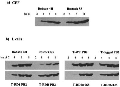
To determine the amount of M1 synthesized during productive and abortive infections, CEF cells or L cells were infected with Dobson 4H, Rostock S3, or transfectant viruses. At 2, 4, 6, and 8 h postinfection cell lysates were prepared and subjected to sodium dodecyl sulfate-polyacrylamide gel electrophoresis (SDS-PAGE) and Western blotting. The accumulation of M1 was followed using a mouse anti-influenza A virus matrix protein monoclonal antibody (Harlan SeraLab) and visualized using enhanced chemiluminescence (ECL) detection (Amersham). Similar amounts of M1 were synthesized in CEF cells infected with Dobson 4H or Rostock S3 (Fig. 3a). However, L cells infected with Rostock made only small amounts of M1 (Fig. 3b). In L cells infected with the rescued transfectant viruses that differ from the Rostock parent only in the origin of their PB2 genes, much more M1 accumulated than in cells infected with Rostock: by 8 h, M1 levels from these infections were similar to those produced by Dobson 4H itself (Fig. 3b). This was also seen from the synthesis of [35S]methionine-labeled polypeptides in cells infected by the different viruses, which revealed that production of M1 protein by Rostock virus infection of L cells was low but production of other viral proteins showed no obvious difference (data not shown).
FIG. 3.
Western blot analysis of expression of M1 in influenza virus-infected CEF (a) and L (b) cells. CEF cells or L cells were infected at high MOI with Dobson 4H, Rostock S3, or transfectant viruses T-WT PB2, T-tagged PB2, T-RD1 PB2, T-RDR, T-RDR1968, or T-RDR2128. At the time points indicated, cells were washed once in ice-cold PBS and incubated with urea-SDS lysis buffer on ice for 10 min. Cell lysates were separated by SDS–15% PAGE and probed for M1 with mouse anti-influenza A virus matrix protein and anti-mouse horseradish peroxidase-conjugated secondary antibodies. Proteins were visualized using an ECL detection system.
To analyze the consequence of altering the PB2 gene on vRNP trafficking, the expression of NP in cells was examined by confocal microscopy. L cells were infected with Dobson 4H, Rostock S3, or transfectant viruses. At 2, 4, 6, and 8 h postinfection, cells were processed for immunofluorescence analysis by fixation with 4% paraformaldehyde in phosphate-buffered saline (PBS) for 10 min at room temperature and then permeabilized with 0.2% Triton X-100–PBS. Cells were stained with mouse anti-influenza A virus nucleoprotein monoclonal antibody (Harlan-SeraLab) and fluorescein isothiocyanate-conjugated goat anti-mouse immunoglobulin. Fluorescence was viewed with a Zeiss confocal microscope.
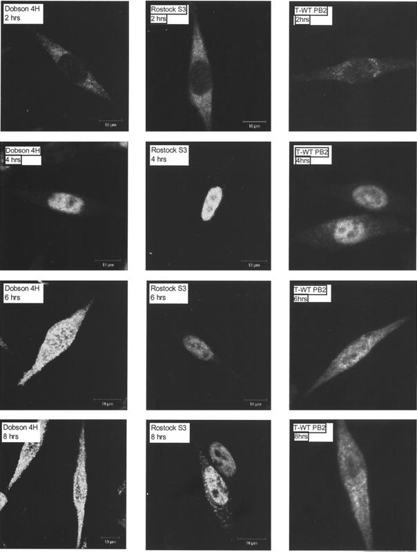
When L cells were infected at a high MOI (Fig. 4), NP was almost exclusively localized to the nucleus in all infected cells at 4 h postinfection. At 6 h or later, NP was present in both the cytoplasm and the nucleus of all cells infected by Dobson 4H and transfectant virus T-WT PB2, but it remained predominantly nuclear in cells infected with Rostock S3. When the distribution of NP for the complete panel of rescued transfectant viruses was analysed at 8 h after infection at low MOI, cytoplasmic NP could be observed. In contrast, no nuclear-cytoplasmic transport was observed for cells infected with Rostock S3 (data not shown). In conclusion, the abortive infection of A/FPV/Rostock in L cells results in a reduction in the synthesis of vRNA segment 7, a reduced synthesis of M1 protein, and a failure to transport vRNPs from the nucleus to the cytoplasm at late stages of replication. Why altering the PB2 gene should overcome this block is still not clear.
FIG. 4.
Localization of NP in L cells during infection. L cells were grown on glass coverslips to 50 to 70% confluency and infected at high MOI with Dobson 4H, Rostock S3, or T-WT PB2. At 2, 4, 6, and 8 h postinfection, cells were processed for immunofluorescence analysis by fixation with 4% paraformaldehyde in PBS for 10 min and then permeablized with 0.2% Triton X-100 in PBS for 5 min. Cells were stained with a mouse anti-influenza A virus nucleoprotein monoclonal antibody and an anti-mouse immunoglobulin G-fluorescein isothiocyanate conjugate. Fluorescence was viewed with a confocal microscope.
Acknowledgments
We thank G. G. Brownlee and E. Fodor (University of Oxford) for discussions. We thank P. Palese (Mount Sinai Medical Center) for pPolICat-RT and human influenza A virus polymerase gene expression plasmids and S. van der Werf and N. Naffakh (Institut Pasteur) for the A/FPV/Rostock/34 plasmids used in the CAT assays. J.W.M. thanks M. Hill for assistance.
Louise Mingay is supported by MRC program grant G9523972 awarded to G. G. Brownlee.
REFERENCES
- 1.Almond J W. A single gene determines the host range of influenza virus. Nature. 1977;270:617–618. doi: 10.1038/270617a0. [DOI] [PubMed] [Google Scholar]
- 2.Almond J W, Barry R D. A single gene controlling the host range of fowl plague virus. In: Mahy B W J, Barry R D, editors. Negative strand viruses and the host cell. New York, N.Y: Academic Press; 1978. pp. 675–684. [Google Scholar]
- 3.Barrett T, Inglis S C. Growth, purification and titration of influenza viruses. In: Mahy B W J, editor. Virology: a practical approach. Oxford, U.K: IRL Press; 1985. pp. 119–150. [Google Scholar]
- 4.Bosch F X, Hay A J, Skehel J J. RNA and protein synthesis in a permissive and an abortive influenza virus infection. In: Mahy B W J, Barry R D, editors. Negative strand viruses and the host cell. New York, N.Y: Academic Press; 1978. pp. 465–474. [Google Scholar]
- 5.Connor R J, Kawaoka Y, Webster R G, Paulson J C. Receptor specificity in human, avian, and equine H2 and H3 influenza virus isolates. Virology. 1994;205:17–23. doi: 10.1006/viro.1994.1615. [DOI] [PubMed] [Google Scholar]
- 6.De Jong J C, Claas E C, Osterhaus A D, Webster R G, Lim W L A. Pandemic warning? Nature. 1997;389:554. doi: 10.1038/39218. [DOI] [PMC free article] [PubMed] [Google Scholar]
- 7.Dybing J K, Schultz-Cherry S, Swayne D E, Suarez D L, Perdue M L. Distinct pathogenesis of Hong Kong-origin H5N1 viruses in mice compared to that of other highly pathogenic H5 avian influenza viruses. J Virol. 2000;74:1443–1450. doi: 10.1128/jvi.74.3.1443-1450.2000. [DOI] [PMC free article] [PubMed] [Google Scholar]
- 8.Fodor E, Devenish L, Engelhardt O G, Palese P, Brownlee G G, Garcia-Sastre A. Rescue of influenza A virus from recombinant DNA. J Virol. 1999;73:9679–9682. doi: 10.1128/jvi.73.11.9679-9682.1999. [DOI] [PMC free article] [PubMed] [Google Scholar]
- 9.Franklin R M, Breitenfeld P M. The abortive infection of Earle's L-cells by fowl plague virus. Virology. 1959;8:293–307. doi: 10.1016/0042-6822(59)90031-5. [DOI] [PubMed] [Google Scholar]
- 10.Gambaryan A S, Tuzikov A B, Piskaev V E, Yamnikova S S, Lvov D K, Robertson J S, Bovin N V, Matrosovich M N. Specification of receptor-binding phenotypes of influenza virus isolates from different hosts using synthetic sialylglycopolymers: non-egg-adapted human H1 and H3 influenza A and influenza B viruses share a common high binding affinity for 6′-sialyl(N-acetyllactosamine) Virology. 1997;232:345–350. doi: 10.1006/viro.1997.8572. [DOI] [PubMed] [Google Scholar]
- 11.Gandhi S S, Bell H B, Burke D C. Abortive infection of L cells by fowl plague virus: comparison of RNA and protein synthesis in infected chick and L cells. J Gen Virol. 1971;13:423–432. doi: 10.1099/0022-1317-13-3-423. [DOI] [PubMed] [Google Scholar]
- 12.Gao P, Watanabe S, Ito T, Goto H, Wells K, McGregor M, Cooley A J, Kawaoka Y. Biological heterogeneity, including systemic replication in mice, of H5N1 influenza A virus isolates from humans in Hong Kong. J Virol. 1999;73:3184–3189. doi: 10.1128/jvi.73.4.3184-3189.1999. [DOI] [PMC free article] [PubMed] [Google Scholar]
- 13.Hiromoto Y, Yamazaki Y, Fukushima T, Saito T, Lindstrom S E, Omoe K, Nerome R, Lim W, Sugita S, Nerome K. Evolutionary characterization of the six internal genes of H5N1 human influenza A virus. J Gen Virol. 2000;81:1293–1303. doi: 10.1099/0022-1317-81-5-1293. [DOI] [PubMed] [Google Scholar]
- 14.Inglis S C, Brown C M. Differences in the control of virus mRNA splicing during permissive or abortive infection with influenza A (fowl plague) virus. J Gen Virol. 1984;65:153–164. doi: 10.1099/0022-1317-65-1-153. [DOI] [PubMed] [Google Scholar]
- 15.Ito T, Couceiro J N, Kelm S, Baum L G, Krauss S, Castrucci M R, Donatelli I, Kida H, Paulson J C, Webster R G, Kawaoka Y. Molecular basis for the generation in pigs of influenza A viruses with pandemic potential. J Virol. 1998;72:7367–7373. doi: 10.1128/jvi.72.9.7367-7373.1998. [DOI] [PMC free article] [PubMed] [Google Scholar]
- 15a.Katz J M, Lu X, Tumpey T M, Smith C B, Shaw M W, Subbarao K. Molecular correlates of influenza A H5N1 virus pathogenesis in mice. J Virol. 2000;74:10807–10810. doi: 10.1128/jvi.74.22.10807-10810.2000. [DOI] [PMC free article] [PubMed] [Google Scholar]
- 16.Kawaoka Y, Krauss S, Webster R G. Avian-to-human transmission of PB1 gene of influenza A virus in the 1957 and 1968 pandemics. J Virol. 1989;63:4603–4608. doi: 10.1128/jvi.63.11.4603-4608.1989. [DOI] [PMC free article] [PubMed] [Google Scholar]
- 17.Lau S C, Scholtissek C. Abortive infection of Vero cells by an influenza A virus (FPV) Virology. 1995;212:225–231. doi: 10.1006/viro.1995.1473. [DOI] [PubMed] [Google Scholar]
- 18.Lohmeyer J, Talens L T, Klenk H D. Biosynthesis of the influenza virus envelope in abortive infection. J Gen Virol. 1979;42:73–88. doi: 10.1099/0022-1317-42-1-73. [DOI] [PubMed] [Google Scholar]
- 19.Ludwig S, Stitz L, Planz O, Van H, Fitch W M, Scholtissek C. European swine virus as a possible source for the next influenza pandemic? Virology. 1995;212:4603–4608. doi: 10.1006/viro.1995.1513. [DOI] [PubMed] [Google Scholar]
- 20.Mahy B W J, Barrett T, Briedis D J, Brownson J M, Wolstenholme A J. Influence of the host cell on influenza virus replication. Phil Trans R Soc Lond B. 1980;288:349–357. doi: 10.1098/rstb.1980.0011. [DOI] [PubMed] [Google Scholar]
- 21.Matrosovich M N, Gambaryan A S, Teneberg S, Piskarev V E, Yamnikova S S, Lvov D K, Robertson J S, Karlsson K A. Avian influenza A viruses differ from human viruses by recognition of sialyloligosaccharides and gangliosides and by a higher conservation of the HA receptor-binding site. Virology. 1997;233:224–234. doi: 10.1006/viro.1997.8580. [DOI] [PubMed] [Google Scholar]
- 22.Matrosovich M, Zhou N, Kawaoka Y, Webster R. The surface glycoproteins of H5 influenza viruses isolated from humans, chickens, and wild aquatic birds have distinguishable properties. J Virol. 1999;73:1146–1155. doi: 10.1128/jvi.73.2.1146-1155.1999. [DOI] [PMC free article] [PubMed] [Google Scholar]
- 23.Naffakh N, Massin P, Escriou N, Crescenzo-Chaigne B, van der Werf S. Genetic analysis of the compatibility between polymerase proteins from human and avian strains of influenza A viruses. J Gen Virol. 2000;81:1283–1291. doi: 10.1099/0022-1317-81-5-1283. [DOI] [PubMed] [Google Scholar]
- 24.Penn C. The role of RNA segment 1 in an in vitro host restriction occurring in an avian influenza virus. Virus Res. 1989;12:349–360. doi: 10.1016/0168-1702(89)90092-0. [DOI] [PubMed] [Google Scholar]
- 25.Percy N, Barclay W S, Sullivan M, Almond J W. A poliovirus replicon containing the chloramphenicol acetyltransferase gene can be used to study the replication and encapsidation of poliovirus RNA. J Virol. 1992;66:5040–5046. doi: 10.1128/jvi.66.8.5040-5046.1992. [DOI] [PMC free article] [PubMed] [Google Scholar]
- 26.Pleschka S, Jaskunas S R, Engelhardt O G, Zurcher T, Palese P, Garcia-Sastre A. A plasmid-based reverse genetics system for influenza A virus. J Virol. 1996;70:4188–4192. doi: 10.1128/jvi.70.6.4188-4192.1996. [DOI] [PMC free article] [PubMed] [Google Scholar]
- 27.Roditi I J, Robertson J S. Nucleotide sequence of the avian influenza virus A/Fowl Plague/Rostock/34 segment 1 encoding the PB2 polypeptide. Virus Res. 1984;1:65–71. [Google Scholar]
- 28.Schultz U, Fitch W M, Ludwig S, Mandler J, Scholtissek C. Evolution of pig influenza viruses. Virology. 1991;183:61–73. doi: 10.1016/0042-6822(91)90118-u. [DOI] [PubMed] [Google Scholar]
- 29.Smith G L, Hay A J. Replication of the influenza-virus genome. Virology. 1982;118:66–108. doi: 10.1016/0042-6822(82)90323-3. [DOI] [PubMed] [Google Scholar]
- 30.Subbarao K, London W, Murphy B R. A single amino acid in the PB2 gene of influenza A virus is a determinant of host range. J Virol. 1993;67:1761–1764. doi: 10.1128/jvi.67.4.1761-1764.1993. [DOI] [PMC free article] [PubMed] [Google Scholar]
- 31.Subbarao K, Klimov A, Katz J, Regnery H, Lim W, Hall H, Perdue M, Swayne D, Bender C, Huang J, Hemphill M, Rowe T, Shaw M, Xu X, Fukuda K, Cox N. Characterization of an avian influenza A (H5N1) virus isolated from a child with a fatal respiratory illness. Science. 1998;279:393–396. doi: 10.1126/science.279.5349.393. [DOI] [PubMed] [Google Scholar]


