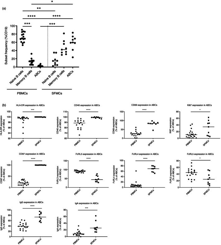
FIGURE 5.

Age‐associated B cells (ABCs) are more abundant in synovial fluid (SF), show a more activated and proliferative phenotype, and their FcRL3, FcRL4 and FcRL5 expression differ from their peripheral blood (PB) counterparts. B‐cell subsets were detected by flow cytometry in the PB or SF of a cross‐sectional cohort of RA patients. The cell subsets were gated as outlined in Figure S1a. PB mononuclear cell (PBMC) (n = 19) and SF mononuclear cell (SFMC) (n = 9). (a) The frequency of each B‐cell subsets in PBMCs and SFMCs is shown as a percentage of total CD19+ B cells. (b) The percentage of positive cells for each marker in the ABC subset is shown. The horizontal lines represent the median value. Statistical significance was assessed using the Mann–Whitney U test; *p < 0.05; **p < 0.01; ***p < 0.001; ****p < 0.0001
