Abstract
Anatomy teachers urge students to come to education sessions at the dissection rooms as well-prepared as possible. To effectuate optimal preparation, assignments are designed to guide the students’ learning processes. These assignment often include the use of anatomical figures in atlases. The role of augmented reality (AR) applications in helping students prepare for body donor-based teaching sessions at the dissection rooms remains quite elusive. Therefore, this study examines the effects of the use of an AR application compared to the use of anatomical atlases in helping students (n = 28) prepare for a neuroanatomy session at the dissection rooms with prosections. The study shows that students from both groups showed a similar improvement in anatomy test scores. The amount of experienced cognitive engagement, however, is higher in the experimental AR group. Based on these results, it can be suggested that an AR application is a valid method to help students prepare and could be an alternative to the use of anatomical atlases. Nevertheless, future studies should re-investigate this research question in larger cohorts. Also, it remains unknown whether cognitive engaged students are indeed the students who are better prepared for educational sessions at the dissection rooms.
Keywords: Augmented reality, Gross anatomy education, Medical education, Preparation
Subject terms: Anatomy, Medical research
Introduction
Understanding the function and temporospatial aspects of the human body is an essential part in the education of becoming a medical professional1. However, for some medical students, this can be a challenging task2. To support this dearth various teaching methods and tools are offered to students. Traditionally anatomical education is based on teaching with body donors, including body donor-based education session at the dissection room and the use of prosected specimens. These aforementioned modalities can help students to understand specific anatomical relations of different structures and organs. Albeit, with world-wide changes in medical curricula, the hours spend on anatomy teaching are decreasing3,4. Additionally, the use of body donors in anatomy education is hampered by logistic, financial and sometimes social-ethical challenges1,5. Therefore, teaching sessions at the dissection rooms are getting more sparse6.
Two dimensional (2D) teaching methods and tools are also often used in anatomy teaching. These methods warrant that static images are mentally rotated by the student to gain insight in spatial properties and relationships of structures and organs6. These insights can be inaccurate as not all students master the capacities needed for the correct mental rotation of static images7. Therefore, students could be supported by providing them different 3D learning materials5.
One of these 3D learning materials concerns AR, in which the digital image is placed on top of the image of the real world by use of a head-mounted device (i.e., AR-glasses) or a screen (e.g., a tablet or a smartphone)1,2,6,8. Preferably, users can interact with elements from the real world and the augmented world, indicating that the AR-model can be enlarged or reduced in size, elements can be removed or isolated and cross-sections can be displayed2,6.
The use of AR models when studying anatomy triggers the interest of students5,9–13, which is a powerful motivator and ensures that students are more cognitively engaged14,15. Students will also put more energy into the subject for a longer period of time in order to reach a higher academic achievement16,17. The academic achievement is the improvement of a student on a course in comparison to the amount of time he or she needed to make that improvement18. To ensure that students get cognitively engaged and therefore recognize the importance of a task, the learning task must be challenging enough and must draw the attention of the students14. Drawing the students attention can be achieved by making sure that the teaching method is presented in a surprising and novel way, whereby the excitement and interests of students are increased19. This ultimately creates a setting that makes students more involved in the subject or task (e.g., a high cognitive engagement) and potentially leading to a higher academic achievement. Due to the novelty of the use of AR models during studying anatomy, students are more interested than when they are using the current teaching methods and tools as 2D education, 3D models and body donor-based education session at the dissection room/prosection6,9,20. The use of AR-models has been suggested to be equally effective as other teaching methods such as 2D teaching methods and 3D models6,21,22. Albeit, the studies included in these reviews mainly compared AR models with more traditional anatomy teaching tools with regard to differences in anatomy test results. However, as reviewed by McBain et al., the literature on the role of AR in anatomical teaching is still in its infancy as many of the studies to date have focused on assessing feasibility, usability, and perceived learning experiences23. The optimal position of AR in anatomy education (e.g., as an additional tool at the dissection rooms, as a replacement of body donor-based education sessions at the dissection rooms or as a preparatory tool) remains understudied.
In The Netherlands, anatomy education is primarily provided to (bio)medical students during the first three years of their individual curricula (Bachelor’s phase) by combining individual assignments, (interactive) lectures, small working group assignments and body donor-based education at the dissection rooms24. At the dissection rooms, prosected specimens, cross-sections, plastinated specimens and (plastic) anatomical models are provided for students to study anatomy. Students are encouraged to visit these sessions well-prepared24,25. To help students prepare, individual assignments are provided which offer guidance for students to browse through different anatomical atlases, pre-recorded movies and/or digital three dimensional (3D) reconstructions. The assignments help students to critically review relevant anatomical figures and reflect on their own perceived knowledge of the specific topic. Students can complete these assignments individually or within small groups, depending on their own preferences. The effectiveness of the use of an AR-application as a preparatory teaching tool prior to body donor-based education sessions at the dissection rooms, however, remains largely elusive. Regarding neuroanatomy education, this is provided to (bio) medical students during the second half of the second Bachelor’s year.
Hence, this study investigated the effectiveness of an AR-application as a preparatory teaching tool for students prior to visiting a body donor-based education session as compared to the use of traditional preparatory methods using anatomical atlases. Next to impact on academic achievement, this study also investigated the effect of AR-models and anatomical atlases on the cognitive engagement of students. Finally, this study investigated the possible impact of cognitive engagement elicited by the different preparatory tools on the anatomy test scores. We hypothesized that the use of an AR-application to prepare for a body-donor based education session was equally effective as using anatomical atlases. However, the use of an AR-application was hypothesized to yield a higher cognitive engagement from the students.
Materials and methods
Participants
This study was conducted at the Faculty of Medical Sciences (Radboud University Medical Center) at the Radboud University in Nijmegen, the Netherlands. First-year and second-year students of Medicine and Biomedical Sciences were recruited as they had received no neuroanatomy education prior to this experiment. Students were allocated over the two groups, based on order of enrolment and additionally based on gender. Participation was voluntarily and students were recruited by means of (online) announcements/advertisements and posters. Students could sign up for the experiment by sending an e-mail to one of the researchers (I.Z.S.), after which they received an information letter containing details of the experiment. Students were invited to come to the university for one extra-curricular evening session (total duration of the experiment: 3.5 h). Prior to the start of this session, all students provided written informed consent.
Ethical approval
Ethical approval
was obtained from the medical ethical review board of the Dutch Society for Medical Education in The Netherlands. Additionally, all students engaged voluntarily and were treated in accordance to the principles outlines in the declaration of Helsinki.
Study design
A pre-test-post-test control group design was used in this study. Students were randomly placed in two groups: one group which prepared the assignment by use of an AR application (the experimental group) and one group of students who prepared the assignment by use of anatomical atlases (the control group). The experimental group prepared for the body donor-based education session at the dissection room using a preparatory assignment with use of the AR application GreyMapp-AR: Brain. GreyMapp-AR is an in-house developed, tablet-based AR application that can be used to study neuroanatomy26. Students in the control group performed the same preparatory assignment using the Sobotta Anatomical Atlas. For this experiment, the Sobotta Anatomical Atlas was chosen as this is the educational standard at our institute.
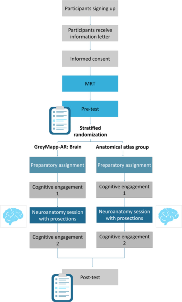
All students received a Mental Rotation Test (MRT), a pre-test, a cognitive involvement questionnaire on two occasions, the body donor-based education session at the dissection room and finally a post-test. Figure 1 depicts a flow chart of the study design. All tests and questionnaires were completed individually. The preparatory assignment and the body donor-based education session were completed in small groups of three to four students. These groups were not formed beforehand but were created at random by the students.
Fig. 1.
Flowchart of the study design.
GreyMapp-AR: brain
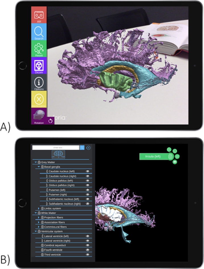
Details with regard to the development of GreyMapp-AR: Brain have been described in detail elsewhere12. In summary, a T1-weighted 7 Tesla post-mortem magnetic resonance imaging scan of the brain of a deceased female body donor was used to manually segment various brain structures (Fig. 2). These segmentations were converted into three-dimensional volumes and mesh-files. Mesh-files were imported in an in-house made application using Unity cross platform game engine, version 2022.2.1 (Unity Technologies ApS, San Francisco, CA). This application, called GreyMapp-AR: Brain, can be exported to tablets and smartphones in order to work with. Figure 3 provides exemplary images of GreyMapp-AR: Brain as displayed on an iPad.
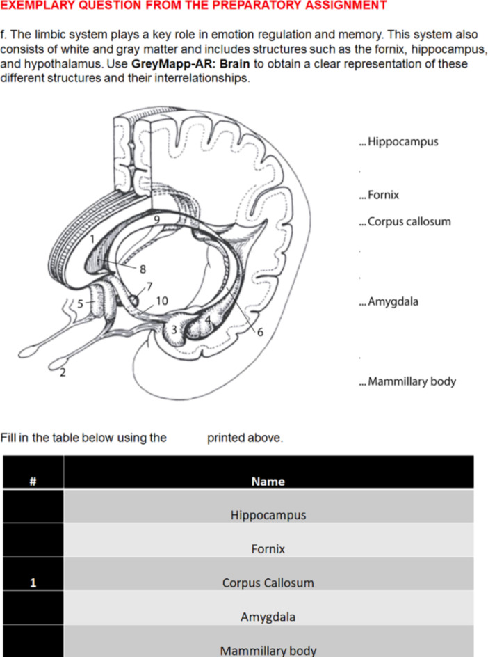
Fig. 2.
Exemplary question from the preparatory assignment.
Fig. 3.
Exemplary images GreyMapp-AR: Brain.
Users can use GreyMapp-AR: Brain on their device by focusing the device’s camera on a flat underground. Standard orthographic views of the model (left lateral, right lateral, frontal, basal, et cetera) can be set by touching the screen. In order to change the user’s perspective of the brain, the user needs to walk around the AR-model. Additionally, structures can be removed from the model or isolated from other structures by touching the screen. Finally, the axial, sagittal and coronal MRI slices could be made visible in order to create a better understanding of the 2D-3D relationship of different structures. Using this feature allows students in both groups to study sectional anatomy as well as gaining a 3D insight in neuroanatomical structures.
Instruments
To ensure that both groups on average had the same level of spatial awareness, an MRT was performed at the beginning of the experiment27. The MRT is used to measure the ability to rotate a given 2D- or 3D-image mentally27. Students were shown an image of different cubes fit together in a particular form. Students were asked to identify which of the following four images were rotated versions of the first image. Only two out of four represent the original form of stashed cubes. Students received three separate parts of the test. The first one was used as a practice and example. The results of second and third part were included in the results. Each part consisted of twelve items, which the students had to fulfil in three minutes. Students did not have to complete the test completely within the allotted time. One point was given for each correct answer. With incomplete or incorrect answers no additional points were assigned. The maximum score for the two parts together was 24 (part 1: 12 points; part 2: 12 points).
Cognitive engagement was measured using four items by Rotgans and Schmidt (2011), which were: (1) “I was engaged with the topic at hand”, (2) “I put in a lot of effort”, (3) I wish we could still continue with the work for a while” and (4) “I was so involved that I forgot everything around me”. The four statements could be answered using a 5-point Likertscale ranging from 1-strongly disagree to 5-strongly agree16.
The pre- and post-test were used to assess the knowledge of students concerning neuro-anatomical structures and their individual relations. The pre- and post-test are derived from Kooloos et al. (2014) and were empirically used in various other studies12,28. The format of the pre- and post-test was identical although both contained variant questions. The pre- and post-test each consisted of three parts: (1) a part with multiple choice question (n = 9), (2) a part with dichotomous questions (n = 11) and (3) a part with cross-sections containing five outlined structures that needed to be named28. Students were first presented with the first two parts of the pre-test/post-test. The last part of the test was not provided until the student submitted the first two parts of the test in order to prevent that students used the cross sections to answer other questions. Students could achieve a maximum score of 35 points for each test (part 1: 9 points; part 2: 11 points; part 3: 15 points).
Preparatory assignment
The students in the experimental group received an assignment using the tablet-based version of the AR application called “GreyMapp-AR: Brain” on an iPad. Students in the control group received an assignment using the Sobotta Atlas. Prior to the assignment, students were given a short introductory training by one of the researchers (L.B. or D.H.) about the use of the tool they were about to use. In comparison to all the previously mentioned tests and questionnaires, the preparation assignment and the body donor-based education session at the dissection room were not used to collect data. The assignment was solely used to prepare students for the body donor-based education session at the dissection room. Both groups received the exact same questions during the preparation assignment, which they needed to answer using the tool they could use, depending on the research group they were placed in. The preparatory assignment involved students to gain a global insight in cortical anatomy and the ventricular system. For this, the students needed to answer questions using either the AR application or the anatomical atlas. When using the AR application, participants were motivated to rotate the model virtually and describe the relationships between structures. When using the atlas, participants were motivated to search for figures depicting different angles and describe the relationships between structures. An exemplary question is provided in Fig. 2. To prevent that any group had an advantage over the other, the figures used in the preparatory assignment were not sourced from the Sobotta Atlas or the AR application.
The body donor-based education session at the dissection room took place the same evening. Both groups worked with similar prosections of brain specimens and plastic models. This education session required active engagement from the participants. All questions were the same for each group. The groups worked simultaneously on these questions, although the groups were separated from each other.
Statistical analyses
The collected data was first pseudonymised and afterwards manually entered into the statistical package SPSS Statistics (v27; IBM SPSS Statistics for Windows, version 22.0, released 2013; IBM Corp., Armonk, NY, U.S.A.). To determine if there is a difference in anatomy test scores between the two groups, a Repeated Measure ANOVA was performed. A sensitivity analysis was performed to calculate the effect size Cohen’s f. A One Way ANOVA was used to determine if there was a significant difference between the average scores in cognitive engagement between the Greymapp-AR: Brain group and the Sobotta Atlas group. The statistical program PROCESS29 was applied to the data to determine if cognitive engagement impacts the relationship between variation of preparatory assignment and anatomy test scores. Bootstrap confidence intervals of the indirect effect is based on 5000 bootstrap samples. A Cronbach’s  of the four items of cognitive engagement was calculated to assess whether the test was internally consistent and therefore reliable to use within this experiment. Internal consistency is generally regarded acceptable when ≥ 0.7. Finally, a t-test was performed to determine whether the scores of cognitive engagement on the two measurements differed from each other. Statistical significance was assumed when P< 0.05. Post-hoc correction using the Bonferroni-correction was carried out.
Results
A total of 28 students were recruited with 17.9% (n = 5) being a first year student and 82.1% (n = 23) being a second year student. The population consisted of 35.7% (n = 10) male students and 64.3% (n = 18) female students, which were divided equally over the experimental and control group. The descriptive statistics from the MRT, cognitive engagement (CE), pre-test and post-test and the score difference between the pre-test and post-test from the experimental and control group can be found in Table 1.
Table 1.
Descriptive statistics mental rotation test (MRT), cognitive engagement (CE), pre-test and post-test per study group.
| Maximum score | AR-group (n = 14) | Sobotta-group (n = 14) | |||
|---|---|---|---|---|---|
| M | SD | M | SD | ||
| Mental rotation test | 24 | 10.79 | 4.17 | 13.64 | 4.29 |
| CE after preparatory assignment | 5 | 3.98 | 0.53 | 3.19 | 0.47 |
| CE after body donor-based education | 5 | 3.70 | 0.67 | 3.57 | 0.45 |
| Pre-test | 35 | 7.21 | 2.94 | 7.29 | 2.05 |
| Post-test | 35 | 15.00 | 3.60 | 13.43 | 4.38 |
| Difference post-test and pre-test | 35 | 7.79 | 3.58 | 6.14 | 5.42 |
Mental rotation test & cognitive engagement
A one-way ANOVA test showed no significant differences between the experimental AR group and the control Sobotta Atlas group in terms of the MRT, F(1,26) = 3.19; P = 0.09. Therefore, it was assumed that both groups have the same degree of spatial awareness. Cronbach’s  of the two tests for cognitive engagement was 0.71 and 0.72, indicating that the test is internally consistent and can be used within this experiment to assess cognitive engagement.
The mean scores of cognitive engagement of the AR group during the preparation assignment are significantly higher than the mean scores in the control group, F(1,25) = 16.62, P < 0.001. In contrast, the AR group’s mean cognitive engagement scores during the body donor-based education session at the dissection room are not significantly different from the mean scores of the control group, F(1,26) = 0.33, P = 0.57. Implying that after the preparation the students in the AR group were more cognitive engaged than students in de control group, but that the amount of experienced cognitive engagement after the body donor-based education session at the dissection room was the same for both groups.
A t-test was performed to determine whether there is a significant decrease or increase in the amount of experienced cognitive involvement in both groups when comparing the two measuring moments. The measuring differences between the two measuring moments on cognitive engagement per study groups is not significant (AR group, t = −1,31, P = 0.21; control group, t = 2.09, P = 0.06). The experienced cognitive engagement at the two measuring moments are the same for both groups.
Pre-test and post-test
Both groups had higher scores on the post-test than on the pre-test and this study found a significant difference in pre-test and post-test per group, F(1,26) = 64.42, P < 0.001. This indicates that all students, independent form the study group they were assigned to, learned from the received education. Furthermore, there is no significant correlation between the pre- and post-test (r = 0.07, P = 0.74, N = 28), which means that they are independent of each other.
A Repeated Measures ANOVA was used to determine whether the level of anatomy test scores of students in the AR group differs from the level of anatomy test scores of students in the control group, as described in the second question. This analysis showed no significant interaction effect between the difference in study group and the difference in anatomy test scores, F(1,26) = 0.90, P = 0.35. This means that students from both groups showed a similar progress when comparing the scores of the pre-test and post-test. A sensitivity analysis showed the Cohen’s maximum detectable effect size f = 0.40, which means that only very large effects can be demonstrated within this sample size.
The impact of cognitive engagement on the relationship between difference in study group and anatomy test scores, was measured using PROCESS. The analysis shows that cognitive engagement during the preparation assignment has no significant impact on the difference in tool used and the difference in anatomy test scores, Bootstrap 95% CI [−1.15, 3.19].
Discussion
The objective of the present study was to examine whether there are differences in anatomy test scores and cognitive engagement of (bio)medical students when using an AR application or an anatomical atlas during a preparatory assignment prior to their participation in a body donor-based education session at the dissection rooms.
The results indicate that a preparatory assignment using an AR application leads to a higher degree of cognitive engagement in comparison with a preparation assignment using a Sobotta Atlas. During the body donor-based education session at the dissection room, there were no differences in the degree of experienced cognitive engagement by the groups of students. In terms of anatomy test scores it can be said that students in both groups performed equally. In addition, it cannot be confirmed that cognitive engagement elicited by the different tools to complete the preparatory assignments impacted the anatomy test scores.
Regarding the effect of AR models and anatomical atlases on students’ cognitive engagement, the results show that students who completed the preparatory assignment using the AR application experienced more cognitive engagement than students who completed the preparatory assignment using the Sobotta Atlas. The results are in line with the studies of Berlyne (1970) and Harackiewicz et al. (2016), which showed that challenging teaching can lead to attracting the student’s attention, which makes the student more interested in the subject and causes the increase of cognitive engagement14,19. Due to their interest in AR, students feel more cognitively engagement during their preparation assignment. In contrast, students who are using an anatomical atlas were less cognitive engaged during their preparation assignment. Therefore, the use of AR can be seen as a valuable addition to the current neuroanatomy education.
The experienced cognitive engagement during the body donor-based education session at the dissection room is equal for every student, regardless of the way they made the preparation assignment. Students find the body donor-based education session at the dissection room, where they can touch and view brains of the human body, interesting and therefor it attracts their attention30. To prepare for body donor-based education session at the dissection room both an AR application and the Sobotta Atlas are good teaching methods.
Based on the research results, the use of an AR application as a preparatory learning tool for students prior to visiting a body donor-based teaching session in the dissection rooms can be seen as an equally effective method as the Sobotta Atlas. Students who made a preparation assignment using the AR application achieved a similar level of anatomy test scores as students who made the same preparation assignment using the Sobotta Atlas. The results of this study suggest that the method of preparation leads to no difference in anatomy test scores. These results contradict former claims of Moro et al. (2017) and Peterson and Mlynarczyk (2016) who see evidence that the addition of AR to anatomy education can lead to higher anatomy test scores levels6,31. A likely explanation is that those two studies used a different alternative to AR in the control group. Moro et al. (2017) used a VR application as alternative to compare with the AR application6. Peterson and Mlynarczyk (2016) provided their experimental group with an extra tool in the form of digital 3D visualisations as an addition of the textbook and the body donor teaching session education. The control group received a textbook and the body donor teaching session education to study anatomy31. The results of the current study, on the other hand, are similar to those reported by a more recent review and meta-analysis by Moro et al. (2021) who found an equally reached academic achievement when comparing AR education to the current 2D and 3D anatomy education21.
An alternative explanation for the lack of difference in anatomy test scores between the use of AR and the use of the Sobotta Atlas is that both teaching methods are a form of multimedia learning. Multimedia learning means that new information is presented to students in different kind of ways32. An AR application in combination with a body donor-based education session at the dissection room and a Sobotta Atlas in combination with a body donor-based education session at the dissection room, are both teaching methods using two different channels to enhance future remembrance. When using a textbook and a body donor-based education session at the dissection room, the two channels are easy to recognize as text with 2D images versus manipulation of the brain. When using an AR application and body donor-based education session at the dissection room the two channels can be more difficult to identify as such. Both AR application and body donor-based education session at the dissection room use 3D visualisation, but in a different way. During the body donor-based education session at the dissection room, students are allowed to touch and lift the human brain, while during working with the AR application students can view and manipulate the human brain digitally. An advantage of the AR application is that text can be added on the screen, parts of the brain can be removed or added and certain parts of the brain can be showed in relation to other parts of the same brain. The two channels used in AR versus body donor-based education session at the dissection room are text with 3D images versus manipulation of the brain. Both use of the AR application and use of the Sobotta Atlas are therefore a form of multimedia learning, which may explain why both forms are equally effective in anatomy test scores.
Limitations of the study
One limitation of this study was the relatively short amount of time that students got to get used to the AR application. Because this took a certain amount of time, less time was left to actually complete the research assignment in comparison to the student who used a Sobotta Atlas. However, it is anticipated that AR will be more often used during class in the near future. The more often students practice with AR, the easier it will be to use during assignments within class. This way, more time will be left to actually learn5,6,33.
A second limitation concerns the limited amount of students participating in this study. Although the study was designed to include more participants, it was not found feasible to achieve this due to the relatively sparse subscriptions. Therefore, it would be recommended to re-investigate the research question in a larger cohort, preferably in a multi-center setting.
Additionally, as the testing method is known to impact outcomes, the use of a paper test might have influenced results. However, this impact is believed to be minimal due to the variety of question types including in this test. The test contained non-spatial multiple choice questions, spatial multiple choice questions and cross-sections and thereby, the majority of the questions have a positive correlation with regard to elucidating the relationship between students’ spatial abilities and anatomy knowledge assessment scores34. Furthermore, the test used in this study was developed by anatomy teachers and has been validated by its use in various education experiments12,27.
Another limitation of this study concerns the lack of quantitative assessment tools to measure the effects of AR technology on the learner. As suggested by McBain et al., future research should incorporate biosensors measuring stress, cognitive load, arousal or measurement methods to analyse the activation of different brain regions during cognitive tasks when working with AR applications to study neuroanatomy23.
Recommendations for future research
It is worth in future research students get to know how to use an AR application so that all students have the same amount of time to spend on the assignment of the research. A second recommendation for follow-up research is therefore to repeat the current research with more students in order to demonstrate a smaller effect. The last recommendation for follow-up research concerns the moment of the second measurement of cognitive engagement. The second measurement took place directly after the body donor-based education session at the dissection room to make it possible to compare with the cognitive engagement experienced during the preparation assignment. For future research it may be interesting to postpone this moment, in order to be able to make statements about the experienced cognitive engagement during the entire education or about the effect of AR over a longer period of time. This is a relatively understudied field of research in the role of AR in education and more information on the long-term recall of information when learning with AR might contribute to the further development of the use of AR in medical education.
Conclusion
The current research suggests that students become more cognitive engaged when they use an AR application-based preparatory assignment prior to an education session at the dissection rooms as compared to their peers who used anatomical atlases to complete the preparatory assignment. This investigation, however, found no significant difference in anatomy test scores between groups. Future research should investigate whether the more cognitively engaged students who worked with an AR application are the better prepared students at education sessions at the dissection rooms. As there was no difference in anatomy test scores between groups, an AR application is considered a valid a method to help students complete a preparatory assignment and could therefore be a alternative to the use of anatomical atlases.
Author contributions
IZ, LB, DH and MH designed the experiment. DH and LB were responsible for the administrative obligations of this study. IZ, DH and LB ran the experiments. IZ and JK performed the statistical analyses and wrote the first versions of the manuscript. JK prepared Figs. 1, 2, 3. All other authors provided feedback on versions of the manuscript and all authors agreed with publication of the manuscript.
Data availability
Data discussed within the manuscript or supplementary information will be provided upon reasonable request by contacting the corresponding author.
Declarations
Competing interests
The authors declare no competing interests.
Footnotes
Publisher’s note
Springer Nature remains neutral with regard to jurisdictional claims in published maps and institutional affiliations.
These authors contributed equally: Dylan Henssen and Lucas L. Boer.
References
- 1.Ma, M. et al. Personalized augmented reality for anatomy education. Clin. Anat. 29 (4), 446–453 (2016). [DOI] [PubMed] [Google Scholar]
- 2.Andrews, C., Southworth, M. K., Silva, J. N. A. & Silva, J. R. Extended reality in Medical Practice. Curr. Treat. Options Cardiovasc. Med. 21 (4), 18 (2019). [DOI] [PMC free article] [PubMed] [Google Scholar]
- 3.Drake, R. L., McBride, J. M., Lachman, N. & Pawlina, W. Medical education in the anatomical sciences: the winds of change continue to blow. Anat. Sci. Educ. 2 (6), 253–259 (2009). [DOI] [PubMed] [Google Scholar]
- 4.McBride, J. M. & Drake, R. L. National survey on anatomical sciences in medical education. Anat. Sci. Educ. 11 (1), 7–14 (2018). [DOI] [PubMed] [Google Scholar]
- 5.Bui, I., Bhattacharya, A., Wong, S. H., Singh, H. R. & Agarwal, A. Role of three-dimensional visualization modalities in medical education. Front. Pediatr. 9, 760363 (2021). [DOI] [PMC free article] [PubMed] [Google Scholar]
- 6.Moro, C., Stromberga, Z., Raikos, A. & Stirling, A. The effectiveness of virtual and augmented reality in health sciences and medical anatomy. Anat. Sci. Educ. 10 (6), 549–559 (2017). [DOI] [PubMed] [Google Scholar]
- 7.Linn, M. C. & Petersen, A. C. Emergence and characterization of sex differences in spatial ability: a meta-analysis. Child. Dev. 56 (6), 1479–1498 (1985). [PubMed] [Google Scholar]
- 8.Munzer, B. W., Khan, M. M., Shipman, B. & Mahajan, P. Augmented reality in emergency medicine: a scoping review. J. Med. Internet Res. 21 (4), e12368 (2019). [DOI] [PMC free article] [PubMed] [Google Scholar]
- 9.Călin, R-A. Virtual reality, augmented reality and mixed reality-trends in pedagogy. Soc. Sci. Educ. Res. Rev. 5 (1), 169–179 (2018). [Google Scholar]
- 10.McGovern, E., Moreira, G. & Luna-Nevarez, C. An application of virtual reality in education: can this technology enhance the quality of students’ learning experience? J. Educ. Bus. 95 (7), 490–496 (2020). [Google Scholar]
- 11.van Deursen, M. et al. Virtual reality and annotated radiological data as effective and motivating tools to help Social Sciences students learn neuroanatomy. Sci. Rep. 11 (1), 12843 (2021). [DOI] [PMC free article] [PubMed] [Google Scholar]
- 12.Henssen, D. et al. Neuroanatomy Learning: augmented reality vs. cross-sections. Anat. Sci. Educ. 13 (3), 353–365 (2020). [DOI] [PMC free article] [PubMed] [Google Scholar]
- 13.Bork, F. et al. The benefits of an augmented reality Magic Mirror System for Integrated Radiology Teaching in Gross anatomy. Anat. Sci. Educ. 12 (6), 585–598 (2019). [DOI] [PMC free article] [PubMed] [Google Scholar]
- 14.Harackiewicz, J. M., Smith, J. L. & Priniski, S. J. Interest matters: the importance of promoting interest in education. Polic. Insights Behav. Brain Sci. 3 (2), 220–227 (2016). [DOI] [PMC free article] [PubMed] [Google Scholar]
- 15.Moreno, R. & Mayer, R. Interactive multimodal learning environments: special issue on interactive learning environments: contemporary issues and trends. Educ. Psychol. Rev. 19, 309–326 (2007). [Google Scholar]
- 16.Rotgans, J. I. & Schmidt, H. G. Cognitive engagement in the problem-based learning classroom. Adv. Health Sci. Educ. Theor. Pract. 16 (4), 465–479 (2011). [DOI] [PMC free article] [PubMed] [Google Scholar]
- 17.Sesmiyanti, S. Student’s Cognitive Engagement in learning process. J. Polingua Sci. J. Linguist. Lit. Lang. Educ. 5 (2), 48–51 (2016). [Google Scholar]
- 18.Bruce, G. S. Learning efficiency goes to college. Evidence-based educational methods: Elsevier; 267 – 75 (2004).
- 19.Berlyne, D. E. Novelty, complexity, and hedonic value. Percept. Psychophys. 8 (5), 279–286 (1970). [Google Scholar]
- 20.Peterson, C. N., Tavana, S. Z., Akinleye, O. P., Johnson, W. H. & Berkmen, M. B. An idea to explore: Use of augmented reality for teaching three-dimensional biomolecular structures. Biochem. Mol. Biol. Educ. 48 (3), 276–282 (2020). [DOI] [PubMed] [Google Scholar]
- 21.Moro, C. et al. Virtual and augmented reality enhancements to medical and science student physiology and anatomy test performance: a systematic review and meta-analysis. Anat. Sci. Educ. 14 (3), 368–376 (2021). [DOI] [PubMed] [Google Scholar]
- 22.Bolek, K. A., De Jong, G. & Henssen, D. The effectiveness of the use of augmented reality in anatomy education: a systematic review and meta-analysis. Sci. Rep. 11 (1), 15292 (2021). [DOI] [PMC free article] [PubMed] [Google Scholar]
- 23.McBain, K. A., Habib, R., Laggis, G., Quaiattini, A. & Noel, N. M. V. Scoping review: the use of augmented reality in clinical anatomical education and its assessment tools. Anat. Sci. Educ. 15 (4), 765–796 (2022). [DOI] [PubMed] [Google Scholar]
- 24.Radboudumc Radboudumc Health Academy Studiecatalogus. In: Academy RH, editor.
- 25.Bergman, E. M. Dissecting anatomy education in the medical curriculum. (2014).
- 26.Henssen, DJHA. et al. Neuroanatomy Learning: Augmented Reality vs. Cross-Sections. Anat. Sci. Educ. 13 (3), 353–365 (2020). 10.1002/ase.1912 [DOI] [PMC free article] [PubMed] [Google Scholar]
- 27.Vandenberg, S. G. & Kuse, A. R. Mental rotations, a group test of three-dimensional spatial visualization. Percept. Mot. Skills 47 (2), 599–604 (1978). [DOI] [PubMed] [Google Scholar]
- 28.Kooloos, J. G., Schepens-Franke, A. N., Bergman, E. M., Donders, R. A. & Vorstenbosch, M. A. Anatomical knowledge gain through a clay-modeling exercise compared to live and video observations. Anat. Sci. Educ. 7 (6), 420–429 (2014). [DOI] [PubMed] [Google Scholar]
- 29.Hayes, A. F. Introduction to Mediation, Moderation, and Conditional Process Analysis: A regression-based Approach (Guilford, 2017).
- 30.Williams, A. D., Greenwald, E. E., Soricelli, R. L. & DePace, D. M. Medical students’ reactions to anatomic dissection and the phenomenon of cadaver naming. Anat. Sci. Educ. 7 (3), 169–180 (2014). [DOI] [PubMed] [Google Scholar]
- 31.Peterson, D. C. & Mlynarczyk, G. S. Analysis of traditional versus three-dimensional augmented curriculum on anatomical learning outcome measures. Anat. Sci. Educ. 9 (6), 529–536 (2016). [DOI] [PubMed] [Google Scholar]
- 32.Mayer, R. The Cambridge Handbook of Multimedia Learning (Cambridge University Press, 2005).
- 33.Hamilton, D., McKechnie, J., Edgerton, E. & Wilson, C. Immersive virtual reality as a pedagogical tool in education: a systematic literature review of quantitative learning outcomes and experimental design. J. Computers Educ. 8 (1), 1–32 (2021). [Google Scholar]
- 34.Langlois, J., Bellemare, C., Toulouse, J. & Wells, G. A. Spatial abilities and anatomy knowledge assessment: a systematic review. Anat. Sci. Educ. 10 (3), 235–241 (2017). [DOI] [PubMed] [Google Scholar]
Associated Data
This section collects any data citations, data availability statements, or supplementary materials included in this article.
Data Availability Statement
Data discussed within the manuscript or supplementary information will be provided upon reasonable request by contacting the corresponding author.


