

Abstract
The aim of the study was to review and determine whether guided endodontic treatment or conventional technique is a better treatment alternative for patients with calcific metamorphosis. The review was done according to the Preferred Reporting Items for Systematic Review and Meta-analysis (PRISMA) statement (PRISMA) guidelines. Databases were searched from 2000 to December 2022 for studies reporting the treatment of calcific metamorphosis through guided endodontic treatment or conventional technique. Quality assessment of the included was evaluated using the critical checklist put forward by the Joanna Briggs Institute for case reports, while for the included in vitro studies, the critical checklist put forward by the Critical Appraisal Skills Program was used. Results were plotted through pooled forest plots and publication bias was explored through funnel plots using RevMan (Review manager) 5.3 version. Summary statistic measure in the form of standardized mean difference (SDM) was used. Five studies were included in qualitative synthesis and three studies for quantitative synthesis. The pooled estimate (SMD) of −0.97 (−1.83–0.10) favors guided endodontic treatment employing a random effect model with an I2 (heterogeneity) value of 83% and a P = 0.03. Publication bias showed symmetric distribution with a systematic heterogeneity. These procedures and techniques are highly promising with better results. Treatments of a minimally invasive nature can be performed, with a reduction in chairside time.
Keywords: Calcific metamorphosis, calcification, guided endodontics, pulp canals
INTRODUCTION
Physiological aging of the tooth leads to pulpal canal calcifications,[1] or conditions resulting from dental trauma (luxation), surgical procedures (intentional replantation), cavities, detrimental habits, extensive orthodontic treatments, direct pulp capping, and regenerative procedures.[2,3,4,5]
Due to dental trauma, the incidence of pulp canal calcification (PCC) has been reported to be approximately 4%–24%.[6] It appears that the occurrence of subsequent pulp necrosis in teeth exhibiting PCC tends to rise as time passes.[7]
Based on the available literature, the likelihood of a calcified tooth developing pulpal issues falls within a range of 1% to 16%.[5,8,9]
The presence of PCC significantly increases the hazard of iatrogenic errors in the treatment of such cases. This is because PCC hinders access to the remaining pulp canal space, particularly after necrosis has occurred.[6]
Cone-beam computed tomography (CBCT) imaging and intraoral scanning of the target area are combined to enable the production of exceptionally significant access guides. To remove as little dental material as possible, the guided endodontic approach was employed to locate and access canals in a calcified maxillary incisor. This strategy is also referred to as minimally invasive access. When identifying badly hardened root canals in extremely complicated instances, where guided endodontic access is advised.[10,11,12]
Accessing root canals through calcifications in anterior teeth using guided techniques has been carried out in the past and documented in writing. The outcomes have been favorable and consistent, as reported.[13,14]
Intentional and CBCT-guided access to calcified roots can aid in the preservation of the dental structure, prevent deviations and perforations, and potentially result in a more favorable long-term prognosis.[12]
This concept being novel also supports clinicians in avoiding unwanted tissue removal, avoiding or minimizing complications, and improving overall treatment prognosis.[13,15]
Going through the evidence, no study has provided a comprehensive, quantitative analysis of comparison of guided endodontics with conventional technique method for calcific metamorphosis on which the best treatment option for calcific metamorphosis could be established. Therefore, we updated our research for related articles and conducted a systematic review to compare guided endodontics with the conventional technique method for calcific metamorphosis through a novel meta-analysis.
METHODOLOGY
Protocol development
This review was done and performed according to the Preferred Reporting Items for Systematic Review and Meta-analysis (PRISMA) statement[16] and registered in PROSPERO-CRD42023441402.
Study design
The research question what is the efficacy of guided endodontic treatment compared to conventional endodontic treatment in patients or teeth with calcific metamorphosis? Was put out in the Participants (P), Intervention (I), Comparison, and Outcome (O) framework.
The Population, Intervention, Comparison, and Outcomes criteria for this review were as follows:
P (population) – Teeth with the presence of calcified canals
I (intervention) – Guided/machine endodontic treatment
C (comparison) – Conventional mode of endodontic treatment
O (outcome) – Assessment of the outcome in terms of better effectiveness in treatment-calcific metamorphosis
S (study designs) – Comparative studies, case reports.
Eligibility criteria
Inclusion criteria
Articles published in the English language
Articles having sufficient data on guided endodontics and conventional techniques in the treatment of calcific metamorphosis
Studies published between 2000 and 2022 and having relevant data on the guided endodontics and conventional techniques in the treatment of calcific metamorphosis
Studies reporting the data in terms of mean and standard deviation
Comparative studies, case reports, and in vitro studies were included
Articles from open-access journals
Articles reporting the study outcomes in terms of mean and standard deviation.
Exclusion criteria
Any studies conducted before 2000
Articles in other than English language
Reviews, abstracts, letters to the editor, editorials, and animal studies were excluded
Articles not from open-access journals
Articles not reporting the study outcomes in terms of mean and standard deviation.
Data extraction
For all included studies, the demographic study characteristics were extracted by two independent reviewing authors through the Microsoft Excel sheet with the following headings included in the final analysis: author(s), country of study, year of study, mean age of the participants, study design, and conclusion.
Search strategy
For research published within the last 22 years (from 2000 to 2022), an electronic search was carried out till December 2022 utilizing the following databases: PubMed, Google Scholar, and EBSCO host to retrieve English language articles.
A manual search of endodontic journals, including the International Endodontic Journal, Journal of Endodontics, Journal of Endodontology, Saudi Endodontic Journal, Journal of Conservative Dentistry, Australian Endodontic Journal, European Endodontic Journal, British Dental Journal, and Journal of American Dental Journal, was also performed.
The proper Boolean operators were used to combine the right key phrases and Medical Subject Heading terms. The keywords and their combinations: guided endodontics OR pulp canals OR (guided technique AND efficacy) OR (endodontic OR pulp calcifications OR root canal) AND (pulp stones OR conventional technique OR calcific OR metamorphosis OR calcification) OR (pulp canal calcification AND (pulp stones OR curved roots).
The reference list of the chosen articles was checked and a hand search was conducted in addition to the electronic search.
Screening process
Search and screening were done by two authors. The process of choosing articles was divided into two phases. Two reviewers looked over the titles and abstracts of every article in the first round. Articles that did not fit into the inclusion were removed. Phase-two involved independent screening and review of a few full papers by the same reviewers. Discussions were held to settle any disputes. A third reviewer was bought in to make the ultimate decision when two reviewers could not agree upon something. All three authors agreed on the choice in the end. When more information was needed, the study’s corresponding authors were contacted by email.
Quality assessment of included studies
For included case reports, the critical checklist by Joanna Briggs Institute (JBI) was used.[17]
For in vitro studies, the checklist by the Critical Appraisal Skills Program (CASP) was used.[18]
Statistical analysis
The standardized mean difference (SMD) was calculated with a random effects model.[19] Cochran’s test was used to assess any discrepancy or heterogeneity.[20] Review manager (RevMan 5.3. version, Copenhagen Denmar. IBM corp.) was used for statistical analysis.
Investigation of publication bias
Publication bias was assessed using Begg’s funnel plot. Funnel plot asymmetry indicates publication bias and other biases.[21]
RESULTS
Study selection
Following the removal of duplicate entries, a reference list of included studies was screened. Of these, five studies were excluded. After this, eligibility of full-text articles was assessed and articles that did not meet inclusion criteria were excluded. Only five articles met the requirements for inclusion and were synthesized qualitatively. Of these, only three articles were synthesized quantitatively. Figure 1 depicts a flowchart for the identification, inclusion, and exclusion of studies.
Figure 1.
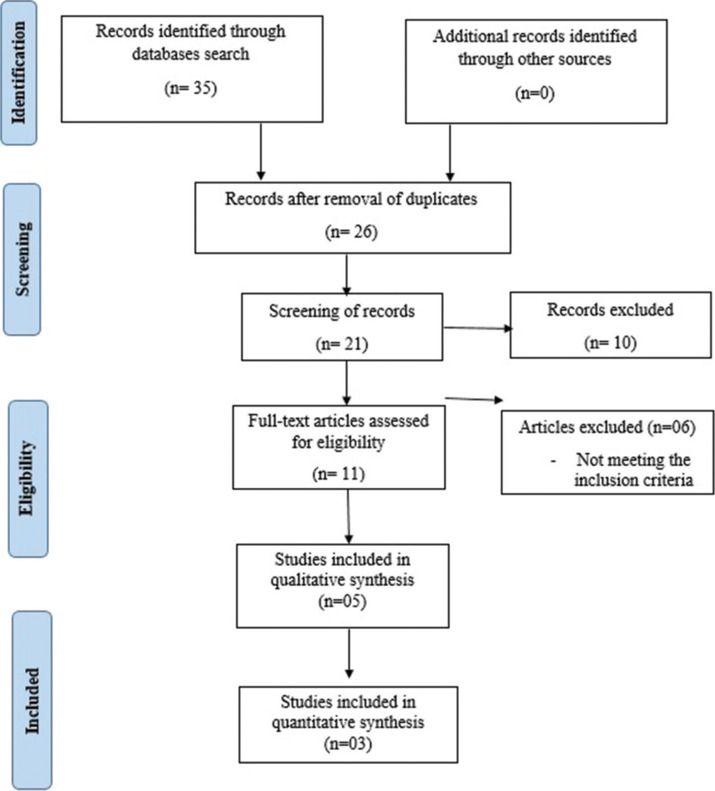
Preferred Reporting Items for Systematic Review and Meta-analysis 2009 flow diagram assessment
Study characteristics
Table 1 displays an overview of the descriptive features of all included studies. Data were evaluated from an aggregate of 104 samples. Two case reports[23,24] and three comparative in vitro studies[22,25,26] were included in this review. All the studies concluded that guided endodontics use causes preservation of large amounts of tooth substance and this approach was found to be safe and fast and can be used as a technique for calcified canals location leading to minimum complications.
Table 1:
An overview of the descriptive features of all included studies
| Authors, study year | Country | Sample size | The mean age of the participant | Study design | Parameters assessed | Outcome |
|---|---|---|---|---|---|---|
| Kostunov et al., 2020[22] | Germany | 10 | Not mentioned | Comparative study | Preservation of the amount of tooth substance to be removed | Guided endodontics use in calcified teeth helps in saving large amounts of tooth substance |
| Llaquet et al., 2020[23] | Spain | 1 | 28 | Case report | Access to root canals and evaluating its accuracy | The approach is safe and the location of calcified canals can be considered a predictable technique with fewer complications |
| Todd et al., 2020[24] | New York | 7 | 40.5 | Case report | Negotiating necrotic calcified root canal | Successful treatment of root canals was done through a “template-guided access technique” with very minimal tooth loss |
| Su et al., 2021[25] | Taiwan | 28 | Not mentioned | Comparative study | Ability on canal accessibility | Preparation of the access cavity with endodontic guides was acceptable |
| Zhang et al., 2022[26] | China | 58 | Not mentioned | Comparative study | Better reliability and feasible accuracy in cavity preparation | Using guided endodontics for the preparation of access cavities has feasible accuracy |
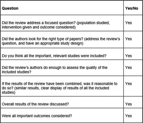
Assessment of methodological quality
Among the included case reports, the overall quality appraisal of the included studies was high as all the questions under the checklist were answered by all the studies as shown in Figure 2.
Figure 2.

Quality appraisal of included case reports using Joanna Briggs Checklist
Overall quality appraisal of the included studies was high as all the questions under the checklist were answered by all the studies, as shown in Figure 3. The overall quality of the included studies was assessed to be high as all the questions under the checklist were answered or informed by authors in the original.
Figure 3.
Quality appraisal of included in vitro studies
Synthesis of result
Three studies containing data on 96 (n = 192) samples, of which (n = 96) samples were evaluated by guided endodontics, and (n = 96) samples were evaluated by conventional techniques for the evaluation or the correction of calcific metamorphosis.
As shown in Figure 4, the standard mean difference is −0.97 (−1.83–0.10) and the pooled estimates favor guided endodontics. This signifies that the correction or treatment of calcific metamorphosis on average is 0.97 times more guided endodontics as compared to conventional techniques (P < 0.05).
Figure 4.
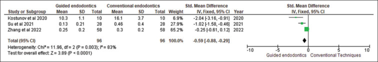
Forest plot showing guided endodontics versus conventional endodontics with regards to the calcific metamorphosis. SD: Standard deviation, CI: Confidence interval
Among all the included studies, Zhang et al. 2022 had the highest weightage at the overall pooled estimate while the lowest weightage was observed by Kostunov et al. 2020 at the pooled estimate.
By employing the random effect model, the I2 statistic showed 83%, the heterogeneity for Tau2 was 0.46, χ2 being P = 0.003, and the overall effect for Z value being 2.19 (P = 0.03).
There was no significant asymmetry seen on the funnel plot, indicating the absence of publication bias, as shown in Figure 5.
Figure 5.

Begg’s Funnel plot with 95% confidence intervals, indicating an absence of publication bias. SE: Standard error, SMD: Standardized mean difference
DISCUSSION
Researchers have introduced the concept of guided endodontics, employing three-dimensional -printed guides for the preparation of access cavities.[2] It involves using CBCT for acquiring volumetric data and using an intraoral scanner for scanning surface data. In addition, also it causes a reduction in chairside time.
This concept being novel also supports clinicians in avoiding unwanted tissue removal, avoiding or minimizing complications, and improving overall treatment prognosis. Furthermore, an overall cumulative review of existing evidence is required to give an overview of the current knowledge about this treatment concept.
No study has provided an analysis of the comparison of guided endodontics with conventional technique methods for calcific metamorphosis on which the best treatment option for calcific metamorphosis could be established. Therefore, we updated our research for related articles and conducted a systematic review to compare guided endodontics with conventional technique methods for calcific metamorphosis through a novel meta-analysis.
Existing literature has described the problems associated with treating teeth with calcifications of the pulp chamber. To overcome this, guided endodontic treatment is an alternative option and also to improve the accuracy of apical surgery.
The present systematic review summarizes evidence from case reports and in vitro studies. Meta-analysis showed that an SDM of −0.97 (−1.83–0.10) favors guided endodontic treatment more than the conventional technique with a significant statistical difference.
Strict following of PRISMA guidelines, unrestricted literature searches, and use of robust methodology for the qualitative synthesis of data are the main strengths of this review. The assessment of the methodological quality of case reports was done by the CASP checklist and the checklist put forward by the JBI was used for in vitro studies. For the quality assessment, all the included studies had a moderate-to-low risk of bias and the overall quality of included studies was high indicating the absence of potential and unavoidable sources of bias with less reporting deficiencies and variability.
However, a few limitations were also present. Going through the evidence, there is a scarcity and paucity of literature on the efficacy of guided endodontic treatment for calcific metamorphosis. Even after going through an unrestricted search and eligibility criteria, the number of included studies for qualitative synthesis was very low. Only five studies were included in our systematic review and only three studies were eligible to be included for meta-analysis. There is a need to conduct more follow-up studies on the efficacy of guided endodontic treatment on the success of calcific metamorphosis. Furthermore, there should be a trial of conducting a systematic review and meta-analysis, for getting an overall pooled estimate of the guided endodontic treatment.
A systematic review is a transparent and reproducible method to identify, select, and critically evaluate published or unpublished literature to answer a well-defined research question. Systematic reviews are often combined with a meta-analysis, which is a statistical analysis that combines numerical data withdrawal from comparable studies. Systematic reviews and meta-analyses are generally considered to be the highest quality evidence. However, the strength of evidence from a systematic review and meta-analysis is related to the quality of the included studies.
CONCLUSION
Predictable outcomes and risk of iatrogenic damages are reduced through guided endodontic procedures and techniques. The accuracy of access cavity preparation through printed templates is increased. The location of root canals in the apical third is done more easily. Minimally invasive treatment can be performed, with a reduction in chairside time. However, evidence is scarce; therefore, further studies with larger population studies with longer follow-up periods are needed.
Financial support and sponsorship
Nil.
Conflicts of interest
There are no conflicts of interest.
Acknowledgement
I would like to express my sincere gratitude to Dr. Amar Shaw for helping me with the statistical analysis of this review. I appreciate his insightful input throughout the research process.
REFERENCES
- 1.Hegde SG, Tawani G, Warhadpande M, Raut A, Dakshindas D, Wankhade S. Guided endodontic therapy: Management of pulp canal obliteration in the maxillary central incisor. J Conserv Dent. 2019;22:607–11. doi: 10.4103/JCD.JCD_21_20. [DOI] [PMC free article] [PubMed] [Google Scholar]
- 2.Goga R, Chandler NP, Oginni AO. Pulp stones: A review. Int Endod J. 2008;41:457–68. doi: 10.1111/j.1365-2591.2008.01374.x. [DOI] [PubMed] [Google Scholar]
- 3.Qassem A, Martins Nda M, da Costa VP, Torriani DD, Pappen FG. Long-term clinical and radiographic follow up of subluxated and intruded maxillary primary anterior teeth. Dent Traumatol. 2015;31:57–61. doi: 10.1111/edt.12135. [DOI] [PubMed] [Google Scholar]
- 4.Andreasen FM, Kahler B. Pulpal response after acute dental injury in the permanent dentition: Clinical implications-a review. J Endod. 2015;41:299–308. doi: 10.1016/j.joen.2014.11.015. [DOI] [PubMed] [Google Scholar]
- 5.Andreasen FM, Zhijie Y, Thomsen BL, Andersen PK. Occurrence of pulp canal obliteration after luxation injuries in the permanent dentition. Endod Dent Traumatol. 1987;3:103–15. doi: 10.1111/j.1600-9657.1987.tb00611.x. [DOI] [PubMed] [Google Scholar]
- 6.Agrawal VS, Kapoor S. Management of root canal stenosis and external inflammatory resorption by surgical root reconstruction using biodentine. J Conserv Dent. 2020;23:102–6. doi: 10.4103/JCD.JCD_128_20. [DOI] [PMC free article] [PubMed] [Google Scholar]
- 7.Sule K, Malik A, Kothari A, Rao V. Management of luxation injuries in line with International Association of Dental Traumatology guidelines – Two case reports. J Conserv Dent Endod. 2024;27:561–3. doi: 10.4103/JCDE.JCDE_191_24. [DOI] [PMC free article] [PubMed] [Google Scholar]
- 8.Panithini DB, Sajjan GS, Kinariwala N, Medicharla UD, Varma KM, Kallepalli M. Real-time guided endodontics: A case report of maxillary central incisor with calcific metamorphosis. J Conserv Dent. 2023;26:113–7. doi: 10.4103/jcd.jcd_506_22. [DOI] [PMC free article] [PubMed] [Google Scholar]
- 9.Jacobsen I, Kerekes K. Long-term prognosis of traumatized permanent anterior teeth showing calcifying processes in the pulp cavity. Scand J Dent Res. 1977;85:588–98. doi: 10.1111/j.1600-0722.1977.tb02119.x. [DOI] [PubMed] [Google Scholar]
- 10.Abella F, Patel S, Durán-Sindreu F, Mercadé M, Bueno R, Roig M. An evaluation of the periapical status of teeth with necrotic pulps using periapical radiography and cone-beam computed tomography. Int Endod J. 2014;47:387–96. doi: 10.1111/iej.12159. [DOI] [PubMed] [Google Scholar]
- 11.Patel S, Durack C, Abella F, Shemesh H, Roig M, Lemberg K. Cone beam computed tomography in endodontics – A review. Int Endod J. 2015;48:3–15. doi: 10.1111/iej.12270. [DOI] [PubMed] [Google Scholar]
- 12.Zehnder MS, Connert T, Weiger R, Krastl G, Kühl S. Guided endodontics: Accuracy of a novel method for guided access cavity preparation and root canal location. Int Endod J. 2016;49:966–72. doi: 10.1111/iej.12544. [DOI] [PubMed] [Google Scholar]
- 13.Krastl G, Zehnder MS, Connert T, Weiger R, Kühl S. Guided endodontics: A novel treatment approach for teeth with pulp canal calcification and apical pathology. Dent Traumatol. 2016;32:240–6. doi: 10.1111/edt.12235. [DOI] [PubMed] [Google Scholar]
- 14.van der Meer WJ, Vissink A, Ng YL, Gulabivala K. 3D computer aided treatment planning in endodontics. J Dent. 2016;45:67–72. doi: 10.1016/j.jdent.2015.11.007. [DOI] [PubMed] [Google Scholar]
- 15.Connert T, Krug R, Eggmann F, Emsermann I, ElAyouti A, Weiger R, et al. Guided endodontics versus conventional access cavity preparation: A comparative study on substance loss using 3-dimensional-printed teeth. J Endod. 2019;45:327–31. doi: 10.1016/j.joen.2018.11.006. [DOI] [PubMed] [Google Scholar]
- 16.Liberati A, Altman DG, Tetzlaff J, Mulrow C, Gøtzsche PC, Ioannidis JP, et al. The PRISMA statement for reporting systematic reviews and meta-analyses of studies that evaluate health care interventions: Explanation and elaboration. PLoS Med. 2009;6:e1000100.. doi: 10.1371/journal.pmed.1000100. [DOI] [PMC free article] [PubMed] [Google Scholar]
- 17.Jun H, Yoon SH, Roh M, Kim SH. Quality assessment and implications for further study of acupotomy: Case reports using the case report guidelines and the Joanna Briggs Institute critical appraisal checklist. J Acupunct Res. 2021;38:122–33. [Google Scholar]
- 18.Nadelson S, Nadelson LS. Evidence-based practice article reviews using CASP tools: A method for teaching EBP. Worldviews Evid Based Nurs. 2014;11:344–6. doi: 10.1111/wvn.12059. [DOI] [PubMed] [Google Scholar]
- 19.DerSimonian R, Laird N. Meta-analysis in clinical trials revisited. Contemp Clin Trials. 2015;45:139–45. doi: 10.1016/j.cct.2015.09.002. [DOI] [PMC free article] [PubMed] [Google Scholar]
- 20.Higgins JP, Thompson SG. Quantifying heterogeneity in a meta-analysis. Stat Med. 2002;21:1539–58. doi: 10.1002/sim.1186. [DOI] [PubMed] [Google Scholar]
- 21.Sterne JA, Egger M. Regression methods to detect publication and other bias in meta-analysis. Publication bias in meta-analysis. Prev Assess Adjustments. 2005;12:99–110. [Google Scholar]
- 22.Kostunov J, Rammelsberg P, Klotz AL, Zenthöfer A, Schwindling FS. Minimization of tooth substance removal in normally calcified teeth using guided endodontics: An in vitro pilot study. J Endod. 2021;47:286–90. doi: 10.1016/j.joen.2020.10.025. [DOI] [PubMed] [Google Scholar]
- 23.Llaquet H, Korkmaz Y, Schneider K, Raab WH. Impact of endodontic treatments on the rigidity of the root. J Dent Res. 2020;85:364–8. doi: 10.1177/154405910608500416. [DOI] [PubMed] [Google Scholar]
- 24.Todd R, Resnick S, Zicarelli T, Linenberg C, Donelson J, Boyd C. Template-guided endodontic access. J Am Dent Assoc. 2021;152:65–70. doi: 10.1016/j.adaj.2020.07.025. [DOI] [PubMed] [Google Scholar]
- 25.Su Y, Chen C, Lin C, Lee H, Chen K, Lin Y, et al. Guided endodontics: Accuracy of access cavity preparation and discrimination of angular and linear deviation on canal accessing ability-an ex vivo study. BMC Oral Health. 2021;21:606.. doi: 10.1186/s12903-021-01936-y. [DOI] [PMC free article] [PubMed] [Google Scholar]
- 26.Zhang C, Zhao X, Chen C, Wang J, Gu P, Ma J, et al. The accuracy of using guided endodontics in access cavity preparation and the temperature changes of root surface: An in vitro study. BMC Oral Health. 2022;22:504.. doi: 10.1186/s12903-022-02548-w. [DOI] [PMC free article] [PubMed] [Google Scholar]