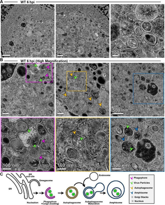
Figure 7. Application of high-angle annular dark-field (HAADF) scanning transmission electron microscopy (STEM) to the study of PV-induced autophagic signals.
(A) HAADF-STEM imaging of WT PV-infected HeLa cells. HeLa cells were infected with WT PV at an MOI of 10 and then fixed in glutaraldehyde at the indicated time points. Fixed samples were dehydrated, stained, embedded, and sectioned in thin micrographs for imaging as described (Fig. S1). Images were collected using a Thermo Scientific Talos F200X G2 (S)TEM operated at 200 kV and a beam current of approximately 0.12 nA. The contrast is also reversed when compared to TEM, with the vacuum appearing dark. WT infection induces virus-containing double membranous vesicles and amphisome-like vesicles with virions in the intra-luminal vesicles. Arrows indicate observed structures. Phagophore (magenta), virus particles (green), autophagosomes (yellow), amphisomes (blue), Golgi (G), nucleus (N). Large outer vesicles with intra-luminal vesicles (100–300 nm diameter) contain ~30 nm particles inside. Double membrane vesicles are located at sites where vesicular-tubular clusters are observed in TEM mode (see Fig. S1). (B) STEM imaging of WT PV-infected HeLa cells (Magnified). In this magnified view of STEM images, 30 nm virus particles were observed inside intra-luminal vesicles. Close-up view of an intra-luminal vesicle that contains 30 nm particles. (C) Autophagic signals during WT PV infection. An ER-derived omegasome buds out and is engaged by multiple autophagy-associated proteins, adaptors, kinases, and protein complexes to yield an autophagophore in preparation for virion loading and maturation of a double-membranous vesicle termed autophagosome. For intact/functional cargo secretion in vesicles, the autophagosome may fuse with endosomes to form a virus-containing amphisome-like vesicle.

