


ABSTRACT
The current studies have shown that the occurrence and development of chronic obstructive pulmonary disease (COPD) are closely related to the changes in gut health and its microenvironment, and even some gut diseases have significant clinical correlation with COPD. The dysbiosis of gut microbiota observed in COPD patients also suggests a potential bidirectional interaction between the gut and lung. Communication between the gut and lung may occur through circulating inflammatory cells, gut microbial metabolites, and circulating inflammatory mediators, but the mechanism of bidirectional communication between the gut and lung in COPD is still under study. Therefore, more research is still needed in this area. In this review, we summarize recent clinical studies and animal models on the role of the gut-lung axis in the occurrence and development of COPD and its mechanisms, so as to provide ideas for further research in this field. In addition, we also summarized the negative effects of COPD medication on gut microbiota and the gut microbiota risk factors for COPD and proposed the potential prevention and treatment strategies.
KEYWORDS: Gut microbiota, gut-lung axis, chronic obstructive pulmonary disease
Introduction
Chronic obstructive pulmonary disease (COPD) is a kind of chronic airway nonspecific inflammatory disease and is characterized by poorly reversible airflow obstruction and alveolar damage well as respiratory symptoms including upper respiratory tract inflammation, emphysema, cough, and increased airway mucus, which is the third leading cause of death globally.1,2 The main treatment of COPD is to optimize lung function and inhibit disease progression through pharmacological and non-pharmacological interventions. Pharmacological interventions mainly include inhaled corticosteroids,3 antibiotics,4 and anticholinergic drugs.5 Non-pharmacological interventions include smoking cessation,6 dietary modification,7 and lung rehabilitation.8 These management measures are effective only in stabilizing disease progression but are suboptimal in preventing or delaying deterioration in lung function. Together with the rising incidence rates and the socioeconomic burden it imposes, has made it a social problem of increasing concern.9 Therefore, the comprehensive investigation of the pathogenic mechanisms underlying COPD is essential to early diagnosis, prevent or slow the progression of COPD and reduce mortality.
The remarkable development of the gut microbiota research in recent years has led to an unprecedented paradigm for disease management.10 The gut system is the largest organ in the human body. In host gut, a large number of bacteria, fungi, archaea and viruses form a vast ecosystem known as the gut microbiota. The human gut microbiota is composed of approximately 1014 microbes, equivalent to 4 × 10⁶ genes, which constitutes the unique gene pool in each human body.11 The significant role of such a vast ecosystem in human health cannot be over looked. With the development of high-throughput sequencing and metagenomics, we can obtain information on the dynamic changes of human gut microbiota, thus revealing the mystery of the interaction between gut microbiota and diseases. The interaction between gut microbiota and host mainly includes immune regulation, microbial metabolites, microbial components and neuromodulation, among which microbial metabolites are a particularly important way.10 The gut microbiota and its derived metabolites have an important impact on host innate immunity, nutrient metabolism, and tissue and organ development.12 The COPD patients are often accompanied by gut microbiota imbalance, which can lead to disorders of the mucosal microenvironment, such as the gut barrier and the immune system, which may have negative effects on the gut and lung.13 A recent study observed increased abundance of Firmicutes, decreased abundance of Bacteroidetes, and increased abundance of Streptococcus and Alistipes at the genus level in feces of COPD patients.14 Streptococcus is one of the common strains that cause lung infection in patients with COPD.15 Gastrointestinal diseases are more prevalent in COPD patients compared to healthy individuals. A retrospective study of 1,228 COPD patients showed that most COPD patients had at least one gastrointestinal symptom.16 Plasma zonulin, a marker of gut leakage, was observed to be elevated in patients with COPD, and higher levels of zonulin were found in plasma of patients with moderate and severe COPD compared with patients with mild COPD,17 which suggests that there is a certain degree of impairment of intestinal barrier function in COPD patients. COPD can not only lead to gut microbiota disorder and gastrointestinal diseases, but also vice versa. When gut microbiota is disordered, it can also affect lung function. For example, patients with gastrointestinal diseases such as inflammatory bowel disease (IBD) experience disruptions in their gut microbiota, leading to decreased lung function in these individuals.18 This suggests a bidirectional communication between the gut and the lung, which is known as the gut-lung axis. Diet, as an important factor affecting gut microbiota and gut health, has an important effect on the gut-lung axis. For example, in a cross-sectional study of 7,486 participants, those who consumed prebiotics and yogurt had a 25% lower incidence of COPD.19 Another study of middle-aged and elderly people found that dietary fiber intake greater than 15.10 g per day was effective in reducing the prevalence of COPD.20 Similarly, a meta-analysis suggested that adding 10 g of total fiber, cereal fiber, or fruit fiber per day reduced the risk of COPD by 26%, 21%, and 37%, respectively.21 The protective effect of dietary fiber on COPD may be due to the gut microbiota metabolizing dietary fiber to form SCFAs, which can improve lung function by regulating immune homeostasis and gut barrier function.22,23
At present, we have made a little progress in the field of the mechanism of the gut microbiota in the pathogenesis of COPD. However, revealing the precise role of actual changes in gut microbiota in the pathogenesis of COPD does not seem to be an easy task due to the limitations of clinical individual heterogeneity. Gut microbiota-derived metabolites seem to be another way to reveal the precise interaction between gut microbiota and disease. So far, some studies have found an association between metabolites of gut microbiota, such as short-chain fatty acids (SCFAs) and trimethylamine N-oxide (TMAO), and COPD.22,24,25 For example, SCFAs can protect lung function by regulating immune homeostasis, enhancing gut barrier function and inhibiting pathogen translocation.22,26–28 However, compared with other diseases, the progress in the relationship between gut microbiota metabolites and COPD is still insufficient. Especially for TMAO, we only stay on its correlation with COPD,24 and there is no direct evidence to reveal the mechanism of action between the two. The rapid development of metagenomics, targeted or non-targeted metabolomics and other technologies has brought important means to reveal the complex relationship between gut microbiota, its metabolites and human health. Now, more and more attention has been paid to the relationship between gut microbiota mediated gut-lung axis and the pathogenesis of COPD. Therefore, this review provides a comprehensive overview of the gut microbiota and its associated metabolites in COPD, highlighting the interaction between gut microbiota and host in the pathogenesis of COPD. We believe that our work can provide guidance for future research in COPD and ultimately contribute to the development of management measures for COPD.
Gut-lung axis and COPD
Gut-lung axis
The distribution and function of the gastrointestinal tract and the respiratory tract are different in humans and animals, but they have the same embryonic origin, so they are similar in structure. The lung, trachea, respiratory epithelium, and gut all originate from the endoderm during embryonic development.29 In addition, a previous study found that hyperactive Wnt signaling in lung progenitor cells expressing lung-specific genes can induce differentiation of lung progenitor cells into gut cell types.30 Therefore, it is not difficult to understand the bidirectional crosstalk between the gut and the lung mediated by the gut-lung axis. The lung inflammation in COPD patients is characterized by changes in the number and function of immune cells, such as reduced antigen presentation capacity of macrophages in the lungs of COPD patients.31,32 Changes in the gut environment are critical for the development and remodeling of lung immunity. Both the respiratory tract and the digestive tract belong to the mucosal immune system, and secreted immunoglobulin A (sIgA) produced by them is the common molecular basis of mucosal immunity in different parts of the body and also an important molecular mediator of the gut-lung axis.33 sIgA is not only involved in the pathogenesis and progression of lung diseases, including COPD, asthma and idiopathic pulmonary fibrosis,34 but also prevent the spread of pathogens in the body and regulate the composition and function of gut microbiota.35 The poor outcome of germ-free mice exposed to acute infection and their susceptibility to allergic airway disease demonstrate the critical role of the gut microbiota in lung homeostasis and immunity.36,37 In addition, the researchers detected the expression of lung function protein pulmonary surfactant protein A in the gut tissue of patients with gut inflammation, which demonstrate the similarity between the lung and gut again.38 These similarities set the stage for bidirectional communication of the gut-lung axis.
Gut microbiota-driven metabolites are important mediators mediating the gut-lung axis. Gut microbiota-driven metabolites circulate in the blood, promoting bidirectional communication between the lung and gut, and exerting different effects on individual organs. SCFAs, as important metabolites of gut microbiota, are essential for the establishment of bidirectional communication between the gut-lung axis.39 SCFAs are mainly derived from the fermentation of dietary fiber by gut microbiota. The levels of circulating SCFAs in high-fiber fed mice are higher than those in low-fiber fed mice, and they show resistance to allergic inflammation in the lung.26 Butyrate, as an important representative of SCFAs, is essential for the maintenance of gut barrier function and the maturation and differentiation of gut immune cells.40,41 Gut-derived butyrate can cross the wall of gut and enter the peripheral blood system, not only regulate the differentiation and maturation of immune cells in the bone marrow cavity, but also reach the lung and directly regulate the activity of regulatory T cells (Treg) through G protein-coupled receptors (GPCRs).42–44 In addition, SCFAs are energy sources of colonic epithelial cells, which maintain the anaerobic environment of the gut lumen, inhibit the proliferation of Proteobacteria, and ultimately maintain the integrity of the gut barrier function by promoting the transition of colonic epithelial cells to β-oxidation.45 The integrity of gut barrier function also inhibits the translocation of pathogens in the gut lumen and indirectly protects lung function.46
Lipopolysaccharide (LPS), as a microbial component, is an important component of the outer membrane of gram-negative bacteria. When the gut barrier function is damaged, LPS will cross the gut barrier and be transported throughout the body through the blood circulation system, promoting bidirectional communication between the gut and the lung. LPS is essential for human health, and its mechanism of immune activation is different from other immune agents. It is the only substance that activates lung phagocytes by binding to TLR4.47 LPS can activate NF-κB and MAPK by binding to TLR4 receptor, leading to the production of pro-inflammatory cytokines (such as IL-1, IL-6 and TNF-α) and ultimately leading to acute lung injury.48,49
In a word, the gut and lung share many similarities in structure, development and immunomodulatory mechanisms, which are the biological and anatomical basis for the gut-lung axis. Furthermore, immune cell interactions, gut microbiota metabolites and microbial components (such as LPS) mediate the bidirectional regulation between gut and lung (Figure 1).
Figure 1.
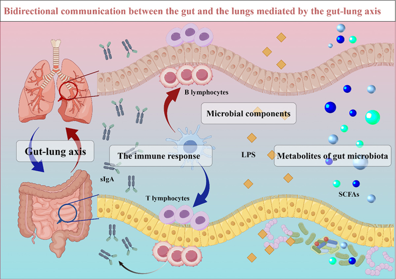
Bidirectional communication between gut and lung mediated by gut-lung axis. The bidirectional interaction between the gut and lungs is mainly achieved through immune responses, microbial composition, and metabolites of the gut microbiota. Dendritic cells receive antigens to activate T-lymphocytes and B-lymphocytes mediated immune responses. LPS, as an important component of gram-negative bacteria in gut microbiota, can regulate lung immune homeostasis through the circulatory system. Gut microbiota metabolites, such as SCFAs, promote bidirectional communication between the gut and lungs through blood circulation. DCs, dendritic cells; LPS, lipopolysaccharide; SCFAs, short-chain fatty acids; sIgA, secretory immunoglobulin A.
Clinical correlation between COPD and gut diseases
Clinical studies have shown an association between gastrointestinal diseases and COPD. Results of people from Taiwan, China-based cohort study show that patients with COPD have a higher risk of functional gastrointestinal disorders than healthy people.50 Similarly, studies have shown that the incidence of Crohn’s disease (CD) and ulcerative colitis (UC) in patients with COPD is 55% and 30% higher than that in the general population, respectively.51 A cohort study showed that IBD was associated with an increased risk of all-cause mortality in COPD patients. In addition, IBD increases the risk of death due to respiratory diseases in COPD patients with asthma.52 First-degree relatives of COPD patients are at increased risk of developing CD. Shared environmental factors may contribute to this, but familial risk factors cannot be excluded.53 Atrophic gastritis and gut nutrient absorption dysfunction exist in COPD patients.54,55 Patients with IBD also have respiratory symptoms such as airway inflammation and sputum production.56 Cigarette smoke exposure is an important risk factor for COPD. During smoking, nicotine accumulates in the gut and leads to the activation of AMPKα.57 In addition, a retrospective cohort study showed that irritable bowel syndrome(IBS) patients had a significantly increased risk of developing COPD.58 In a retrospective cohort study, the cumulative incidence of COPD increased significantly in patients with IBS over more than 10 years of follow-up, and those who were managed for IBS had significantly lower risk for developing COPD.58 There is also a potential association between COPD and colorectal cancer. A Mendelian randomization analysis showed that COPD can increase the risk of colorectal cancer, and fatty acid metabolism is a potential mediator of the two diseases.59 Other subtypes of gastrointestinal diseases such as gastroesophageal reflux disease showed a higher risk of COPD.60,61 A data review of 141,057 COPD patients in the Korean national Health Insurance Database showed that gastroesophageal reflux disease is one of the most common comorbidities of COPD.60 Cigarette smoke is a risk factor for COPD, and nicotine may lead to an increased probability of reflux events.62 Gastroesophageal reflux may increase the severity of acute COPD by irritating the airways and vagal.63
Mechanism of the association between COPD and gut disease
The gut microbiota is an important mediator of the gut-lung bidirectional communication mediated by the gut-lung axis. According to Global Initiative for Chronic Obstructive Lung Disease (GOLD) guidelines, one study recruited 73 healthy controls, 67 patients with COPD severity stages I and II (COPD I-II) and 32 COPD severity stages IIII and IV (COPD III-IV) patients, and transplanted fecal microbiota from these three groups into mice, the results show that fecal microbiota from COPD III-IV patients resulted in significantly higher levels of IL-1β and TNF-α in the plasma of mice. And the percentage of leukocytes in bronchial lavage fluid increased. Compared with mice receiving fecal microbiota from healthy people, the total number of CD3+T lymphocytes, auxiliary T lymphocytes and cytotoxic T lymphocytes in peripheral blood of mice receiving fecal microbiota from COPD I-II and COPD III-IV significantly increased, and the total number of B lymphocytes decreased.64 In addition, the α smooth-muscle actin and matrix metalloproteinase 2 levels were significantly elevated in mice receiving the fecal microbiota of COPD III-IV patients, which led to airway remodeling and increased lung mucus secretion in the mice.64 A recent study has shown that Pediococcus pentosaceus SMM914 can regulate the transformation of gut microbiota in COPD mice into a community structure characterized by the production of SCFAs and antioxidant metabolites, and activate the tryptophane-melatonin pathway of mice to increase the content of 6-hydroxymelatonin and hypotaurine in lung tissue, which increase the secretion of anti-inflammatory cytokines, and ultimately alleviate the inflammatory response of COPD mice.65 Nucleotide-binding oligomerization domain containing 2 (NOD2) is closely related to the occurrence and development of CD. Mutations in the NOD2 gene have been reported in 15% of CD patients.66 The defect of NOD2 leads to increased levels of the serum cytokines IL-1β and IL-12, which can lead to impaired gut barrier function.67,68 And in COPD patients and mouse models, IL-12 was associated with activation of CD8 cytotoxic T-cell and natural killer (NK) cells.69,70 Plasma levels of C-reactive protein (CRP) and IL-6 are higher in COPD patients than in healthy individual, and circulating IL-6 and CRP were associated with the risk of IBD in a prospective case-control study.71,72 Pro-inflammatory cytokines such as TNF-α and IL-1β, which are excessively elevated in some gastrointestinal diseases, are also important risk factors for exacerbating COPD progression.73,74 Group 2 innate lymphoid cells (ILC2) are the innate counterparts of adaptive T lymphocytes, which strongly modulate COPD pathogenesis. The elevated levels of ILC2 and IL-5, a cytokine that favors ILC2 activation and is partly produced by macrophages, were observed in peripheral blood of patients with COPD.75 Studies have shown that ILC2, as a tissue-resident cell, can also migrate between tissues, and IL-25-induced inflammatory ILC2 are circulating cells produced by resting ILC2 residing in the lamina propria of the gut. Inflammatory ILC2 can migrate to different tissues, including the lungs,76 which may explain the interaction between gut disease and COPD. However, there is still a lack of direct evidence of bidirectional communication between COPD and gut diseases mediated by ILC2.
Changes of gut microbiota in COPD
The gastrointestinal tract contains hundreds of species of bacteria, which are closely involved in the physiological functions of the body through metabolites, immune regulation, microbial components, and neuro modulation. Gut microbiota plays an important role in metabolism, genetics and immune regulation. So far, studies have found that alterations and dysbiosis of gut microbiota is closely related to acute pancreatitis, gut diseases, cardiovascular diseases and cancer.41,77–79 In recent years, many studies have found that gut microbiota is involved in the pathogenesis of COPD, and changes in gut microbiota have been observed in both animal models and COPD patients (Figure 2).80–82 We summarize the overview of gut microbiota in COPD, which lays a foundation for further exploration of the role and mechanism of gut microbiota in COPD (Table 1).
Figure 2.
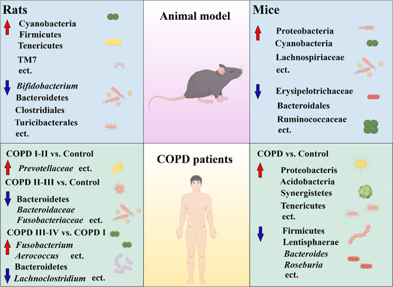
Changes of gut microbiota composition in COPD. Changes in gut microbiota were observed in both animal models and clinical studies. Clinically, the dominant bacteria in the gut microbiota of COPD patients at different stages of development were different. COPD, chronic obstructive pulmonary disease; COPD I, COPD severity stages I; COPD I-II, COPD severity stages I and II; COPD II-III, COPD severity stages II and III; COPD III-IV, COPD severity stages III and IV.
Table 1.
Changes of gut microbiota in COPD.
| References | Sample (sample size) | Differential taxa feature in COPD |
|---|---|---|
| Naijian Li et al.64 | COPD I-II (67) COPD III-IV (32) Healthy controls (73) |
COPD I-II vs. healthy controls ↑:Prevotellaceae COPD III-IV vs. healthy controls ↓:Bacteroidetes, Bacteroidaceae, Fusobacteriaceae. |
| Mei Wang et al.82 | COPD patients (40) Healthy controls (40) |
↑:Proteobacteria, Acidobacteria, Synergistetes, Tenericutes, Ruminococcus, Blautia, Eubacterium, Sphingomonas, Turicibater, Enterobacter, Enterococcus ↓:observed otus, chao1, shannon, simpson, Firmicutes, Lentisphaerae, Cyanobacteria, Subdoligranulum, Bilophila, Faecalibacterium, Phenylobacterium, Barnesiella, Brevundimonas, Lachnospira, Anaerostipes, Bifidobacterium |
| Kate L. Bowerman et al.2 | COPD patients (28) Healthy controls (29) |
↑:Streptococcus, Rothia, Romboutsia, Intestinibacter, Streptococcus spp., Romboutsia timonensis, Intestinibacter bartlettii. ↓:Bacteroides, Roseburia, Lachnospira, Coprobacter fastidiosus, Coprobacter secundus, Rikenellaceae genus RC9, Christensenellales family CAG-74 |
| Yu-Chi Chiu et al.83 | COPD I (20) COPD II (20) COPD III-IV (20) |
COPD III-IV vs. COPD I ↑:Fusobacterium, Aerococcus, Tyzzerella 4, Dialister ↓:Bacteroidetes, Ruminococcaceae NK4A214 group, Lachnoclostridium COPD III-IV vs. COPD II ↑:Veillonella, Corynebacterium 1, Romboutsia, Aerococcus COPD I vs. COPD II ↓:Megasphaera |
| Yanhong Yang et al.84 | CS-induced mice (10) Control mice (10) |
↑:Proteobacteria, Cyanobacteria |
| K. Tomoda et al.85 | CS-induced ale albino rats (5) Control ale albino rats (5) |
↓:Bifidobacterium |
| Lai, HC et al.81 | CS-induced mice (10) Control mice (10) |
↑: Lachnospiriaceae ↓: Erysipelotrichaceae, Bacteroidales, Ruminococcaceae |
| X. Wang et al.86 | Active smoking rats (9) Control rats (10) |
↑:Cyanobacteria, Firmicutes, Tenericutes,TM7 ↓: Bacteroidetes, Clostridiales, Turicibacterales, Clostridiaceae, Turicibacteraceae, Clostridium, Turicibacter, Clostridium perfringens |
Abbreviations: COPD, chronic obstructive pulmonary disease; COPD III-IV, COPD severity stages III and IV; COPD I-II, COPD severity stages I and II; COPD I, COPD severity stages I; COPD II, COPD severity stages II; CS, cigarette smoke.
COPD patients
In recent years, more and more studies have observed gut microbiota dysbiosis in COPD patients.87 A cohort study showed that the relative abundance of Bacteroidetes, Bacteroidaceae, Fusobacteriaceae were lower but that of Proteobacteria, Acidobacteria, Synergistetes, Acidobacteria, Tenericutes, Ruminococcus, Blautia, Eubacterium, Sphingomonas, Turicibater, Enterobacter, Enterococcus were higher in COPD III-IV patients than in healthy population. The abundance of Prevotellaceae was higher in COPD I-II group compared to the healthy population.64 Furthermore, transplantation of gut microbiota from COPD patients resulted in decreased lung function and emphysema in mice, and isolated commensal bacterium Parabacteroides goldsteinii could ameliorate COPD.64,81 In another study, chao1, shannon, simpson and observed_otus were significantly lower in COPD patients than in healthy controls.82 Compared with healthy individuals, the abundance of Proteobacteria, Acidobacteria, Synergistetes, Acidobacteria, Tenericutes, Ruminococcus, Blautia, Eubacterium, Sphingomonas, Turicibater, Enterobacter, Enterococcus increased in COPD patients. The abundance of Firmicutes, Lentisphaerae, Cyanobacteria, Subdoligranulum, Bilophila, Faecalibacterium, Phenylobacterium, Barnesiella, Brevundimonas, Lachnospira, Anaerostipes, and Bifidobacterium decreased. Enterococcus, Ruminococcus, Blautia and Enterobacter were associated with the immune status of COPD patients.82 In a study of 28 COPD patients and 29 healthy controls, several bacteria, including Streptococcus and multiple members of the family Lachnospiraceae, were also associated with reduced lung function.2 Streptococcus was positively correlated with bilirubin, allo-threonine, N-acetyl-cadaverine, and 2-acetamidobutanoate. And negatively correlated with N-carbamoyl glutamate, N-acetylglutamate, 2’-deoxyadenosine, harmane, suberate, sebacate, undecanedioate, dodecanedipate.2 In a clinical study involving 60 patients with COPD, according to GOLD guidelines, COPD patients were divided into COPD I patients, COPD II patients, and COPD III-IV patients. The composition of gut microbiota in COPD I patients and COPD II patients was similar. Compared with COPD I patients, Fusobacterium, Aerococcus, Tyzzerella 4, and Dialister were enriched in the gut microbiota of patients with COPD III-IV. Compared with COPD II, Veillonella, Corynebacterium 1, Romboutsia, and Aerococcus were enriched in the gut microbiota of patients with COPD III-IV.83
Animal models
The model of COPD in C57BL/6 mice induced by cigarette smoke exposure are common animal models of COPD. The abundance of Proteobacteria and Cyanobacteria in gut microbiota of COPD mice induced by cigarette smoke exposure is increased.84 KEGG pathway enrichment analysis showed that cigarette smoke exposure induced changes in metabolic pathways in the liver of COPD mice, such as bile acid metabolism, amino acid metabolism, and energy metabolism.84 In rats, cigarette smoke exposure resulted in a decrease in the abundance of Bifidobacterium.85 In addition, the reduction of Bifidobacterium in the gut microbiota of COPD mice has also been observed in other studies and Bifidobacterium are also potential probiotic prevention and treatment strategies for COPD.88,89 Studies have shown that Bifidobacterium longum subsp.longum can alleviate the expression of inflammatory factors and adhesion factors, and reduce lung inflammation. The mutant strain with impaired acetic acid production also alleviated inflammation in COPD mice, suggesting that the therapeutic effect of Bifidobacterium longum subsp. longum on COPD is independent of acetic acid.88 The transfer of gut microbiota from cigarette smoke-induced COPD in Nlrp6−/− mice to wild-type mice through co-housing also led to airway inflammation in wild-type mice,90 which indicated that cigarette smoke-regulated gut microbiota may be the cause of COPD morbidity. Depletion of gut microbiota by antibiotic cocktail can significantly alleviate emphysema related characteristics such as alveolar wall destruction in COPD mice. Transplantation of gut microbiota from the control group could also alleviate emphysema features in mice, and Parabacteroides goldsteinii may play a dominant role.81 Most studies have only looked at the composition of the microbiota in the cecal contents or feces of COPD mice/rats. Another study investigated the changes of microbiota in multiple regions of the digestive tract in active smoking rat model. The abundance of Bacteroidetes was reduced in the oral cavity of active smoking rat model, whereas that of Cyanobacteria was increased. In the cecal region, the abundance of Firmicutes was reduced in active smoking rat model, whereas the relative abundance of Tenericutes and TM7 was increased in the colon.81
There is no doubt that COPD can lead to gut microbiota disorder and COPD-regulated gut microbiota can promote the development of COPD. However, most of the current research only stays in the changes of microbiota, and the pathogenesis of COPD promoted by such changes and the dominant bacteria in this process are still unclear. Therefore, it is necessary to conduct in-depth research on this.86
Potential mechanisms of gut microbiota in the pathogenesis of COPD
Usually, Bacteroidetes, Firmicutes, Proteobacteria and Actinobacteria are dominant in the gut microbiota of healthy population.91 The disorder of gut microbiota may affect the respiratory system by causing gut mucosal damage.10 Mucins secreted by gut epithelial cells limit the migration of harmful bacteria in the gut to epithelial tissues. Once the gut mucosa is damaged, this restriction will be lifted, and the risk of bacterial translocation from the gut lumen to distant organs will increase, which may lead to severe systemic reactions.92 Gut microbiota translocation may play a role in the gut-lung axis in COPD patients (Figure 3). The presence of Enterobacteriaceae in the lung during acute lung injury demonstrated the presence of translocation of gut microbiota in the gut-lung axis.93,94 Increased zonulin, a marker of gut leakage, was observed in the plasma of COPD patients,17 which indicates that the gut barrier function of COPD patients is impaired. A decrease in the abundance of Clostridium and Turicibacter has been observed in smoker-induced COPD rat model,86 Clostridium and Turicibacter are important commensal bacteria in the gut with the ability to regulate gut barrier function and inflammatory and immune responses associated with gut diseases.95,96 The impairment of gut barrier function caused by COPD can cause the translocation of bacteria and their products, in which bacterial toxins such as enterotoxins and LPS can promote the development of COPD, and the inflammation associated with gut microbiota may be systemic.97–100 In addition, gut commensal bacteria, including Bacteroides and Bifidobacterium, can induce the production of antimicrobial peptides and sIgA,93 Clostridium can also promote anti-inflammatory Treg cell responses.101 In the respiratory tract, Streptococcus activates host p38 mitogen-activated protein kinase (MAPK) in TLR-dependent manner to amplify the inflammatory response.102 Enrichment of Streptococcus in gut microbiota and depletion of Bacteroides and Bifidobacterium are frequently observed in COPD patients and animal models.2,14,82,85 In addition, Proteobacteria dominated dysbiosis has been observed in COPD patients and animal models,82,84 which may be related to the expression of inflammatory genes in lung leukocytes caused by Proteobacteria, while Bacteroidetes are associated with gene expression profiles characteristic of lung tissue remodeling.103 Microbial components also play a role in regulating inflammation. Polysaccharide A (PSA) derived from Bacteroides fragilis can prevent allergic airway inflammation in mice by inducing IL-10 production by T cells.104 SCFAs can directly or indirectly protect lung function by regulating lung immune homeostasis and gut barrier function. Reduced SCFAs content was observed in the feces of COPD patients,64 which may be related to the depletion of SCFAs producing bacteria (such as Clostridium, Bifidobacterium and Ruminococcaceae) caused by COPD.82,85,86
Figure 3.
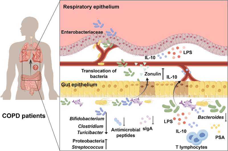
Potential mechanisms of gut microbiota in the pathogenesis of COPD. the disorder of gut microbiota in COPD patients can lead to increased gut permeability, increase the risk of gut bacterial translocation and LPS transfer, and thus promote lung inflammation. The decrease in the abundance of Bacteroides and bifidobacterium can affect the host immune homeostasis by inhibiting the production of sIgA and antimicrobial peptides. Bacteroidetes derived PSA can also prevent airway inflammation by regulating the activity of T lymphocytes to produce IL-10. LPS, Lipopolysaccharide; sIgA, secreted immunoglobulin A; PSA, polysaccharide A.
Although we have observed the changes of gut microbiota in patients with COPD, the results from different studies are quite different and even contradictory.82,86 This is mainly due to the influence of confounding factors such as individual differences, sampling methods, environment, genetics and living habits. Although animal models avoid the above confounding factors to a certain extent, due to the differences in lung anatomy and physiology between mice and humans, it is difficult to capture the changes of chronic bronchitis and emphysema simultaneously in a single model. An ideal animal model should recapitulate the main features of the human disease, namely chronic bronchitis, mucus hypersecretion, small airway remodeling, and emphysema. However, all described animal models exhibit only a subset of these features.13 Therefore, in the future, large-scale clinical cohort studies are warranted to investigate the characteristics of gut microbiota in COPD and the role of landmark bacteria in the pathogenesis of COPD.
The relationship between gut microbiota metabolites and the pathogenesis of COPD
Metabolites derived from the gut microbiota are a major mode of crosstalk between gut microbiota and host. At present, studies have revealed the role and mechanism of metabolites driven by gut microbiota in a variety of diseases. Here, we summarize the metabolites driven by gut microbiota in COPD (Figure 4), which provides innovative ideas for the subsequent exploration of the pathogenesis of COPD and the development of clinical diagnosis and treatment strategies.
Figure 4.
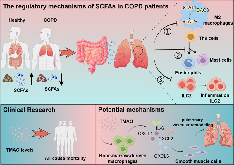
Effects and mechanisms of gut microbiota metabolites on lung health. the content of SCFAs in feces of COPD patients is decreased. ① SCFAs can inhibit the phosphorylation of STAT1 by inhibiting the binding of STAT1 and HDAC3, and promote the transformation of pulmonary macrophages into the anti-inflammatory M2 phenotype; ② SCFAs can alleviate lung inflammation by inhibiting the recruitment and activation of mast cells and eosinophils mediated by Th9 cells; ③ SCFAs can inhibit the transition of ILC2 to inflammatory ILC2, thereby alleviating lung inflammation. The increased plasma TMAO levels in COPD patients are associated with all-cause mortality. TMAO stimulates bone-marrow-derived macrophages to secrete chemokines and inflammatory factors, leading to excessive proliferation and migration of pulmonary vascular smooth muscle cells, which is a potential mechanism of pulmonary vascular remodeling. STAT1, signal transducer and activator of transcription 1; HDAC3, histone deacetylase 3; ILC2, group 2 innate lymphoid cells; TMAO, trimethylamine N-oxide.
SCFAs
SCFAs are the most intensively studied bacterial metabolites in the gastrointestinal tract, which are mainly derived from the microbial fermentation of indigestible foods in the gastrointestinal tract. The effects of microbiota-derived SCFAs are not only limited to the gastrointestinal tract, but also disseminate into the bloodstream and then circulate to various organs throughout the body, including the maintenance of healthy homeostasis in the lung.105 SCFAs are essential for host health homeostasis and immune regulation. The regulation of host immunity by SCFAs is mainly through binding to GPCRs, which in addition couple to different downstream effector molecules, leading to different outcomes of cell functions and responses in different cell types.106,107 The pleiotropic functions of SCFAs in the gut have been well studied,108–111 but the question of how commensal-derived SCFAs link the gut to the lung is only beginning to be revealed.112 There is no doubt that SCFAs have a good protective effect on lung function. A positive correlation between higher fecal acetate levels and forced expiratory volume in first second was observed in patients with emphysema, and exogenous acetate supplementation reduced alveolar destruction and proinflammatory cytokine production in mice model of emphysema.113 One study found that compared to healthy controls, COPD patients showed a Prevotella-dominated gut type and lower SCFAs in feces, including acetic acid, isobutyric acid and isovaleric acid, and that the severity of COPD patients was associated with reduced SCFAs concentrations in feces.64 This could be an adverse subsequent events of gut microbiota disturbance, which is again demonstrated by antibiotic-induced gut microbiota imbalance leading to SCFAs reduction and aggravating the development of emphysema in mice.114 Another study found that gavage of 3 × 10⁸ CFU/mL acetate-producing Bifidobacterium longum subsp. longum was found to alleviate lung inflammation and butyrate depletion in the cecum of mice in a COPD model induced by 8 weeks of cigarette smoke exposure.88 Gut microbiota-derived SCFAs could directly or indirectly regulate the immune homeostasis of the lung, thereby alleviating the development of COPD.115–117 One study showed that bacterial pneumonia caused by methicillin-resistant Staphylococcus aureus (MRSA) infection in mice resulted in a significant decrease in the abundance of butyrate producer unclassified_f__Lachnospiraceae in the gut, as well as a significant decrease in the levels of butyrate in gut and serum, while exogenous butyrate supplementation decreased the binding of histone deacetylase 3 (HDAC3) to signal transducer and activator of transcription 1 (STAT1), inhibited the phosphorylation of STAT1, and thus altered the polarization of lung macrophages toward the anti-inflammatory M2 phenotype. Ultimately, the colonization of the lung by MRSA is reduced and the lung tissue morphology is improved.118 Th9 cells can promote allergic lung inflammation by recruiting and activating eosinophils and mast cells. One study observed that supplementing mice with butyrate alleviated lung inflammation and mucus production in ovalbumin (OVA)-induced type 2 airway inflammation and hypersensitivity mice, while adoptive transfer of Th9 cells restored lung inflammation, suggesting that butyrate may inhibit lung inflammation by suppressing Th9-mediated immune responses.119 In a model of acute exacerbation COPD (AECOPD) induced by cigarette smoke and LPS, the amount of inflammatory ILC2 cells in the lungs and colon of mice was significantly increased, and the contents of SCFAs in the gut was significantly decreased. Butyrate treatment reduced the proportion of inflammatory ILC2 cells in the colon and lung tissue of AECOPD mice and inhibited inflammatory ILC2 cells phenotypes and the secretion of inflammatory factors from the colon and lung of normal mice.120 Propionate and butyrate can also alleviate the inflammatory response in COPD patients by inhibiting the activity of histone deacetylase (HDAC) and regulating the generation of Treg.115 In addition, SCFAs, as an important energy source of colonic epithelial cells, can enhance the gut barrier function, thereby effectively inhibiting the spread of pathogens and endotoxin in the gut lumen.26–28 There is no doubt that SCFAs are closely related to the occurrence and development of COPD, but further studies are needed to clarify the mechanism.
TMAO
TMAO is a related metabolite of the gut microbiota. Nearly a decade ago, TMAO was thought to predict the risk of cardiovascular disease.121 In recent years, its relationship with lung disease has gradually been revealed. Some bacteria in the gut microbiota can convert choline from food into trimethylamine (TMA), which can be circulated through the portal vein to the liver and produced by flavin-containing monooxygenases (FMOs) to TMAO.122 TMAO is produced in the liver and transported through the bloodstream to various organs in the body, including the heart and lungs.123 Results of a prospective follow-up study showed that elevated circulating TMAO was associated with all-cause mortality in patients with COPD. And the patients who died had a significantly higher median admission TMAO compared to the survivors.24 TMAO can lead to inflammation, resulting in structural damage to lung blood vessels. Studies have shown that the increase of TMAO concentration in the blood can lead to damage of vascular endothelial function, induce oxidative stress, and eventually lead to vascular inflammation.124,125 One study found that TMAO significantly increased the expression levels of Kng1, Cxcl1, Cxcl2, Cxcl6, and IL-6 in bone-marrow-derived macrophages. In addition, the macrophage conditioned medium after TMAO treatment increased the excessive proliferation and migration of pulmonary artery smooth muscle cells, but the treatment of TMAO itself did not have this effect, suggesting that TMAO may lead to pulmonary vascular remodeling by stimulating the production of chemokines and cytokines by macrophages.126 Compared with SCFAs, our knowledge of TMAO in the pathogenesis of lung diseases such as COPD is still insufficient, which suggests that we should pay more attention to TMAO in order to comprehensively and deeply explain the adverse effects and molecular mechanism of TMAO on COPD.
Other metabolites
In addition to SCFAs and TMAO, other gut microbiota-dependent metabolites have been less explored in COPD. A study found that Pediococcus pentosaceus SMM914 synthesizes L-tryptophanamide, 5-hydroxy-L-tryptophane, and 3-sulfino-L-alanine to activate the tryptophane-melatonin pathway, increasing the amount of 6-hydroxymelatonin and hypotaurine in the lungs, which reduced the polarization of macrophages and enhanced the anti-inflammatory and antioxidant capacity of COPD mice.65 Hypotaurine is the precursor of taurine, and it can also show excellent antioxidant properties under physiological conditions.127 It has been found that Bacteroides uniformis in gut microbiota can promote the production of taurine, thereby inhibiting the activation of NF-κB and IL-17 signaling pathway in neutrophils, and ultimately controlling the inflammatory response of pancreatitis.128 However, there is a lack of research about the effect and mechanism of taurine on lung related diseases. Currently, little is known about the impact of gut microbiota metabolites on lung diseases such as COPD, so it is necessary to further reveal the relationship between gut microbiota metabolites and the pathogenesis of COPD.
Gut virome and COPD
The virus content in human feces reaches 109-1010 virus-like particles (VLPs)/g, which is an important component of gut microbiota with high abundance, temporal persistence and remarkable diversity.129 The human gut virome is less characterized than the gut bacteriome and is often referred to as the “dark matter” in the human gut microbiota.130 Recent studies suggest that gut virome may play an important role in the ecological processes of the gut microbiota and further influence human health and disease. At present, specific changes of gut virome have been found in IBD, diabetes, hypertension and other diseases.131 Bacteriophages have shown promising therapeutic potential in a variety of diseases by precisely regulating the composition of gut microbiota.132–134 Thus, improving our understanding of the gut virome is critical to reveal the complex interplay between the gut microbiota and human health.
The cross-kingdom interactions between bacteriophages and bacteria and between viruses/bacteriophages and the host immune system are the main ways in which the human gut virome regulates the host.131 Bacteriophages, as natural parasites of bacteria, can change the composition of bacteria in the gut and regulate the metabolism of bacteria in the gut.135 The ability of bacteriophages to shape gut bacterial communities depends largely on their life cycle (including lytic, temperate/lysogenic, pseudolysogenic, and bacterial budding life cycles). Lytic bacteriophages inject their genomes into bacterial cells to produce a large number of viral particles, resulting in bacterial death, which can effectively regulate the composition of the gut bacteriome.136 In addition, the large number of viruses/bacteriophages present in the mucosal layer of the gastrointestinal tract is essential for the establishment and regulation of host innate and adaptive immunity. Variable Ig-like proteins of a set of bacteriophages capsids have been reported to bind to mucin glycoproteins on the host gut mucous layer through the interaction between Ig-like proteins and mucin-displayed glycans.137 Bacteriophages that adhere to the mucous layer of the gut can help build an innate immune barrier by providing an antimicrobial upfront defense against pathogens in the gut.135 In addition, bacteriophages can interact with the human immune system to maintain host immune homeostasis and influence disease progression. For example, there was increased infiltration of interferon gamma (IFN-γ)-producing CD4+ and CD8+ T cells in the gut mucosa of germ-free mice fed with E. coli bacteriophage or T4 bacteriophage. In a mice model of UC induced by dextran sodium sulfate, E. coli bacteriophages can aggravate UC by activating TLR9 and IFN-γ-dependent pathways,138 which suggests that bacteriophages play an important role in the pathogenesis of UC and may well be potential therapeutic targets.
In addition to gut diseases, the relationship between extra-intestinal diseases and gut virome has also been gradually revealed. A study analyzed the gut virome of 674 children aged 1 year from the Copenhagen Prospective Studies on Asthma in Childhood2010 (COPSAC2010) mother – child cohort and conducted a longitudinal assessment of asthma diagnoses. The results showed significant differences in the relative abundance of caudoviruses and microviruses between children with and without asthma before age 5. In addition, the relative abundance of bacteriophage life cycle was shown to be associated with asthma.139 Another study that conducted deep metagenomics sequencing of fecal virus-like particles in 92 subjects from China found that the abundance and diversity of gut virome were lower in COPD patients compared to healthy individuals. Among them, the abundance of Clostridium phage, Myoviridae sp., and Synechococcus phage decreased significantly in COPD patients, and they were significantly positively correlated with pulmonary ventilation function. In addition, virus-bacterial interactions were weakened in COPD patients compared to healthy individuals.140 These results suggest that the gut virome plays an important role in the development of COPD and other lung diseases. However, the mechanism of action of the gut virome in COPD is still unclear, which suggests that we should still pay more attention to the point in the future.
Gut mycobiome and COPD
Fungi, as eukaryotes, have a long history of life origin. For a long time, because of the public impression of fungal infections, human fungi play a negative role in the vast majority of cases. In addition, because gut fungi account for less than 0.1% of the total microbial cells in the gut microecosystem, they have been neglected as a research area.141 Thanks to the continuous progress and development of deep sequencing technology, more and more fungal species in the human gut have been paid attention to by researchers, including key fungal species that are critical to human health or have an important impact on the development of diseases.142 For example, candida spp can obtain available nutrients, secondary metabolites, and influence the production of antimicrobial peptides through cell contact, competition, or cooperation, with complex effects on the composition and function of gut bacteria.143–146
Over the past few decades, researchers have studied the gut mycobiome genome by targeting fungal 18S rRNA gene, or ribosomal DNA (rDNA) internal transcribed spacer (ITS) regions, which has greatly advanced our understanding of the human gut mycobiome genome.147 For example, 18S rRNA and 18S rDNA sequencing techniques observed an increase in gut fungal diversity in CD patients. ITS2 sequencing results showed that the ratio of Basidiomycota/Ascomycota increased, the proportion of Candida albicans increased, and the level of Saccharomyces cerevisiae decreased in IBD patients.148 The gut mycobiome were characterized by ITS2 sequencing of CD patients and their non-CD first-degree relatives from 9 familial clusters living in northern France-Belgium, as well as healthy individuals from 4 families living in the same region. The results showed an increase in the total fungal load and a higher prevalence of Cystofilobasidiaceae family and C. glabrata species during CD. In addition, the abundance of Candida tropicalis increased in patients with CD compared to non-CD relatives.149 Further animal studies have shown that long-term oral antifungals aggravate the severity of UC leading to gut mycobiome dysregulation, characterized by Candida spp. decreased and Aspergillus, Wallemia and Epicoccum spp. increase.150 In addition to gastrointestinal diseases, the gut mycobiome is also closely related to the occurrence of diseases of extra-intestinal organs. One study found that patients with alcoholic hepatitis had a low diversity of gut mycobiome and an overgrowth of Candida spp.. In addition, the increase of serum anti-Saccharomyces cerevisiae antibodies was associated with increased mortality in patients with alcoholic hepatitis.151 In mice, chronic alcohol management was found to increase the number of fungi in the gut as well as the transport of fungal β-glucan into the systemic circulation, β-glucan can induce liver inflammation via the C-type lectin-like receptor CLEC7A on Kupffer cells.152 The gut mycobiome is also closely related to the occurrence and development of lung-related diseases. Antibiotic-induced disruption of the gut microbiota in mice leads to overgrowth of Candida albicans in the gut and T-cell-dependent airway inflammation, but the mechanism was not elucidated in this study.153 Another study delves deeper into this mechanism, the results show that Candida albicans overproliferates in antibiotic-treated mice, and that Candida albicans can promote M2 macrophage polarization by synthesizing prostaglandin E-2 (PGE2), thereby exacerbating allergic airway inflammation in mice.154 Oral fluconazole (0.5 mg/mL) was used to clear the Candida albicans of mice in gut, followed by oral Candida albicans to establish a mouse model of Candida albicans overgrowth. Compared with the control group (oral fluconazole only and no Candida albicans treatment), oral administration of Candida albicans can accelerate the progression of bleomycin-induced pulmonary fibrosis. IL-17A neutralizing antibody treatment slowed the severity of Candida albicans accelerated pulmonary fibrosis, but this was not observed in the control group. This suggests that IL-17A may be involved in pulmonary fibrosis aggravated by Candida albicans.155 However, while reducing the abundance of Candida albicans, fluconazole could also increase the abundance of Aspergillus amstelodami, Epicoccum nigrum, and Wallemia mellicola. Single intragastric administration of live conidia from these three fungi or conidia from Wallemia mellicola alone to mice with allergic airway disease was found to increase the severity of the disease.150,156 In addition, Candida albicans can increase airway inflammation in mice and humans by activating the Th17 response.157,158 The expansion of Th17 cells leads to an increased chronic immune response against Aspergillus fumigatus in the airways of patients with inflammatory respiratory diseases, including asthma and COPD.157 However, the role of gut mycobiome in the occurrence and development of COPD remains unclear.
Although advances in deep sequencing-based technologies have led to our understanding of the role of gut mycobiome in the gut and extra-intestinal organs, this understanding is still very limited compared to our understanding of the gut bacterial community. A major reason for the lack of understanding of the role of gut mycobiome in disease pathogenesis is the limited availability of reference genomes. Although the Unified Human Gastrointestinal Genome (UHGG) has collected more than 230,000 gut microbial genomes from more than 5,400 species,159 these resources lack information on gut fungi. Although some measures have been taken to culture gut fungi and construct fungal genomes from fecal metagenomes,160,161 these measures only cover a small fraction of gut fungi. The limited availability of reference genomes leads to inaccurate quantification of gut fungal species in high-throughput sequencing concentrations, which greatly limit our understanding and knowledge of this important part of the gut mycobiome. A recent study established a cultivated gut fungi (CGF) catalog, which included 760 fungal genomes from 206 species obtained from stool samples of healthy individuals, including 69 species previously unidentified. In addition, the 7,660,447 protein-coding genes encoded by the CGF genome were organized into 643,717 non-redundant protein clusters, 77% of which were not present in respiratory/digestive tract fungi in the human-associated fungi (PHF) catalog, indicating a 247% increase in human intestinal fungal protein sequences.162 The establishment of the CGF genome greatly expands the genomic library of gut mycobiome, provides new insights into the biological significance of the human gut fungal group, and contributes a valuable resource for exploring the composition and functional diversity of the human gut microbiota and the role that gut fungal groups play in human health and disease.
Effects of COPD medication on gut microbiota
The interaction between gut microbiota and drugs is complex and bidirectional. The use of drugs, especially antibiotics, has a strong effect on the composition and function of the gut microbiota. Vice versa, the gut microbiota can also influence an individual’s response to a drug by enzymatically altering its structure, bioavailability, bioactivity, or toxicity. For patients with COPD, antibiotics, corticosteroids and anticholinergic are the main medication, and there is an interaction between these drugs and gut microbiota.
Several important antibiotics, including penicillins, cephalosporins and tetracycline, are clinically used in the treatment of COPD. A randomized controlled trial comparing azithromycin, clarithromycin, erythromycin, roxithromycin and moxifloxacin in the treatment of COPD, the results showed that azithromycin and erythromycin were the most effective in COPD patients.163 Some reports also reflects a negative effect of antibiotic treatment. For example, a course of antibiotics lasting 7 days or longer was associated with more overall adverse events than a shorter course of antibiotics (shorter than 6 days).164 In a study of smoking-induced emphysema models in mice, antibiotic treatment was found to worsen emphysema by causing disruption to the gut microbiota, showing higher IL-6 and IFN-γ levels and an increase in inflammatory cells in bronchial lavage fluid.114 At present, the main concern about antibiotic is the spread of antibiotic-resistant bacteria and disruption of the gut microbiota. In one study, COPD patients treated with long-term antibiotics (median duration of antibiotic treatment was 40 days) had significantly higher abundance of antibiotic resistance genes (ARGs) compared to healthy individuals. The main categories are aminoglycoside, beta-lactam, and macrolide-lincosamide-streptogramin resistance genes and the abundances of Enterococcus and Escherichia were positively correlated with ARGs and antibiotic treatment days.165 After 30 weeks of antibiotic treatment in nude mice, Klebsiella was found to grow and become one of the most common microbes.166 These microorganisms can be potentially harmful to the health of the host. We cannot deny the great role of antibiotics in the treatment of COPD, but we cannot ignore the potential risks of antibiotics. The effect of antibiotic use may be related to the treatment dose, route and duration, so we should use antibiotics more cautiously in the treatment of COPD patients.
Anticholinergic and steroids are considered to be the main therapeutic agents for COPD, and can be used individually or in combination in clinical practice.167 But these drugs affect the composition and function of the gut microbiota. A case-control study observed that anticholinergic was associated with a decrease in peripheral blood butyric acid concentration.168 One study conducted an association analysis of 51 drugs in the TwinsUK cohort and their relationship to gut microbiota. The results showed that inhaled anticholinergic were negatively correlated with the abundance and α diversity of Ruminococcaceae and Peptococcaceae, which suggest that non-oral medications may indirectly affect the gut microbiota.169 A retrospective study found that patients who used inhaled corticosteroids prior to an acute exacerbation of COPD had a significantly elevated risk of developing bacterial lung infections, and patients who used systemic corticosteroids had a higher risk of developing bacterial lung infections than those who used inhaled corticosteroids.170 Prenatal corticosteroid therapy is an effective treatment to reduce the incidence of neonatal respiratory distress syndrome by promoting fetal lung maturation.171 However, a recent study found that prenatal dexamethasone exposure (PDE) can lead to increased susceptibility to various diseases in adulthood, possibly due to the effect of PDE on the composition of the gut microbiota. PDE led to a decrease in the abundance of 13 bacteria genera including Akkermansia, Bacteroides and Parabacteroides, and an increase in the relative abundance of 11 bacteria genera including Klebsiella in the gut of girls at 6 months of age. The abundance of 8 species including Parabacteroides, Lachnoclostridium and Bacteroides decreased in male infants, while the abundance of 9 species including Atopobium increased. PDE was also found in the offspring of rats to lead to a decreased abundance of Lactobacillus reuteri and an increased abundance of Klebsiella pneumonia 6 weeks after birth, while cholestatic liver injury was observed. Fecal microbiota transplantation (FMT) results showed that rats receiving fecal bacteria from the offspring of PDE also exhibited cholestatic liver injury, suggesting that PDE-induced changes in the offspring gut microbiota may increase their susceptibility to cholestatic liver injury.172 Notably, increased Klebsiella abundance in the gut was observed in both rats and humans in this study, and Klebsiella infection plays an important role in COPD and COPD exacerbation.173,174 In addition, gut microbiota also has an impact on drug metabolism. The hepatic cytochrome P450 3A (CYP3A) is a major corticosteroid metabolizing enzyme in the liver, which is closely related to the gut microbiota.175,176 In particular, the human gut microbiota has a significant influence on the activity of CYP3A through the regulation of Cyp3a11 gene expression.177 Therefore, differences in gut microbiota between different individuals may affect the metabolism of drugs such as corticosteroids by affecting host CYP3A, and ultimately lead to differences in drug efficacy between different individuals.
These findings suggest that the interaction between the gut microbiota and drugs is complex and bidirectional: the use of drugs can affect the composition and function of the gut microbiota, and thus the health of the body. Conversely, the gut microbiota can also influence drug metabolism, resulting in different individuals showing different responses to specific drugs. All in all, uncovering the complex interactions between the gut microbiota and drugs is not an easy task, and each drug seems to interact with the gut microbiota in a unique way, making it difficult to come up with a common mechanism. Therefore, understanding how the gut microbiota metabolizes drugs and influences the effectiveness of medications will open up the possibility of regulating the gut microbiota to improve medications.
Gut microbiota risk factors for COPD
Smoking
Smoking is a detrimental habit that is prevalent all over the world. Epidemiological studies have shown that smoking is a major risk factor for COPD and gastrointestinal diseases.178 Smoking cessation can effectively halt the advancement of COPD. A study using the Korean National Health Insurance Service (NHIS) database showed that patients who quit smoking within two years of their COPD diagnosis had a 17% lower risk of all-cause mortality and a 44% lower risk of cardiovascular death than COPD patients who continued to smoke.179 Another multicentre cohort study using the Korean COPD Subgroup Study (KOCOSS) database, which recruited patients from 54 medical centers in Korea, compared the clinical characteristics of smoking COPD patients with nonsmoking COPD and showed that smoking COPD patients had a higher incidence of emphysema according to the radiological findings, and over 5 years of follow-up data, smoking patients with COPD have a more rapid decline in lung function.180 In addition, the effects of smoking behavior on COPD differ by sex, with female smokers having a higher risk of COPD than male smokers.181
However, recent research suggests that the imbalance in gut microbiota caused by smoking plays a key role in this process. Various components in smoke, such as nicotine, interact with the gut microbiota in the gut, resulting in adverse effects on the health of the host.85,182 Numerous studies have shown that smoking can remodels the gut microbiota. For example, compared with nonsmokers, the abundance of Prevotella, Veillonella, Bacteroides, Acidaminococcus and Oscillospira increased in the fecal of smokers and the abundance of Firmicutes and Proteobacteria was reduced.183,184 Another study in healthy males showed that smoking led to a high abundance of Actinobacteria and a low abundance of Bacteroides, and an increased proportion of Gram-negative bacteria in the gut.185 LPS is an important component of the outer membrane of most Gram-negative bacteria, as well as an important virulence factor, which can interact with the host pattern recognition receptors (such as toll-like receptors, TLRs) to produce immune inflammatory effects in the human body. When LPS activates TLR4, it causes a series of intracellular signal transmission that releases inflammatory factors such as IL-1β and TNF-α, ultimately leading to COPD,186,187 and LPS can also activate neutrophil-derived extracellular vesicles to induce COPD-like disease.188 In addition, smoking causes a decrease in the amount of SCFAs in the gut, which may be related to a decrease in the abundance of SCFAs producing bacteria.184 It has been found that SCFAs content and population of Bifidobacterium in cecum of rats exposed to cigarette smoke for 4 weeks are significantly reduced, and the pH of cecum contents is increased.85 In mice, the intragastric administration of cigarette smoke condensate inhibited the production of antimicrobial peptides and reduced bactericidal capacity, resulting in a dysregulation of gut microbiota, an increase in the abundance of Erysipellaceae, and a decrease in the abundance of Rikenellaceae and Eisenbergiella.189 These results suggest that smoking may alter the host gut environment, thereby affecting the composition of the gut microbiota.
Due to the difficulty of sample collection, most current studies analyze the composition of gut microbiota by detecting stool samples of subjects, which does not reflect the authentic gut microbiota. The gut is not a uniform mix of species and environments, and different regions and microenvironments in the gut have unique physical and biochemical conditions, which leads to differences in the composition of the gut microbiota at different spatial scales.190 The results of duodenal mucosal microbiome analysis of humans undergoing upper gastrointestinal endoscopy showed that Streptococcus and Veillonella were more abundant in smokers, while Neisseria and Prevotella were less abundant.191 Another study found that compared with nonsmokers, smokers had a relatively high abundance of Enterobacteriaceae, mainly Escherichia-Shigella and Klebsiella, and the abundance of Escherichia-Shigella was 20.51-fold higher than that of nonsmokers.192 Streptococcus, as an opportunistic pathogen, is associated with decreased lung function and can lead to the deterioration of COPD.193 In addition, in a prospective cohort study, Klebsiella was detected in sputum culture in both stable COPD and AECOPD patients, and the isolation rate of Klebsiella was higher in acute exacerbation than in stable COPD patients, Klebsiella is a common clinical pathogen of pulmonary inflammation,174 which suggested that Klebsiella plays a pathogenic role in acute exacerbation of COPD.173 In conclusion, smoking may affect the composition of gut microbiota (such as excessive proliferation of pathogenic bacteria) by changing the gut microenvironment, thereby regulating the host immune inflammatory response and the production of SCFAs, and ultimately leading to the deterioration of COPD.
Diet
In the past few years, studies have found that dietary patterns have an important impact on the development of COPD.194 Insufficient intake of fiber and other nutrients is related to the occurrence and development of COPD.195 A meta-analysis showed that the risk of COPD was significantly increased in Western dietary patterns (high intake of red or processed meat, refined grains, saturated fatty acids, and sweets).196 On the other hand, the Mediterranean diet pattern (characterized by a high intake of vegetables, fruits, and whole grains) helps maintain lung function.197,198 Epidemiological studies from cross-sectional and longitudinal studies consistently show that high intake of fruits, vegetables, whole grains, and dietary fiber is negatively associated with the risk of COPD.199–207 In addition, a meta-analysis showed that every 50 g/week increase in processed red meat intake was associated with an 8% increase in COPD risk.208 The Western diet pattern is rich in choline, which is converted by the gut microbiota into TMA, which is converted in the liver to form TMAO.121,209 TMAO is associated with all-cause mortality in patients with COPD. After 6 years of follow-up in 189 patients with COPD exacerbations, all-cause mortality was 55.6%, and patients who died showed higher circulating TMAO levels compared to those who survived.24 In animal studies, it has been found that long-term Western diet can cause the mitochondrial activity of mouse intestinal epithelial cells to decline, which further destroys the hypoxia of the gut environment. The increase of oxygen creates favorable conditions for the proliferation of E. coli, and ultimately strengthens the respiration-dependent choline catabolism of E. coli, which also increases circulating TMAO levels.210 This may be related to the increase in E. coli abundance caused by the Western diet disrupting the hypoxic state of the gut. A healthy gut is rich in SCFAs producing bacteria such as Prevotella, Bacteroides, Ruminococcaceae and Lachnospiraceae. These SCFAs help maintain an anaerobic environment in the gut.211 However, long-term Western diet feeding can inhibit the proliferation of SCFAs producing bacteria such as Bifidobacterium, Akkermansia and Bacteroides, resulting in the decrease of SCFAs content,212,213 which may be related to the disruption of intestinal hypoxia caused by long-term Western diet. COPD III-IV patients had lower levels of SCFAs in stools compared to COPD I-II patients and healthy volunteers.64 In addition, patients with digestive disorders have lower levels of SCFAs in plasma and stool and are more likely to develop lung disease.105 In general, the long-term Western diet pattern will lead to the destruction of gut hypoxia, resulting in the proliferation of aerobic bacteria and facultative anaerobic bacteria, thus destroying the composition of gut microbiota, resulting in the inhibition of SCFAs production. In addition, choline, which is rich in the Western diet, is metabolized into TMA by the gut microbiota and enters the liver, thereby increasing the level of circulating TMAO and increasing the risk of COPD.
Most patients with severe COPD also present with thin body mass and often in a state of malnutrition or undernutrition, termed “pulmonary cachexia syndrome” (PCS), which is mainly characterized by muscle wasting. Muscle wasting in COPD patients not only leads to decreased exercise capacity, but also is a major mortality factor independent of airflow obstruction.214,215 Nutritional supplementation therapy can effectively maintain and improve muscle and exercise capacity in patients with COPD. It has been shown that a low-carbohydrate diet can significantly improve lung function in patients with COPD compared to a conventional high-carbohydrate diet.216 Fruits and vegetables are rich in antioxidants, minerals, vitamins, flavonoids, and fiber, which are beneficial for acute and chronic respiratory diseases.217 Vitamin D deficiency is associated with the early development of COPD, which may be related to the immunomodulatory effect of vitamin D, while vitamin D also alleviates the myopathy/muscle weakness.218,219 One study found that colonization of the gut by a protective Escherichia coli strain during pneumonia can alter signaling of the insulin-like growth factor 1/phosphatidylinositol 3-kinase/AKT pathway without affecting host metabolism and energy intake, which is required for the prevention of muscle wasting.220 This finding suggests that gut microbiota may play a role in muscle wasting caused by cachexia. Nutrient intake is an important factor affecting the composition of gut microbiota. For example, low serum vitamin D level can lead to a decrease in the diversity of gut microbiota and the abundance of Bacteroides, Prevotella and Clostridiales.221 However, we still lack a systematic understanding of how nutrient intake affects cachexia induced muscle wasting by influencing gut microbiota composition.
Aging
COPD is a disease associated with aging, and older individuals are at higher risk of developing it than younger individuals. As the world populations ages, about 20% of the world’s population is expected to be over the age of 65 years old by 2050, and 80% of these older people will live in low- and middle-income countries.222 The susceptibility of older people to multiple diseases, including COPD, will pose a significant public health challenge. A cross-sectional study comparing disease severity in older and younger COPD patients found that older COPD patients, especially those 75 years and older, had significantly increased rates of dyspnea, exercise tolerance, and severe exacerbations compared to younger people. The prevalence of comorbidities, including cardiovascular disease and cancer, was significantly higher in older COPD patients.223 Many lung diseases in the elderly are associated with changes in the gut microbiota.224,225 People over 65 years old showed greater individual differences in their gut microbiota compared to younger people,226 but numerous studies consistently show that a decline in the abundance of probiotics like Bifidobacterium and Lactobacillus in the gut of the elderly, while the abundance of certain pathogenic bacteria, like Clostridium difficile and Enterobacteriaceae, has increased.227–230 Colonization of the lungs by Enterobacteriaceae was observed during acute lung injury, possibly as a result of increased permeability of the gut and lung epithelium.231 Numerous studies consistently show that the increased epithelial permeability in both the lung and gut in elderly or senescent animal individuals.232–234 The “leakage” of epithelial barrier function makes it easier for microbes and microbial metabolites to be transported between the two organs, providing more opportunities for pathogens and endotoxins to enter the lungs through lymph and blood. COPD is closely related to immune imbalance mediated by Th17/Treg ratio.235 Several mucosal associated bacteria in the gut microbiota (such as E. coli) and LPS can stimulate Tregs and drive the pro-inflammatory Th17 response to exacerbates COPD.236–238 This enhanced bidirectional communication results in a loss of compartmentalization between the gut and lungs, which may be responsible for the increased COPD susceptibility. As a potential therapeutic target susceptible to intervention, exploring the role of gut microbiota in COPD and its mechanisms in older adults is of great significance to alleviate the global public health burden.
Particulate matter
Traditionally, smoking is widely recognized as the most important risk factor for COPD, but it is now recognized that about one quarter to one third of cases cannot be explained by smoking alone.239 In a report by the American Thoracic Society, the population attribution ratio of smoking to COPD in most clinical studies was < 80%, indicating that other risk factors besides smoking also play an important role in the development and development of COPD.240 Globally, particulate matter (PM) air pollution is a major threat to environmental governance and public health, and working and living in an environment with high concentrations of particulate matter for long periods of time can have serious health consequences. PM2.5, as an important component of PM, is closely related to the incidence of COPD. One meta-analysis found that every 10 ug/m3 increase in PM2.5 was associated with a 1.4–2.5% increase in COPD hospitalizations.241 Studies have found that long-term exposure to PM2.5 can lead to reduced movement of respiratory cilia, increased mucus, and ultimately reduced cilia’s ability to clear the airway. It has been found that PM2.5 can inhibit the expression level of 8-oxyguanine DNA glycosylase 1 (OGG1) in type II alveolar epithelial cells, thereby inhibiting its proliferation and self-renewal.242,243 In addition, microbiome imbalances caused by PM2.5 are also associated with the development of COPD.244 One study found that for every 10 ug/m3 increase in PM2.5 concentration in the air, α-diversity in COPD patients decreased by 2.16%. Moreover, Clostridium sp. CAG127, Enterococcus faecium, and Ruthenibacterium lactatiformans showed a correlation with PM2.5.245 An animal study showed that exposure to particulate matter for 24 weeks resulted in an inflammatory response in the lungs of rats and pathological changes characteristic of COPD. At the same time, the gut microbiota diversity of rats decreased, the serum endotoxin content increased, and the SCFAs content decreased.246 Another study showed that low levels of air pollution could increase the level of Fusobacteria and Verrucomicrobia in the lungs of rats, increase the abundance of Bacteroidetes and Proteobacteria and reduce Firmicutes in the gut microbiota.247 There is a symbiotic relationship between the hosts of Fusobacteria, which may lead to opportunistic infection in the host.248 At present, more and more studies have shown that Proteobacteria can play a role in gut diseases (such as IBD) by regulating the production of pro-inflammatory interleukin and immunoglobulin.249 However, in recent years, studies have found that Proteobacteria also plays a role in lung diseases, but the mechanism of action is still unclear.250
Prevention and treatment strategies
Dietary regulation
Prevention and treatment strategies based on nutritional regulation can not only change the innate immunity of the body and reduce systemic inflammatory response, but also be a potential non-pharmacological method without side effects to prevent and treat COPD, respiratory diseases and smoking-related cancers.18 A prospective study showed that a higher Alternate Healthy Eating Index 2010 (AHEI-2010) (reflecting high intakes of whole grains, polyunsaturated fatty acids, nuts, and long chain omega-3 fats and low intakes of red/processed meats, refined grains, and sugar sweetened drinks) is associated with a lower risk of COPD and is critical in COPD multi-intervention programs.251 In a population-based prospective study of men, a strong negative association was found between total fiber intake and grain fiber intake and COPD among smokers and ex-smokers. Vegetable and fruit fiber intake only reduced COPD risk in current smokers.205 Another prospective study of Swiss women found that long-term high dietary fiber intake was associated with a 30% lower risk of COPD. High intakes of grain and fruit fiber were associated with a lower risk of COPD, but not with plant fiber intake.207 A high-fiber diet mainly results in an increase in circulating SCFAs by reducing the Firmicutes/Bacteroides ratio and fiber metabolism in the gut microbiota,252 which can enhance gut barrier function, regulate body immunity, improve lung function and inhibit inflammatory response by the gut-lung axis.253 Increasing dietary fiber intake, as a nutritional regulation strategy that is easy to intervene and has no side effects, is of great significance for the prevention and alleviation of COPD and the enhancement of lung function.
Probiotics
Probiotics can positively regulate host immunity and pathogen microbiota. The intake of probiotics alone or supplementation with functional foods containing probiotics can reduce oxidative damage, free radical clearance rate, and local or systemic inflammatory response in humans.254–256 A study have found that oral administration of Bifidobacterium longum can improve the immune response and survival rate of mice infected with Klebsiella pneumoniae.257 The combination of multi strain probiotic significantly reduced plasma C-reactive protein and 8-isoprostane levels, and also alleviated gut barrier function in patients with COPD.258 Lactobacillus rhamnosus can alleviate the balance of pro-inflammatory and anti-inflammatory factors in human bronchial epithelial cells induced by cigarette smoke (CS) exposure, and can alleviate the inflammatory response in the airway and lung parenchyma of CS-induced COPD mice, reduce inflammatory cell infiltration and the production of pro-inflammatory cytokines.259 Supplementation with acetate-producing Bifidobacterium longum subsp. longum alleviated lung inflammation, the expression of inflammatory cytokines and adhesion factors, and attenuated cigarette smoke-induced depletion of cecal butyrate.88 A prospective, multi-center, randomized trial showed that long-term oral administration of Lactobacillus rhamnosus GG significantly slowed the progression from moderate to severe COPD.260 The above studies suggest that the development of probiotic strains and their functional components has the potential to become alternative drugs for the prevention or treatment of COPD. However, these mechanisms must be confirmed in large-scale randomized controlled trials before clinical application.
FMT
FMT has systemic effects on the human body and has been shown to have significant therapeutic effects on a variety of diseases.261–263 One study found that the gut microbiota of mice with COPD induced by cigarette smoke exposure was sufficient to increase lung inflammation in antibiotic-treated mice while suppressing colonic immune responses.2 This suggests that gut microbiota is involved in the pathogenesis of COPD. FMT increased the abundance of Bacteroidetes and Lachnospiraceae, which can catabolize fibers into SCFAs, thereby suppressing local and systemic inflammation and attenuating the development of emphysema.252 The disease characteristics of COPD were correlated with the relative abundance of members of the Muribaculaceae, Desulfovibrionaceae and Lachnospiraceae family members, the microbiota from CS-induced COPD was associated with down-regulated glucose and starch metabolism. The disease outcomes can be improved with complex carbohydrate supplementation.2 In conclusion, gut microbiota may be a potential therapeutic target for COPD, but there are no clinical studies of FMT in the treatment of COPD. In the future, more animal experiments and clinical trials are needed to explore the effect and mechanism of FMT in the treatment of COPD before large-scale clinical application.
Future perspectives
With the in-depth understanding of the “gut – lung axis”, the bidirectional crosstalk between the gut and the lung has been gradually revealed, indicating that the gut microenvironment and the homeostasis of gut microbiota play an important role in maintaining lung immune homeostasis. Therefore, the development of COPD prevention and treatment strategies targeting gut microbiota can be considered in future studies. The disruption of the gut microenvironment (including impairment of gut barrier function, gut inflammation, and oxidative stress) and disruption of gut microbiota may influence the degree of lung inflammation and immune homeostasis through the “gut-lung axis”. As described earlier, the presence of gut-associated bacteria in the lungs was observed during acute lung injury, suggesting that there is also a migration mechanism between the gut and lungs. However, this mechanism still needs to be further understood to better elucidate the interaction mechanism between gut microbiota and lung microbiota and to provide possible targets for prevention and treatment of COPD to improve the clinical outcome of patients with COPD. The use of fluorescently labeled bacteria could provide more solid evidence for the hypothesis of gut origin of lung bacteria.264 Up to now, the role and mechanism of gut microbiota in regulating local and systemic immune responses in a variety of diseases have been extensively and deeply studied, but the research in COPD is still limited to correlation studies at the phylum and genus levels. In future studies, it is necessary to use multi-omics combined analysis to further reveal the bacteria that play a key regulatory role in the occurrence of COPD and the target of gut microbiota on the host immune response and metabolic pathway, so as to provide a solid theoretical basis for the clinical prognosis of COPD patients. Metagenomics could be used to more accurately locate the key species and functional genes in the gut microbiota, so as to provide accurate diagnosis and treatment strategy for COPD patients.
In addition, the medication has brought light to COPD patients, but many studies have also observed its negative effects on gut microbiota, which leads to increased susceptibility of COPD patients to other adverse events, such as lung infection and emphysema. Due to the diversity of the interaction mechanisms between drugs and gut microbiota, it is difficult to conclude the common mechanism of the action of different drugs and gut microbiota. Therefore, we should pay more attention to carry out large-scale clinical cohort studies to systematically study the interaction between common drugs and gut microbiota in the future research, so as to provide a reliable theoretical basis for establishing a more scientific, standardized and rigorous clinical medication strategy for COPD patients.
Abbreviations
- COPD
Chronic obstructive pulmonary disease
- IBD
inflammatory bowel disease
- SCFAs
short-chain fatty acids
- TMAO
trimethylamine N-oxide
- TMA
trimethylamine
- sIgA
secreted immunoglobulin A
- Treg
regulatory T cells
- LPS
Lipopolysaccharide
- CD
Crohn’s disease
- UC
ulcerative colitis
- IBS
irritable bowel syndrome
- NOD2
Nucleotide-binding oligomerization domain containing 2
- NK
natural killer
- CRP
C-reactive protein
- ILC2
Group 2 innate lymphoid cells
- HDAC3
histone deacetylase 3
- STAT1
signal transducer and activator of transcription 1
- AECOPD
acute exacerbation COPD
- ARGs
antibiotic resistance genes
- FMT
Fecal microbiota transplantation
- CYP3A
cytochrome P450 3A
- PM
particulate matter
- CS
cigarette smoke
Funding Statement
The work was supported by the National Natural Science Foundation of China [32072746]; Innovation Foundation for Doctor Dissertation of Northwestern Polytechnical University [CX2023021].
Disclosure statement
No potential conflict of interest was reported by the author(s).
References
- 1.Guo L, Wu XL, Wu XL.. Aspergillus infection in chronic obstructive pulmonary diseases. Clin Respir J. 2023;17(3):129–33. doi: 10.1111/crj.13585. [DOI] [PMC free article] [PubMed] [Google Scholar]
- 2.Bowerman KL, Rehman SF, Vaughan A, Lachner N, Budden KF, Kim RY, Wood DLA, Gellatly SL, Shukla SD, Wood LG, et al. Disease-associated gut microbiome and metabolome changes in patients with chronic obstructive pulmonary disease. Nat Commun. 2020;11(1):5886. doi: 10.1038/s41467-020-19701-0. [DOI] [PMC free article] [PubMed] [Google Scholar]
- 3.Stolbrink M, Thomson H, Hadfield RM, Ozoh OB, Nantanda R, Jayasooriya S, Allwood B, Halpin DMG, Salvi S, de Oca MM, et al. The availability, cost, and affordability of essential medicines for asthma and COPD in low-income and middle-income countries: a systematic review. Lancet Global Health. 2022;10(10):E1423–E1442. doi: 10.1016/S2214-109X(22)00330-8. [DOI] [PMC free article] [PubMed] [Google Scholar]
- 4.Lou Y, Jiang G. Associations between TLRs 2 and 4, and β-lactam antibiotics in COPD patients complicated with pulmonary infections. J Infect Dev Ctries. 2024;18(6):950–956. doi: 10.3855/jidc.18918. [DOI] [PubMed] [Google Scholar]
- 5.King PJ, Ramic L, Wilson J, Aaron S, Stiell IG. Evaluation of the quality of ED management for patients with COPD. Clin Exp Emerg Med. 2024;11(3):268–275. doi: 10.15441/ceem.24.197. [DOI] [PMC free article] [PubMed] [Google Scholar]
- 6.Schadegg MJ, Dixon LJ, Lee AA. Emotion regulation difficulties and smoking behavior among adults with chronic obstructive pulmonary diseases. Psychiatr Q. 2024;95(3):433–445. doi: 10.1007/s11126-024-10080-z. [DOI] [PMC free article] [PubMed] [Google Scholar]
- 7.Sun TY, Wang J, Zheng M, Cai CS, Yu JJ, Fu LN, Duan L. Assessment of the relationship between genetic determinants of obesity, unhealthy eating habits and chronic obstructive pulmonary disease: a Mendelian randomisation study. COPD-J Chronic Obstr Pulm Disease. 2024;21(1). doi: 10.1080/15412555.2024.2309236. [DOI] [PubMed] [Google Scholar]
- 8.Ding KX, Chen JL, Zhan WL, Zhang SP, Chen Y, Long SP, Lei M. Microbiome links cigarette smoke-induced chronic obstructive pulmonary disease and dietary fiber via the gut-lung axis: a narrative review. COPD-J Chronic Obstr Pulm Disease. 2021;19(1):10–17. doi: 10.1080/15412555.2021.2019208. [DOI] [PubMed] [Google Scholar]
- 9.Zhao XN, Liu GG, Liu DW, Zou L, Huang Q, Chen M, Li DM, Wu B, Wu H, Huang D, et al. Clinical and economic burden of anxiety/depression among older adult COPD patients: evidence from the COPD-AD china registry study. Front Psychiatry. 2024;14:1221767. doi: 10.3389/fpsyt.2023.1221767. [DOI] [PMC free article] [PubMed] [Google Scholar]
- 10.Yang Y, Zhang H, Wang Y, Xu J, Shu S, Wang P, Ding S, Huang Y, Zheng L, Yang Y, et al. Promising dawn in the management of pulmonary hypertension: the mystery veil of gut microbiota. iMeta. 2024;3(2). doi: 10.1002/imt2.159. [DOI] [PMC free article] [PubMed] [Google Scholar]
- 11.Dehhaghi M, Panahi HKS, Heng B, Guillemin GJ. The gut microbiota, Kynurenine pathway, and immune system interaction in the development of brain cancer. Front Cell Dev Biol. 2020;8:562812. doi: 10.3389/fcell.2020.562812. [DOI] [PMC free article] [PubMed] [Google Scholar]
- 12.Liu XJ, Cheng Y, Zang D, Zhang M, Li XH, Liu D, Gao B, Zhou H, Sun JZ, Han X, et al. The role of gut microbiota in lung cancer: from carcinogenesis to immunotherapy. Front Oncol. 2021;11:11. doi: 10.3389/fonc.2021.720842. [DOI] [PMC free article] [PubMed] [Google Scholar]
- 13.Wang L, Cai Y, Garssen J, Henricks PAJ, Folkerts G, Braber S. The bidirectional gut–lung axis in chronic obstructive pulmonary disease. Am J Respir Crit Care Med. 2023;207(9):1145–1160. doi: 10.1164/rccm.202206-1066TR. [DOI] [PMC free article] [PubMed] [Google Scholar]
- 14.Yan JL, Wu ZH, Deng L, Huang CZ, Jing YT, Chen XY, Xu YJ. Comprehensive analysis of the gut microbiota in patients with chronic obstructive pulmonary disease of varying severity-A prospective, observational study. Heliyon. 2024;10(11):e31512. doi: 10.1016/j.heliyon.2024.e31512. [DOI] [PMC free article] [PubMed] [Google Scholar]
- 15.Dao DT, Le HY, Nguyen MH, Thi TD, Nguyen XD, Bui TT, Tran THT, Pham V, Do HN, Horng JT, et al. Spectrum and antimicrobial resistance in acute exacerbation of chronic obstructive pulmonary disease with pneumonia: a cross-sectional prospective study from Vietnam. Bmc Infect Dis. 2024;24(1). doi: 10.1186/s12879-024-09515-6. [DOI] [PMC free article] [PubMed] [Google Scholar]
- 16.Rutten EPA, Spruit MA, Franssen FME, Buurman WA, Wouters EFM, Lenaerts K. GI symptoms in patients with COPD. Chest. 2014;145(6):1437–1438. doi: 10.1378/chest.14-0285. [DOI] [PubMed] [Google Scholar]
- 17.Qaisar R, Hussain S, Karim A, Muhammad T, Ustrana S, Hussain MA, Ahmad F. A leaky gut contributes to postural imbalance in male patients with chronic obstructive pulmonary disease. Clin Nutr Espen. 2024;62:157–163. doi: 10.1016/j.clnesp.2024.05.018. [DOI] [PubMed] [Google Scholar]
- 18.Song W, Yue Y, Zhang Q. Imbalance of gut microbiota is involved in the development of chronic obstructive pulmonary disease: a review. Biomedicine & pharmacotherapy. 2023: 165. [DOI] [PubMed]
- 19.Hong Y, Luo T. The potential protective effects of probiotics, prebiotics, or yogurt on chronic obstructive pulmonary disease: results from NHANES 2007-2012. Food Sci Nutr. 2024; doi: 10.1002/fsn3.4332. [DOI] [Google Scholar]
- 20.Jin J, Bian YM, Gu ZY, Lin ME. Association between dietary fiber intake and prevalence of chronic obstructive pulmonary disease in a middle-aged and elderly population: a study based on the national health and nutrition examination survey database. Chronic Obstr Pulm Dis-J COPD Foundation. 2024;11(2):216–228. doi: 10.15326/jcopdf.2023.0457. [DOI] [PMC free article] [PubMed] [Google Scholar]
- 21.Valisoltani N, Ghoreishy SM, Imani H, Harsini AR, Jowshan M, Travica N, Mohammadi H. Fiber intake and risk of chronic obstructive pulmonary disease: a systematic review and dose response meta-analysis. Food Sci Nutr. 2023;11(11):6775–6788. doi: 10.1002/fsn3.3640. [DOI] [PMC free article] [PubMed] [Google Scholar]
- 22.Jiang M, Wang J, Li Z, Xu D, Jing J, Li FS, Ding JB, Li QF, Fröde TS. Dietary fiber-derived microbial butyrate suppresses ILC2-dependent airway inflammation in COPD. Media Inflam. 2024;2024(1). doi: 10.1155/2024/6263447. [DOI] [PMC free article] [PubMed] [Google Scholar]
- 23.Tang Q, Shen D, Dai PY, Liu JZ, Zhang MY, Deng KD, Li CM. Pectin alleviates the pulmonary inflammatory response induced by PM2.5 from a pig house by modulating intestinal microbiota. Ecotoxicol Environ Saf. 2023;261:261. doi: 10.1016/j.ecoenv.2023.115099. [DOI] [PubMed] [Google Scholar]
- 24.Ottiger M, Nickler M, Steuer C, Bernasconi L, Huber A, Christ-Crain M, Henzen C, Hoess C, Thomann R, Zimmerli W, et al. Gut, microbiota-dependent trimethylamine-N-oxide is associated with long-term all-cause mortality in patients with exacerbated chronic obstructive pulmonary disease. Nutrition. 2018;45:135–141. doi: 10.1016/j.nut.2017.07.001. [DOI] [PubMed] [Google Scholar]
- 25.Wang TX, Su WT, Li L, Wu HY, Huang H, Li ZJ. Alteration of the gut microbiota in patients with lung cancer accompanied by chronic obstructive pulmonary diseases. Heliyon. 2024;10(9):e30380. doi: 10.1016/j.heliyon.2024.e30380. [DOI] [PMC free article] [PubMed] [Google Scholar]
- 26.Trompette A, Gollwitzer ES, Yadava K, Sichelstiel AK, Sprenger N, Ngom-Bru C, Blanchard C, Junt T, Nicod LP, Harris NL, et al. Gut microbiota metabolism of dietary fiber influences allergic airway disease and hematopoiesis. Nat Med. 2014;20(2):159–166. doi: 10.1038/nm.3444. [DOI] [PubMed] [Google Scholar]
- 27.Maslowski KM, Vieira AT, Ng A, Kranich J, Sierro F, Yu D, Schilter HC, Rolph MS, Mackay F, Artis D, et al. Regulation of inflammatory responses by gut microbiota and chemoattractant receptor GPR43. Nature. 2009;461(7268):1282–U1119. doi: 10.1038/nature08530. [DOI] [PMC free article] [PubMed] [Google Scholar]
- 28.Fukuda S, Toh H, Hase K, Oshima K, Nakanishi Y, Yoshimura K, Tobe T, Clarke JM, Topping DL, Suzuki T, et al. Bifidobacteria can protect from enteropathogenic infection through production of acetate. Nature. 2011;469(7331):543–U791. doi: 10.1038/nature09646. [DOI] [PubMed] [Google Scholar]
- 29.Tichelaar JW, Lim L, Costa RH, Whitsett JA. HNF-3/forkhead homologue-4 influences lung morphogenesis and respiratory epithelial cell differentiation in vivo. Dev Biol. 1999;213(2):405–417. doi: 10.1006/dbio.1999.9380. [DOI] [PubMed] [Google Scholar]
- 30.Okubo T, Hogan BLM. Hyperactive wnt signaling changes the developmental potential of embryonic lung endoderm. J Biol. 2004;3(3):11. doi: 10.1186/jbiol3. [DOI] [PMC free article] [PubMed] [Google Scholar]
- 31.Bassler K, Fujii W, Kapellos TS, Dudkin E, Reusch N, Horne A, Reiz B, Luecken MD, Osei-Sarpong C, Warnat-Herresthal S, et al. Alveolar macrophages in early stage COPD show functional deviations with properties of impaired immune activation. Front Immunol. 2022;13:917232. doi: 10.3389/fimmu.2022.917232. [DOI] [PMC free article] [PubMed] [Google Scholar]
- 32.Chen ZJ, Liu YQ, Huang WZ. Alveolar macrophage modulation via the gut–lung axis in lung diseases. Front Immunol. 2023;14:1279677. doi: 10.3389/fimmu.2023.1279677. [DOI] [PMC free article] [PubMed] [Google Scholar]
- 33.Brown EL, Essigmann HT, Hoffman KL, Palm NW, Gunter SM, Sederstrom JM, Petrosino JF, Jun G, Aguilar D, Perkison WB, et al. Impact of diabetes on the gut and salivary IgA microbiomes. Infect Immun. 2020;88(12):e00301–00320. doi: 10.1128/IAI.00301-20. [DOI] [PMC free article] [PubMed] [Google Scholar]
- 34.Wang XT, Zhang J, Wu Y, Xu YC, Zheng JX. SIgA in various pulmonary diseases. Eur J Med Res. 2023;28(1). doi: 10.1186/s40001-023-01282-5. [DOI] [PMC free article] [PubMed] [Google Scholar]
- 35.Bunker JJ, Flynn TM, Koval JC, Shaw DG, Meisel M, McDonald BD, Ishizuka IE, Dent AL, Wilson PC, Jabri B, et al. Innate and adaptive humoral responses coat distinct commensal bacteria with immunoglobulin a. Immunity. 2015;43(3):541–553. doi: 10.1016/j.immuni.2015.08.007. [DOI] [PMC free article] [PubMed] [Google Scholar]
- 36.Fagundes CT, Amaral FA, Vieira AT, Soares AC, Pinho V, Nicoli JR, Vieira LQ, Teixeira MM, Souza DG. Transient TLR activation restores inflammatory response and ability to control pulmonary bacterial infection in germfree mice. The J Immunol. 2012;188(3):1411–1420. doi: 10.4049/jimmunol.1101682. [DOI] [PubMed] [Google Scholar]
- 37.Olszak T, An DD, Zeissig S, Vera MP, Richter J, Franke A, Glickman JN, Siebert R, Baron RM, Kasper DL, et al. Microbial exposure during early life has persistent effects on natural killer T cell function. Science. 2012;336(6080):489–493. doi: 10.1126/science.1219328. [DOI] [PMC free article] [PubMed] [Google Scholar]
- 38.J-M L, Z-Q L, Eugene CY. Overexpression of pulmonary surfactant protein a like molecules in inflammatory bowel disease tissues. Zhong nan da xue xue bao yi xue ban =. J Central South Univ Med Sci. 2008;33(11):979–986. [PubMed] [Google Scholar]
- 39.Rastogi S, Mohanty S, Sharma S, Tripathi P. Possible role of gut microbes and host’s immune response in gut–lung homeostasis. Front Immunol. 2022;13:954339. doi: 10.3389/fimmu.2022.954339. [DOI] [PMC free article] [PubMed] [Google Scholar]
- 40.Koh A, De Vadder F, Kovatcheva-Datchary P, Bäckhed F. From dietary fiber to host physiology: short-chain fatty acids as key bacterial metabolites. Cell. 2016;165(6):1332–1345. doi: 10.1016/j.cell.2016.05.041. [DOI] [PubMed] [Google Scholar]
- 41.van den Berg FF, van Dalen D, Hyoju SK, van Santvoort HC, Besselink MG, Wiersinga WJ, Zaborina O, Boermeester MA, Alverdy J. Western-type diet influences mortality from necrotising pancreatitis and demonstrates a central role for butyrate. Gut. 2021;70(5):915–927. doi: 10.1136/gutjnl-2019-320430. [DOI] [PMC free article] [PubMed] [Google Scholar]
- 42.Cait A, Hughes MR, Antignano F, Cait J, Dimitriu PA, Maas KR, Reynolds LA, Hacker L, Mohr J, Finlay BB, et al. Microbiome-driven allergic lung inflammation is ameliorated by short-chain fatty acids. Mucosal Immunol. 2018;11(3):785–795. doi: 10.1038/mi.2017.75. [DOI] [PubMed] [Google Scholar]
- 43.Potgieter M, Bester J, Kell DB, Pretorius E. Pretorius E: the dormant blood microbiome in chronic, inflammatory diseases. FEMS Microbiol Rev. 2015;39(4):567–591. doi: 10.1093/femsre/fuv013. [DOI] [PMC free article] [PubMed] [Google Scholar]
- 44.Tan J, McKenzie C, Potamitis M, Thorburn AN, Mackay CR, Macia L. The role of short-chain fatty acids in health and disease. In: Alt F. editor. Advances in immunology. Vol. 121. 2014. p. 91–119. doi: 10.1016/B978-0-12-800100-4.00003-9. [DOI] [PubMed] [Google Scholar]
- 45.Byndloss MX, Olsan EE, Rivera-Chávez F, Tiffany CR, Cevallos SA, Lokken KL, Torres TP, Byndloss AJ, Faber F, Gao YD, et al. Microbiota-activated ppar-γ signaling inhibits dysbiotic enterobacteriaceae expansion. Science. 2017;357(6351):570±. doi: 10.1126/science.aam9949. [DOI] [PMC free article] [PubMed] [Google Scholar]
- 46.Ziaka M, Exadaktylos A. Gut-derived immune cells and the gut-lung axis in ARDS. Crit Care. 2024;28(1). doi: 10.1186/s13054-024-05006-x. [DOI] [PMC free article] [PubMed] [Google Scholar]
- 47.Yang RB, Mark MR, Gray A, Huang A, Xie MH, Zhang M, Goddard A, Wood WI, Gurney AL, Godowski PJ. Toll-like receptor-2 mediates lipopolysaccharide-induced cellular signalling. Nature. 1998;395(6699):284–288. doi: 10.1038/26239. [DOI] [PubMed] [Google Scholar]
- 48.Yamamoto M, Sato S, Hemmi H, Hoshino K, Kaisho T, Sanjo H, Takeuchi O, Sugiyama M, Okabe M, Takeda K, et al. Role of adaptor TRIF in the MyD88-independent toll-like receptor signaling pathway. Science. 2003;301(5633):640–643. doi: 10.1126/science.1087262. [DOI] [PubMed] [Google Scholar]
- 49.Han KJ, Su XQ, Xu LG, Bin LH, Zhang J, Shu HB. Mechanisms of the trif-induced interferon-stimulated response element and nf-κB activation and apoptosis pathways. J Biol Chem. 2004;279(15):15652–15661. doi: 10.1074/jbc.M311629200. [DOI] [PubMed] [Google Scholar]
- 50.Chiu YC, Chang WP, Tang GJ, Lan TY, Lee KY, Vyf S. Chronic obstructive pulmonary disease is associated with a higher risk of functional gastrointestinal disorders. Respir Med. 2022;197:106833. doi: 10.1016/j.rmed.2022.106833. [DOI] [PubMed] [Google Scholar]
- 51.Brassard P, Vutcovici M, Ernst P, Patenaude V, Sewitch M, Suissa S, Bitton A. Increased incidence of inflammatory bowel disease in Québec residents with airway diseases. Eur Respir J. 2015;45(4):962–968. doi: 10.1183/09031936.00079414. [DOI] [PubMed] [Google Scholar]
- 52.Vutcovici M, Bitton A, Ernst P, Kezouh A, Suissa S, Brassard P. Inflammatory bowel disease and risk of mortality in COPD. Eur Respir J. 2016;47(5):1357–1364. doi: 10.1183/13993003.01945-2015. [DOI] [PubMed] [Google Scholar]
- 53.Ekbom A, Brandt L, Granath F, Löfdahl CG, Egesten A. Increased risk of both ulcerative colitis and Crohn’s disease in a population suffering from COPD. Lung. 2008;186(3):167–172. doi: 10.1007/s00408-008-9080-z. [DOI] [PubMed] [Google Scholar]
- 54.Keely S, Talley NJ, Hansbro PM. Pulmonary-intestinal cross-talk in mucosal inflammatory disease. Mucosal Immunol. 2012;5(1):7–18. doi: 10.1038/mi.2011.55. [DOI] [PMC free article] [PubMed] [Google Scholar]
- 55.Bueno-Sacristán D, Pintado B, Durán-Barata D, Navarro-Cantero A, Mirambeaux RM, Palacios J, Barrios D, Benito A. Acute dyspnea and Hemoptysis in an 84-year-old man with multiple comorbidities. Chest. 2022;161(2):E97–E101. doi: 10.1016/j.chest.2021.09.011. [DOI] [PubMed] [Google Scholar]
- 56.Ji XQ, Wang LX, Lu DG. Pulmonary manifestations of inflammatory bowel disease. World J Gastroenterol. 2014;20(37):13501–13511. doi: 10.3748/wjg.v20.i37.13501. [DOI] [PMC free article] [PubMed] [Google Scholar]
- 57.Chen B, Sun LL, Zeng GY, Shen Z, Wang K, Yin LM, Xu F, Wang PC, Ding Y, Nie QX, et al. Gut bacteria alleviate smoking-related NASH by degrading gut nicotine. Nature. 2022;610(7932):562–568. doi: 10.1038/s41586-022-05299-4. [DOI] [PMC free article] [PubMed] [Google Scholar]
- 58.Lai HC, Lin HJ, Kao YW, Wang KH, Chou JW, Shia BC, Huang ST. Irritable bowel syndrome increases the risk of chronic obstructive pulmonary disease: a retrospective cohort study. Sci Rep. 2020;10(1):10008. doi: 10.1038/s41598-020-66707-1. [DOI] [PMC free article] [PubMed] [Google Scholar]
- 59.Zhou YT, Lin ZK, Xie SJ, Gao Y, Zhou HB, Chen FZ, Fu YW, Yang CY, Ke CF. Interplay of chronic obstructive pulmonary disease and colorectal cancer development: unravelling the mediating role of fatty acids through a comprehensive multi-omics analysis. J Transl Med. 2023;21(1):587. doi: 10.1186/s12967-023-04278-1. [DOI] [PMC free article] [PubMed] [Google Scholar]
- 60.Kim J, Lee JH, Kim Y, Kim K, Oh YM, Yoo KH, Rhee CK, Yoon HK, Kim YS, Park YB, et al. Association between chronic obstructive pulmonary disease and gastroesophageal reflux disease: a national cross-sectional cohort study. BMC Pulm Med. 2013;13(1):51. doi: 10.1186/1471-2466-13-51. [DOI] [PMC free article] [PubMed] [Google Scholar]
- 61.Kim SW, Lee JH, Sim YS, Ryu YJ, Chang JH. Prevalence and risk factors for reflux esophagitis in patients with chronic obstructive pulmonary disease. Korean J Intern Med. 2014;29(4):466–473. doi: 10.3904/kjim.2014.29.4.466. [DOI] [PMC free article] [PubMed] [Google Scholar]
- 62.Pandolfino JE, Kahrilas PJ. Smoking and gastro-oesophageal reflux disease. Eur J Gastroenterol Hepatol. 2000;12(8):837–842. doi: 10.1097/00042737-200012080-00002. [DOI] [PubMed] [Google Scholar]
- 63.Gaude GS. Pulmonary manifestations of gastroesophageal reflux disease. Ann Thorac Med. 2009;4(3):115–123. doi: 10.4103/1817-1737.53347. [DOI] [PMC free article] [PubMed] [Google Scholar]
- 64.Li NJ, Dai ZL, Wang Z, Deng ZS, Zhang JH, Pu JD, Cao WT, Pan TH, Zhou YM, Yang ZW, et al. Gut microbiota dysbiosis contributes to the development of chronic obstructive pulmonary disease. Respir Res. 2021;22(1):274. doi: 10.1186/s12931-021-01872-z. [DOI] [PMC free article] [PubMed] [Google Scholar]
- 65.Liu YT, Li LJ, Feng J, Wan B, Tu Q, Cai W, Jin F, Tang GY, Rodrigues LR, Zhang XW, et al. Modulation of chronic obstructive pulmonary disease progression by antioxidant metabolites from pediococcus pentosaceus: enhancing gut probiotics abundance and the tryptophan-melatonin pathway. Gut Microbes. 2024;16(1):2320283. doi: 10.1080/19490976.2024.2320283. [DOI] [PMC free article] [PubMed] [Google Scholar]
- 66.van der Heide F, Nolte IM, Kleibeuker JH, Wijmenga C, Dijkstra G, Weersma RK. Differences in genetic background between active smokers, passive smokers, and non-smokers with Crohn’s disease. Am J Gastroenterol. 2010;105(5):1165–1172. doi: 10.1038/ajg.2009.659. [DOI] [PubMed] [Google Scholar]
- 67.Eckmann L, Karin M. NOD2 and Crohn’s disease: loss or gain of function? Immunity. 2005;22(6):661–667. doi: 10.1016/j.immuni.2005.06.004. [DOI] [PubMed] [Google Scholar]
- 68.Strober W, Kitani A, Fuss I, Asano N, Watanabe T. The molecular basis of NOD2 susceptibility mutations in Crohn’s disease. Mucosal Immunol. 2008;1:S5–S9. doi: 10.1038/mi.2008.42. [DOI] [PMC free article] [PubMed] [Google Scholar]
- 69.Freeman CM, Han MK, Martinez FJ, Murray S, Liu LX, Chensue SW, Polak TJ, Sonstein J, Todt JC, Ames TM, et al. Cytotoxic potential of lung CD8(+) T cells increases with chronic obstructive pulmonary disease severity and with in vitro stimulation by IL-18 or IL-15. J Immunol. 2010;184(11):6504–6513. doi: 10.4049/jimmunol.1000006. [DOI] [PMC free article] [PubMed] [Google Scholar]
- 70.Motz GT, Eppert BL, Wortham BW, Amos-Kroohs RM, Flury JL, Wesselkamper SC, Borchers MT. Chronic cigarette smoke exposure primes NK cell activation in a mouse model of chronic obstructive pulmonary disease. The J Immunol. 2010;184(8):4460–4469. doi: 10.4049/jimmunol.0903654. [DOI] [PubMed] [Google Scholar]
- 71.Lochhead P, Khalili H, Ananthakrishnan AN, Richter JM, Chan AT. Association between circulating levels of C-Reactive protein and interleukin-6 and risk of inflammatory bowel disease. Clin Gastroenterol Hepatol. 2016;14(6):818±. doi: 10.1016/j.cgh.2016.01.016. [DOI] [PMC free article] [PubMed] [Google Scholar]
- 72.Yanbaeva DG, Dentener MA, Spruit MA, Houwing-Duistermaat JJ, Kotz D, Passos VL, Wouters EFM. IL6 and CRP haplotypes are associated with COPD risk and systemic inflammation: a case-control study. BMC Med Genet. 2009;10(1):23. doi: 10.1186/1471-2350-10-23. [DOI] [PMC free article] [PubMed] [Google Scholar]
- 73.MacNee W. Systemic inflammatory biomarkers and co-morbidities of chronic obstructive pulmonary disease. Ann Med. 2013;45(3):291–300. doi: 10.3109/07853890.2012.732703. [DOI] [PubMed] [Google Scholar]
- 74.Matera MG, Calzetta L, Cazzola M. Tnf-α inhibitors in asthma and COPD: we must not throw the baby out with the bath water. Pulm Pharmacol Ther. 2010;23(2):121–128. doi: 10.1016/j.pupt.2009.10.007. [DOI] [PubMed] [Google Scholar]
- 75.Lahire S, Fichel C, Rubaszewski O, Lerévérend C, Audonnet S, Visneux V, Perotin JM, Deslée G, Le Jan S, Potteaux S, et al. Elastin-derived peptides favor type 2 innate lymphoid cells in chronic obstructive pulmonary disease. Am J Physiol-Lung Cellular Mol Physiol. 2024;326(6):L812–L820. doi: 10.1152/ajplung.00306.2023. [DOI] [PubMed] [Google Scholar]
- 76.Huang YF, Mao K, Chen X, Sun MA, Kawabe T, Li WZ, Usher N, Zhu J, Urban JF, Paul WE, et al. S1P-dependent interorgan trafficking of group 2 innate lymphoid cells supports host defense. Science. 2018;359(6371):114–119. doi: 10.1126/science.aam5809. [DOI] [PMC free article] [PubMed] [Google Scholar]
- 77.Sun YQ, Wang XR, Li L, Zhong C, Zhang Y, Yang XD, Li MY, Yang C. The role of gut microbiota in intestinal disease: from an oxidative stress perspective. Front Microbiol. 2024;15:1328324. doi: 10.3389/fmicb.2024.1328324. [DOI] [PMC free article] [PubMed] [Google Scholar]
- 78.Shi BZ, Li HY, He XM. Advancing lifelong precision medicine for cardiovascular diseases through gut microbiota modulation. Gut Microbes. 2024;16(1):2323237. doi: 10.1080/19490976.2024.2323237. [DOI] [PMC free article] [PubMed] [Google Scholar]
- 79.Pandey M, Bhattacharyya J. Gut microbiota and epigenetics in colorectal cancer: implications for carcinogenesis and therapeutic intervention. Epigenomics. 2024;16(6):403–418. doi: 10.2217/epi-2023-0382. [DOI] [PubMed] [Google Scholar]
- 80.Wei Y, Lu XC, Liu C. Gut microbiota and chronic obstructive pulmonary disease: a Mendelian randomization study. Front Microbiol. 2023;14:1196751. doi: 10.3389/fmicb.2023.1196751. [DOI] [PMC free article] [PubMed] [Google Scholar]
- 81.Lai HC, Lin TL, Chen TW, Kuo YL, Chang CJ, Wu TR, Shu CC, Tsai YH, Swift S, Lu CC. Gut microbiota modulates COPD pathogenesis: role of anti-inflammatory Parabacteroides goldsteinii lipopolysaccharide. Gut. 2022;71(2):309–321. doi: 10.1136/gutjnl-2020-322599. [DOI] [PubMed] [Google Scholar]
- 82.Wang M, Song J, Yang H, Wu X, Zhang J, Wang S. Gut microbiota was highly related to the immune status in chronic obstructive pulmonary disease patients. Aging. 2024;16(4):3241–3256. doi: 10.18632/aging.205532. [DOI] [PMC free article] [PubMed] [Google Scholar]
- 83.Chiu YC, Lee SW, Liu CW, Lin RCJ, Huang YC, Lan TY, Lsh W. Comprehensive profiling of the gut microbiota in patients with chronic obstructive pulmonary disease of varying severity. PLOS ONE. 2021;16(4):e0249944. doi: 10.1371/journal.pone.0249944. [DOI] [PMC free article] [PubMed] [Google Scholar]
- 84.Yang YH, Yang CY, Lei ZL, Rong HD, Yu SP, Wu HJ, Yang LX, Lei YT, Liu WW, Nie Y, et al. Cigarette smoking exposure breaks the homeostasis of cholesterol and bile acid metabolism and induces gut microbiota dysbiosis in mice with different diets. Toxicology. 2021;450:152678. doi: 10.1016/j.tox.2021.152678. [DOI] [PubMed] [Google Scholar]
- 85.Tomoda K, Kubo K, Asahara T, Andoh A, Nomoto K, Nishii Y, Yamamoto Y, Yoshikawa M, Kimura H. Cigarette smoke decreases organic acids levels and population of bifidobacterium in the caecum of rats. J Toxicological Sci. 2011;36(3):261–266. doi: 10.2131/jts.36.261. [DOI] [PubMed] [Google Scholar]
- 86.Wang X, Ye P, Fang L, Ge S, Huang F, Polverini PJ, Heng WW, Zheng LC, Hu QA, Yan FH, et al. Active smoking induces aberrations in digestive tract microbiota of rats. Front Cellular Infect Microbiol. 2021;11:737204. doi: 10.3389/fcimb.2021.737204. [DOI] [PMC free article] [PubMed] [Google Scholar]
- 87.Marsland BJ, Trompette A, Gollwitzer ES. The gut–lung axis in respiratory disease. Ann ATS. 2015;12(Supplement 2):S150–S156. doi: 10.1513/AnnalsATS.201503-133AW. [DOI] [PubMed] [Google Scholar]
- 88.Budden KF, Gellatly SL, Vaughan A, Amorim N, Horvat JC, Hansbro NG, Wood DLA, Hugenholtz P, Dennis PG, Wark PAB, et al. Probiotic Bifidobacterium longum subsp. longum protects against cigarette smoke-induced inflammation in mice. Int J Mol Sci. 2023;24(1):252. doi: 10.3390/ijms24010252. [DOI] [PMC free article] [PubMed] [Google Scholar]
- 89.Mortaz E, Adcock IM, Ricciardolo FLM, Varahram M, Jamaati H, Velayati AA, Folkerts G, Garssen J, Thatcher TH. Anti-inflammatory effects of Lactobacillus Rahmnosus and Bifidobacterium Breve on cigarette smoke activated human macrophages. PLOS ONE. 2015;10(8):e0136455. doi: 10.1371/journal.pone.0136455. [DOI] [PMC free article] [PubMed] [Google Scholar]
- 90.Nascimento M, Huot-Marchand S, Fanny M, Straube M, Le Bert M, Savigny F, Apetoh L, Van Snick J, Trovero F, Chamaillard M, et al. NLRP6 controls pulmonary inflammation from cigarette smoke in a gut microbiota-dependent manner. Front Immunol. 2023;14:1224383. doi: 10.3389/fimmu.2023.1224383. [DOI] [PMC free article] [PubMed] [Google Scholar]
- 91.Wypych TP, Wickramasinghe LC, Marsland BJ. The influence of the microbiome on respiratory health. Nat Immunol. 2019;20(10):1279–1290. doi: 10.1038/s41590-019-0451-9. [DOI] [PubMed] [Google Scholar]
- 92.Haglund U, Falk A. Myrvold H: Pulmonary vascular reactions in experimental septicemia, A preliminary report. Acta Chir Scand Suppl. 1980;499:31–43. [PubMed] [Google Scholar]
- 93.Budden KF, Gellatly SL, Wood DLA, Cooper MA, Morrison M, Hugenholtz P, Hansbro PM. Emerging pathogenic links between microbiota and the gut–lung axis. Nat Rev Microbiol. 2017;15(1):55–63. doi: 10.1038/nrmicro.2016.142. [DOI] [PubMed] [Google Scholar]
- 94.Lim EY, Song EJ, Shin HS. Gut microbiome as a possible cause of occurrence and therapeutic target in chronic obstructive pulmonary disease. J Microbiol Biotechnol. 2023;33(9):1111–1118. doi: 10.4014/jmb.2301.01033. [DOI] [PMC free article] [PubMed] [Google Scholar]
- 95.Cai Y, Folkerts J, Folkerts G, Maurer M, Braber S. Microbiota-dependent and -independent effects of dietary fibre on human health. Br J Pharmacol. 2020;177(6):1363–1381. doi: 10.1111/bph.14871. [DOI] [PMC free article] [PubMed] [Google Scholar]
- 96.Zhong YD, Nyman M, Fåk F. Modulation of gut microbiota in rats fed high-fat diets by processing whole-grain barley to barley malt. Mol Nutr Food Res. 2015;59(10):2066–2076. doi: 10.1002/mnfr.201500187. [DOI] [PubMed] [Google Scholar]
- 97.Kish L, Hotte N, Kaplan GG, Vincent R, Tso R, Gänzle M, Rioux KP, Thiesen A, Barkema HW, Wine E, et al. Environmental particulate matter induces murine intestinal inflammatory responses and alters the gut microbiome. PLOS ONE. 2013;8(4):e62220. doi: 10.1371/journal.pone.0062220. [DOI] [PMC free article] [PubMed] [Google Scholar]
- 98.Zuo LG, Li Y, Wang HG, Wu R, Zhu WM, Zhang W, Cao L, Gu LL, Gong JF, Li N, et al. Cigarette smoking is associated with intestinal barrier dysfunction in the small intestine but not in the large intestine of mice. J Crohns Colitis. 2014;8(12):1710–1722. doi: 10.1016/j.crohns.2014.08.008. [DOI] [PubMed] [Google Scholar]
- 99.Huvenne W, Lanckacker EA, Krysko O, Bracke KR, Demoor T, Hellings PW, Brusselle GG, Joos GF, Bachert C, Maes T. Exacerbation of cigarette smoke-induced pulmonary inflammation by staphylococcus aureus enterotoxin B in mice. Respir Res. 2011;12(1):12. doi: 10.1186/1465-9921-12-69. [DOI] [PMC free article] [PubMed] [Google Scholar]
- 100.Brass DM, Hollingsworth JW, Cinque M, Li ZW, Potts E, Toloza E, Foster WM, Schwartz DA. Chronic LPS inhalation causes emphysema-like changes in mouse lung that are associated with apoptosis. Am J Respir Cell Mol Biol. 2008;39(5):584–590. doi: 10.1165/rcmb.2007-0448OC. [DOI] [PMC free article] [PubMed] [Google Scholar]
- 101.Atarashi K, Tanoue T, Oshima K, Suda W, Nagano Y, Nishikawa H, Fukuda S, Saito T, Narushima S, Hase K, et al. Treg induction by a rationally selected mixture of clostridia strains from the human microbiota. Nature. 2013;500(7461):232±. doi: 10.1038/nature12331. [DOI] [PubMed] [Google Scholar]
- 102.Ratner AJ, Lysenko ES, Paul MN, Weiser JN. Synergistic proinflammatory responses induced by polymicrobial colonization of epithelial surfaces. Proc Natl Acad Sci USA. 2005;102(9):3429–3434. doi: 10.1073/pnas.0500599102. [DOI] [PMC free article] [PubMed] [Google Scholar]
- 103.Bernasconi E, Pattaroni C, Koutsokera A, Pison C, Kessler R, Benden C, Soccal PM, Magnan A, Aubert JD, Marsland BJ, et al. Airway microbiota determines innate cell inflammatory or tissue remodeling profiles in lung transplantation. Am J Respir Crit Care Med. 2016;194(10):1252–1263. doi: 10.1164/rccm.201512-2424OC. [DOI] [PubMed] [Google Scholar]
- 104.Johnson JL, Jones MB, Cobb BA. Bacterial capsular polysaccharide prevents the onset of asthma through T-cell activation. Glycobiology. 2015;25(4):368–375. doi: 10.1093/glycob/cwu117. [DOI] [PMC free article] [PubMed] [Google Scholar]
- 105.Dang AT, Marsland BJ. Microbes, metabolites, and the gut-lung axis. Mucosal Immunol. 2019;12(4):843–850. doi: 10.1038/s41385-019-0160-6. [DOI] [PubMed] [Google Scholar]
- 106.Husted AS, Trauelsen M, Rudenko O, Hjorth SA, Schwartz TW. GPCR-Mediated signaling of metabolites. Cell Metab. 2017;25(4):777–796. doi: 10.1016/j.cmet.2017.03.008. [DOI] [PubMed] [Google Scholar]
- 107.Thorburn AN, Macia L, Mackay CR. Diet, metabolites, and “western-lifestyle” inflammatory diseases. Immunity. 2014;40(6):833–842. doi: 10.1016/j.immuni.2014.05.014. [DOI] [PubMed] [Google Scholar]
- 108.Jiang WX, Wu JL, Zhu SF, Xin LY, Yu CH, Shen Z. The role of short chain fatty acids in irritable bowel syndrome. J Neurogastroenterol Motil. 2022;28(4):540–548. doi: 10.5056/jnm22093. [DOI] [PMC free article] [PubMed] [Google Scholar]
- 109.Fusco W, Lorenzo MB, Cintoni M, Porcari S, Rinninella E, Kaitsas F, Lener E, Mele MC, Gasbarrini A, Collado MC, et al. Short-chain fatty-acid-producing bacteria: key components of the human gut microbiota. Nutrients. 2023;15(9):2211. doi: 10.3390/nu15092211. [DOI] [PMC free article] [PubMed] [Google Scholar]
- 110.Shin Y, Han S, Kwon J, Ju S, Choi TG, Kang IS, Kim SS. Roles of short-chain fatty acids in inflammatory bowel disease. Nutrients. 2023;15(20):4466. doi: 10.3390/nu15204466. [DOI] [PMC free article] [PubMed] [Google Scholar]
- 111.Cheng JB, Hu JL, Geng F, Nie SP. Bacteroides utilization for dietary polysaccharides and their beneficial effects on gut health. Food Sci Hum Wellness. 2022;11(5):1101–1110. doi: 10.1016/j.fshw.2022.04.002. [DOI] [Google Scholar]
- 112.Sharon G, Garg N, Debelius J, Knight R, Dorrestein PC, Mazmanian SK. Specialized metabolites from the microbiome in health and disease. Cell Metab. 2014;20(5):719–730. doi: 10.1016/j.cmet.2014.10.016. [DOI] [PMC free article] [PubMed] [Google Scholar]
- 113.Lee SH, Kim J, Kim NH, Kim OH, Shon CH, Kim SJ, Jang Y, Yun S, Lim SE, Jung SY, et al. Gut microbiota composition and metabolite profiling in smokers: a comparative study between emphysema and asymptomatic individuals with therapeutic implications. Thorax. 2023;78(11):1080–1089. doi: 10.1136/thorax-2021-217923. [DOI] [PubMed] [Google Scholar]
- 114.Kim NH, Choi BY, Kim ES, Kim SJ, Hong JY, Heo SH, Jeong JY, Kim K, Yoo HJ, Sul WJ, et al. Systemic antibiotics cause deterioration of emphysema associated with exaggerated inflammation and autophagy. Exp Mol Med. 2023;55(10):2260–2268. doi: 10.1038/s12276-023-01099-6. [DOI] [PMC free article] [PubMed] [Google Scholar]
- 115.DeMarini DM. Genotoxicity of tobacco smoke and tobacco smoke condensate: a review. Mutat Res-Rev Mutat Res. 2004;567(2–3):447–474. doi: 10.1016/j.mrrev.2004.02.001. [DOI] [PubMed] [Google Scholar]
- 116.Kotlyarov S. Role of short-chain fatty acids produced by gut microbiota in innate lung immunity and pathogenesis of the heterogeneous course of chronic obstructive pulmonary disease. Int J Mol Sci. 2022;23(9):4768. doi: 10.3390/ijms23094768. [DOI] [PMC free article] [PubMed] [Google Scholar]
- 117.Zhou YX, Liu MJ, Liu KX, Wu GJ, Tan YR. Lung microbiota and potential treatment of respiratory diseases. Microb Pathogen. 2023;181:106197. doi: 10.1016/j.micpath.2023.106197. [DOI] [PubMed] [Google Scholar]
- 118.Zhao Y, Sun HM, Chen YW, Niu Q, Dong YT, Li M, Yuan Y, Yang XJ, Sun QZ, Pier GB. Butyrate protects against MRSA pneumonia via regulating gut-lung microbiota and alveolar macrophage M2 polarization. Mbio. 2023;14(5). doi: 10.1128/mbio.01987-23. [DOI] [PMC free article] [PubMed] [Google Scholar]
- 119.Vieira RD, Castoldi A, Basso PJ, Hiyane MI, Câmara NOS, Almeida RR. Butyrate attenuates lung inflammation by negatively modulating Th9 cells. Front Immunol. 2019;10:10. doi: 10.3389/fimmu.2019.00067. [DOI] [PMC free article] [PubMed] [Google Scholar]
- 120.Jiang M, Li ZW, Zhang FB, Li Z, Xu D, Jing J, Li FS, Wang J, Ding JB. Butyrate inhibits iILC2-mediated lung inflammation via lung-gut axis in chronic obstructive pulmonary disease (COPD). BMC Pulm Med. 2023;23(1). doi: 10.1186/s12890-023-02438-z. [DOI] [PMC free article] [PubMed] [Google Scholar]
- 121.Wang ZN, Klipfell E, Bennett BJ, Koeth R, Levison BS, Dugar B, Feldstein AE, Britt EB, Fu XM, Chung YM, et al. Gut flora metabolism of phosphatidylcholine promotes cardiovascular disease. Nature. 2011;472(7341):57–63. doi: 10.1038/nature09922. [DOI] [PMC free article] [PubMed] [Google Scholar]
- 122.Yin J, Liao SX, He Y, Wang S, Xia GH, Liu FT, Zhu JJ, You C, Chen Q, Zhou L, et al. Dysbiosis of gut microbiota with reduced trimethylamine-N-Oxide level in patients with large-artery atherosclerotic stroke or transient ischemic attack. J Am Heart Assoc. 2015;4(11). doi: 10.1161/JAHA.115.002699. [DOI] [PMC free article] [PubMed] [Google Scholar]
- 123.Velasquez MT, Ramezani A, Manal A, Raj DS. Trimethylamine N-Oxide: the good, the bad and the unknown. Toxins. 2016;8(11):326. doi: 10.3390/toxins8110326. [DOI] [PMC free article] [PubMed] [Google Scholar]
- 124.Boini KM, Hussain T, Li PL, Koka S. Trimethylamine-N-Oxide instigates NLRP3 inflammasome activation and endothelial dysfunction. Cellular Physiol Biochem. 2017;44(1):152–162. doi: 10.1159/000484623. [DOI] [PMC free article] [PubMed] [Google Scholar]
- 125.Sun XL, Jiao XF, Ma YR, Liu Y, Zhang L, He YZ, Chen YH. Trimethylamine N-oxide induces inflammation and endothelial dysfunction in human umbilical vein endothelial cells via activating ROS-TXNIP-NLRP3 inflammasome. Biochem Biophys Res Commun. 2016;481(1–2):63–70. doi: 10.1016/j.bbrc.2016.11.017. [DOI] [PubMed] [Google Scholar]
- 126.Huang YH, Lin FJ, Tang RD, Bao CL, Zhou QX, Ye KW, Shen Y, Liu CL, Hong C, Yang K, et al. Gut microbial metabolite trimethylamine N-Oxide aggravates pulmonary hypertension. Am J Respir Cell Mol Biol. 2022;66(4):452–460. doi: 10.1165/rcmb.2021-0414OC. [DOI] [PubMed] [Google Scholar]
- 127.Nishimura T, Duereh M, Sugita Y, Yoshida Y, Higuchi K, Tomi M, Nakashima E. Protective effect of hypotaurine against oxidative stress-induced cytotoxicity in rat placental trophoblasts. Placenta. 2015;36(6):693–698. doi: 10.1016/j.placenta.2015.02.014. [DOI] [PubMed] [Google Scholar]
- 128.Li GQ, Liu LW, Lu TQ, Sui YH, Zhang C, Wang YW, Zhang T, Xie Y, Xiao P, Zhao ZJ, et al. Gut microbiota aggravates neutrophil extracellular traps-induced pancreatic injury in hypertriglyceridemic pancreatitis. Nat Commun. 2023;14(1). doi: 10.1038/s41467-023-41950-y. [DOI] [PMC free article] [PubMed] [Google Scholar]
- 129.Hoyles L, McCartney AL, Neve H, Gibson GR, Sanderson JD, Heller KJ, van Sinderen D. Characterization of virus-like particles associated with the human faecal and caecal microbiota. Res Microbiol. 2014;165(10):803–812. doi: 10.1016/j.resmic.2014.10.006. [DOI] [PubMed] [Google Scholar]
- 130.Shkoporov AN, Hill C. Bacteriophages of the human gut: the “known unknown’’ of the microbiome. Cell Host Microbe. 2019;25(2):195–209. doi: 10.1016/j.chom.2019.01.017. [DOI] [PubMed] [Google Scholar]
- 131.Liang GX, Bushman FD. The human virome: assembly, composition and host interactions. Nat Rev Microbiol. 2021;19(8):514–527. doi: 10.1038/s41579-021-00536-5. [DOI] [PMC free article] [PubMed] [Google Scholar]
- 132.Duan Y, Llorente C, Lang S, Brandl K, Chu HK, Jiang L, White RC, Clarke TH, Nguyen K, Torralba M, et al. Bacteriophage targeting of gut bacterium attenuates alcoholic liver disease. Nature. 2019;575(7783):505–511. doi: 10.1038/s41586-019-1742-x. [DOI] [PMC free article] [PubMed] [Google Scholar]
- 133.Fujimoto K, Kimura Y, Shimohigoshi M, Satoh T, Sato S, Tremmel G, Uematsu M, Kawaguchi Y, Usui Y, Nakano Y, et al. Metagenome data on intestinal phage-bacteria associations aids the development of phage therapy against pathobionts. Cell Host Microbe. 2020;28(3):380±. doi: 10.1016/j.chom.2020.06.005. [DOI] [PubMed] [Google Scholar]
- 134.Hsu BB, Gibson TE, Yeliseyev V, Liu Q, Lyon L, Bry L, Silver PA, Gerber GK. Dynamic modulation of the gut microbiota and metabolome by bacteriophages in a mouse model. Cell Host Microbe. 2019;25(6):803±. doi: 10.1016/j.chom.2019.05.001. [DOI] [PMC free article] [PubMed] [Google Scholar]
- 135.Popescu M, Van Belleghem JD, Khosravi A, Bollyky PL. Bacteriophages and the immune system. In: Enquist L. editor. Annual review of virology. Vol. 8. 2021. p. 415–435. doi: 10.1146/annurev-virology-091919-074551. [DOI] [PubMed] [Google Scholar]
- 136.Federici S, Nobs SP. Elinav E: phages and their potential to modulate the microbiome and immunity. Cell Mol Immunol. 2021;18(4):889–904. doi: 10.1038/s41423-020-00532-4. [DOI] [PMC free article] [PubMed] [Google Scholar]
- 137.Barr JJ, Auro R, Furlan M, Whiteson KL, Erb ML, Pogliano J, Stotland A, Wolkowicz R, Cutting AS, Doran KS, et al. Bacteriophage adhering to mucus provide a non–host-derived immunity. Proc Natl Acad Sci USA. 2013;110(26):10771–10776. doi: 10.1073/pnas.1305923110. [DOI] [PMC free article] [PubMed] [Google Scholar]
- 138.Gogokhia L, Buhrke K, Bell R, Hoffman B, Brown DG, Hanke-Gogokhia C, Ajami NJ, Wong MC, Ghazaryan A, Valentine JF, et al. Expansion of bacteriophages is linked to aggravated intestinal inflammation and colitis. Cell Host Microbe. 2019;25(2):285±. doi: 10.1016/j.chom.2019.01.008. [DOI] [PMC free article] [PubMed] [Google Scholar]
- 139.Rodríguez CL, Shah SA, Rasmussen MA, Thorsen J, Boulund U, Pedersen CET, Castro-Mejia JL, Poulsen CE, Poulsen CS, Deng L, et al. The infant gut virome is associated with preschool asthma risk independently of bacteria. Nat Med. 2024;30(1):138–148. doi: 10.1038/s41591-023-02685-x. [DOI] [PubMed] [Google Scholar]
- 140.Liu Y, Huang QS, Zhuang ZH, Yang HJ, Gou XL, Xu T, Liu K, Wang J, Liu B, Gao PY, et al. Gut virome alterations in patients with chronic obstructive pulmonary disease. Microbiol Spectr. 2024;12(7). doi: 10.1128/spectrum.04287-23. [DOI] [PMC free article] [PubMed] [Google Scholar]
- 141.Nash AK, Auchtung TA, Wong MC, Smith DP, Gesell JR, Ross MC, Stewart CJ, Metcalf GA, Muzny DM, Gibbs RA, et al. The gut mycobiome of the human microbiome project healthy cohort. Microbiome. 2017;5(1):5. doi: 10.1186/s40168-017-0373-4. [DOI] [PMC free article] [PubMed] [Google Scholar]
- 142.Hoggard M, Vesty A, Wong G, Montgomery JM, Fourie C, Douglas RG, Biswas K, Taylor MW. Characterizing the human mycobiota: a comparison of small subunit rRNA, ITS1, ITS2, and large subunit rRNA genomic targets. Front Microbiol. 2018;9:9. doi: 10.3389/fmicb.2018.02208. [DOI] [PMC free article] [PubMed] [Google Scholar]
- 143.Peleg AY, Hogan DA. Medically important bacterial–fungal interactions. Nat Rev Microbiol. 2010;8(5):340–349. doi: 10.1038/nrmicro2313. [DOI] [PubMed] [Google Scholar]
- 144.Frey-Klett P, Burlinson P, Deveau A, Barret M, Tarkka M, Sarniguet A. Bacterial-fungal interactions: hyphens between agricultural, clinical, environmental, and food microbiologists. Microbiol Mol Biol Rev. 2011;75(4):583–609. doi: 10.1128/MMBR.00020-11. [DOI] [PMC free article] [PubMed] [Google Scholar]
- 145.Rao CT, Coyte KZ, Bainter W, Geha RS, Martin CR, Rakoff-Nahoum S. Multi-kingdom ecological drivers of microbiota assembly in preterm infants. Nature. 2021;591(7851):633–638. doi: 10.1038/s41586-021-03241-8. [DOI] [PMC free article] [PubMed] [Google Scholar]
- 146.Zuo T, Wong SH, Cheung CP, Lam K, Lui R, Cheung K, Zhang F, Tang W, Ching JYL, Jcy W, et al. Gut fungal dysbiosis correlates with reduced efficacy of fecal microbiota transplantation in clostridium difficile infection. Nat Commun. 2018;9(1):9. doi: 10.1038/s41467-018-06103-6. [DOI] [PMC free article] [PubMed] [Google Scholar]
- 147.Wu XY, Xia YY, He F, Zhu CR, Ren WK. Intestinal mycobiota in health and diseases: from a disrupted equilibrium to clinical opportunities. Microbiome. 2021;9(1). doi: 10.1186/s40168-021-01024-x. [DOI] [PMC free article] [PubMed] [Google Scholar]
- 148.Sokol H, Leducq V, Aschard H, Pham HP, Jegou S, Landman C, Cohen D, Liguori G, Bourrier A, Nion-Larmurier I, et al. Fungal microbiota dysbiosis in IBD. Gut. 2017;66(6):1039–1048. doi: 10.1136/gutjnl-2015-310746. [DOI] [PMC free article] [PubMed] [Google Scholar]
- 149.Hoarau G, Mukherjee PK, Gower-Rousseau C, Hager C, Chandra J, Retuerto MA, Neut C, Vermeire S, Clemente J, Colombel JF, et al. Bacteriome and mycobiome interactions underscore microbial dysbiosis in familial Crohn’s disease. Mbio. 2016;7(5). doi: 10.1128/mBio.01250-16. [DOI] [PMC free article] [PubMed] [Google Scholar]
- 150.Wheeler ML, Limon JJ, Bar AS, Leal CA, Gargus M, Tang J, Brown J, Funari VA, Wang HL, Crother TR, et al. Immunological consequences of intestinal fungal dysbiosis. Cell Host Microbe. 2016;19(6):865–873. doi: 10.1016/j.chom.2016.05.003. [DOI] [PMC free article] [PubMed] [Google Scholar]
- 151.Lang S, Duan Y, Liu JY, Torralba MG, Kuelbs C, Ventura-Cots M, Abraldes JG, Bosques-Padilla F, Verna EC, Brown RS, et al. Intestinal fungal dysbiosis and systemic immune response to fungi in patients with alcoholic hepatitis. Hepatology. 2020;71(2):522–538. doi: 10.1002/hep.30832. [DOI] [PMC free article] [PubMed] [Google Scholar]
- 152.Yang AM, Inamine T, Hochrath K, Chen P, Wang LR, Llorente C, Bluemel S, Hartmann P, Xu J, Koyama Y, et al. Intestinal fungi contribute to development of alcoholic liver disease. J Clin Investigation. 2017;127(7):2829–2841. doi: 10.1172/JCI90562. [DOI] [PMC free article] [PubMed] [Google Scholar]
- 153.Noverr MC, Falkowski NR, McDonald RA, McKenzie AN, Huffnagle GB. Development of allergic airway disease in mice following antibiotic therapy and fungal microbiota increase: role of host genetics, antigen, and interleukin-13. Infect Immun. 2005;73(1):30–38. doi: 10.1128/IAI.73.1.30-38.2005. [DOI] [PMC free article] [PubMed] [Google Scholar]
- 154.Kim YG, Udayanga KGS, Totsuka N, Weinberg JB, Núñez G, Shibuya A. Gut dysbiosis promotes M2 macrophage polarization and allergic airway inflammation via fungi-induced PGE2. Cell Host Microbe. 2014;15(1):95–102. doi: 10.1016/j.chom.2013.12.010. [DOI] [PMC free article] [PubMed] [Google Scholar]
- 155.Yamada T, Nakashima T, Masuda T, Sakamoto S, Yamaguchi K, Horimasu Y, Miyamoto S, Iwamoto H, Fujitaka K, Hamada H, et al. Intestinal overgrowth of Candida albicans exacerbates bleomycin-induced pulmonary fibrosis in mice with dysbiosis. The J Pathol. 2023;261(2):227–237. doi: 10.1002/path.6169. [DOI] [PubMed] [Google Scholar]
- 156.Skalski JH, Limon JJ, Sharma P, Gargus MD, Nguyen C, Tang J, Coelho AL, Hogaboam CM, Crother TR, Underhill DM, et al. Expansion of commensal fungus wallemia mellicola in the gastrointestinal mycobiota enhances the severity of allergic airway disease in mice. PLOS Pathog. 2018;14(9):e1007260. doi: 10.1371/journal.ppat.1007260. [DOI] [PMC free article] [PubMed] [Google Scholar]
- 157.Bacher P, Hohnstein T, Beerbaum E, Röcker M, Blango MG, Kaufmann S, Röhmel J, Eschenhagen P, Grehn C, Seidel K, et al. Human anti-fungal Th17 immunity and pathology rely on cross-reactivity against Candida albicans. Cell. 2019;176(6):1340–1355.e15. doi: 10.1016/j.cell.2019.01.041. [DOI] [PubMed] [Google Scholar]
- 158.Shao TY, Ang WXG, Jiang TT, Huang FS, Andersen H, Kinder JM, Pham G, Burg AR, Ruff B, Gonzalez T, et al. Commensal Candida albicans positively calibrates systemic Th17 immunological responses. Cell Host Microbe. 2019;25(3):404±. doi: 10.1016/j.chom.2019.02.004. [DOI] [PMC free article] [PubMed] [Google Scholar]
- 159.Almeida A, Nayfach S, Boland M, Strozzi F, Beracochea M, Shi ZJ, Pollard KS, Sakharova E, Parks DH, Hugenholtz P, et al. A unified catalog of 204,938 reference genomes from the human gut microbiome. Nat Biotechnol. 2021;39(1):105–114. doi: 10.1038/s41587-020-0603-3. [DOI] [PMC free article] [PubMed] [Google Scholar]
- 160.Huseyin CE, Rubio RC, O’Sullivan O, Cotter PD, Scanlan PD. The fungal frontier: a comparative analysis of methods used in the study of the human gut mycobiome. Front Microbiol. 2017;8:8. doi: 10.3389/fmicb.2017.01432. [DOI] [PMC free article] [PubMed] [Google Scholar]
- 161.Hamad I, Ranque S, Azhar EI, Yasir M, Jiman-Fatani AA, Tissot-Dupont H, Raoult D, Bittar F. Culturomics and amplicon-based metagenomic approaches for the study of fungal population in human gut microbiota. Sci Rep. 2017;7(1):7. doi: 10.1038/s41598-017-17132-4. [DOI] [PMC free article] [PubMed] [Google Scholar] [Retracted]
- 162.Yan QL, Li SH, Yan QS, Huo XK, Wang C, Wang XF, Sun Y, Zhao WY, Yu ZL, Zhang Y, et al. A genomic compendium of cultivated human gut fungi characterizes the gut mycobiome and its relevance to common diseases. Cell. 2024;187(12):2969–2989.e24. doi: 10.1016/j.cell.2024.04.043. [DOI] [PubMed] [Google Scholar]
- 163.Huckle AW, Fairclough LC, Todd I. Prophylactic antibiotic use in COPD and the potential anti-inflammatory activities of antibiotics. Respir Care. 2018;63(5):609–619. doi: 10.4187/respcare.05943. [DOI] [PubMed] [Google Scholar]
- 164.Stolbrink M, Amiry J, Blakey JD. Does antibiotic treatment duration affect the outcomes of exacerbations of asthma and COPD? A systematic review. Chronic Respir Disease. 2018;15(3):225–240. doi: 10.1177/1479972317745734. [DOI] [PMC free article] [PubMed] [Google Scholar]
- 165.Cho Y, Kim J, Pai H, Rho M. Deciphering resistome in patients with chronic obstructive pulmonary diseases and clostridioides difficile infections. Front Microbiol. 2022;13:13. doi: 10.3389/fmicb.2022.919907. [DOI] [PMC free article] [PubMed] [Google Scholar]
- 166.Hansen AK. Antibiotic-treatment of nude rats and ITS impact on the aerobic bacterial-flora. Lab Anim. 1995;29(1):37–44. doi: 10.1258/002367795780740410. [DOI] [PubMed] [Google Scholar]
- 167.Gomes F, Cheng SL. Pathophysiology, therapeutic targets, and future therapeutic alternatives in COPD: focus on the importance of the cholinergic system. Biomolecules. 2023;13(3):476. doi: 10.3390/biom13030476. [DOI] [PMC free article] [PubMed] [Google Scholar]
- 168.Shin C, Lim Y, Lim H, Ahn TB. Plasma short-chain fatty acids in patients with parkinson’s disease. Mov Disord. 2020;35(6):1021–1027. doi: 10.1002/mds.28016. [DOI] [PubMed] [Google Scholar]
- 169.Jackson MA, Verdi S, Maxan ME, Shin CM, Zierer J, Bowyer RCE, Martin T, Williams FMK, Menni C, Bell JT, et al. Gut microbiota associations with common diseases and prescription medications in a population-based cohort. Nat Commun. 2018;9(1):9. doi: 10.1038/s41467-018-05184-7. [DOI] [PMC free article] [PubMed] [Google Scholar]
- 170.Sim YS, Lee JH, Lee EG, Choi JY, Lee CH, An TJ, Park Y, Yoon YS, Park JH, Yoo KH. COPD exacerbation-related pathogens and previous COPD treatment. J Clin Med. 2023;12(1):111. doi: 10.3390/jcm12010111. [DOI] [PMC free article] [PubMed] [Google Scholar]
- 171.Bonanno C, Wapner RJ. Antenatal corticosteroid treatment: what’s happened since Drs Liggins and Howie? Am J Obstet Gynecol. 2009;200(4):448–457. doi: 10.1016/j.ajog.2008.12.011. [DOI] [PubMed] [Google Scholar]
- 172.Lu XQ, Chen BD, Xu D, Hu W, Wang X, Dai YG, Wang Q, Peng Y, Chen KQ, Zhao DC, et al. Epigenetic programming mediates abnormal gut microbiota and disease susceptibility in offspring with prenatal dexamethasone exposure. Cell Rep Med. 2024;5(2):101398. doi: 10.1016/j.xcrm.2024.101398. [DOI] [PMC free article] [PubMed] [Google Scholar]
- 173.Bari MR, Hiron MM, Zaman SM, Rahman MM, Ganguly KC. Microbes responsible for acute exacerbation of COPD. Mymensingh Med J. 2010;19(4):576–585. [PubMed] [Google Scholar]
- 174.van der Windt GJW, Hoogerwerf JJ, de Vos AF, Florquin S, van der Poll T. Osteopontin promotes host defense during Klebsiella pneumoniae-induced pneumonia. Eur Respir J. 2010;36(6):1337–1345. doi: 10.1183/09031936.00002710. [DOI] [PubMed] [Google Scholar]
- 175.Cubero-Leon E, Puinean AM, Labadie P, Ciocan C, Itoh N, Kishida M, Osada M, Minier C, Hill EM, Rotchell JM. Two CYP3A-like genes in the marine mussel mytilus edulis: mRNA expression modulation following short-term exposure to endocrine disruptors. Mar Environ Res. 2012;74:32–39. doi: 10.1016/j.marenvres.2011.11.012. [DOI] [PubMed] [Google Scholar]
- 176.Collins SL, Patterson AD. The gut microbiome: an orchestrator of xenobiotic metabolism. Acta Pharmaceutica Sin B. 2020;10(1):19–32. doi: 10.1016/j.apsb.2019.12.001. [DOI] [PMC free article] [PubMed] [Google Scholar]
- 177.Togao M, Kurakawa T, Tajima S, Wagai G, Ohta-Takada Y, Otsuka J, Kurita A, Kawakami K. Human gut microbiota influences drug-metabolizing enzyme hepatic Cyp3a: A human flora-associated mice study. J Toxicological Sci. 2023;48(6):333–343. doi: 10.2131/jts.48.333. [DOI] [PubMed] [Google Scholar]
- 178.Russo C, Colaianni V, Ielo G, Valle MS, Spicuzza L, Malaguarnera L. Impact of lung microbiota on COPD. Biomedicines. 2022;10(6):1337. doi: 10.3390/biomedicines10061337. [DOI] [PMC free article] [PubMed] [Google Scholar]
- 179.Doo JH, Kim SM, Park YJ, Kim KH, Oh YH, Kim JS, Park SM. Smoking cessation after diagnosis of COPD is associated with lower all-cause and cause-specific mortality: a nationwide population-based cohort study of South Korean men. Bmc Pulm Med. 2023;23(1). doi: 10.1186/s12890-023-02533-1. [DOI] [PMC free article] [PubMed] [Google Scholar]
- 180.Choi JY, Kim JW, Kim YH, Yoo KH, Jung KS, Lee JH, Um SJ, Lee WY, Park D, Yoon HK. Clinical characteristics of non-smoking chronic obstructive pulmonary disease patients: findings from the KOCOSS cohort. COPD-J Chronic Obstr Pulm Disease. 2022;19(1):174–181. doi: 10.1080/15412555.2022.2053088. [DOI] [PubMed] [Google Scholar]
- 181.Chung C, Lee KN, Han K, Shin DW, Lee SW. Effect of smoking on the development of chronic obstructive pulmonary disease in young individuals: a nationwide cohort study. Front Med. 2023;10:10. doi: 10.3389/fmed.2023.1190885. [DOI] [PMC free article] [PubMed] [Google Scholar]
- 182.Miao L, Targher G, Byrne CD, Zheng MH. The gut microbiome and nicotine metabolism in NAFLD. Sci Bull. 2023;68(3):233–235. doi: 10.1016/j.scib.2023.01.023. [DOI] [PubMed] [Google Scholar]
- 183.Lee SH, Yun Y, Kim SJ, Lee EJ, Chang Y, Ryu S, Shin H, Kim HL, Kim HN, Lee JH. Association between cigarette smoking status and composition of gut microbiota: population-based cross-sectional study. J Clin Med. 2018;7(9):282. doi: 10.3390/jcm7090282. [DOI] [PMC free article] [PubMed] [Google Scholar]
- 184.Lin RB, Zhang YW, Chen LY, Qi YD, He JM, Hu MJ, Zhang Y, Fan LN, Yang T, Wang L, et al. The effects of cigarettes and alcohol on intestinal microbiota in healthy men. J Microbiol. 2020;58(11):926–937. doi: 10.1007/s12275-020-0006-7. [DOI] [PubMed] [Google Scholar]
- 185.Yoon H, Lee DH, Lee JH, Kwon JE, Shin CM, Yang SJ, Park SH, Lee JH, Kang SW, Lee JS, et al. Characteristics of the gut microbiome of healthy young male soldiers in South Korea: the effects of smoking. Gut Liver. 2021;15(2):243–252. doi: 10.5009/gnl19354. [DOI] [PMC free article] [PubMed] [Google Scholar]
- 186.Kim HM, Park BS, Kim JI, Kim SE, Lee J, Oh SC, Enkhbayar P, Matsushima N, Lee H, Yoo OJ, et al. Crystal structure of the TLR4-MD-2 complex with bound endotoxin antagonist eritoran. Cell. 2007;130(5):906–917. doi: 10.1016/j.cell.2007.08.002. [DOI] [PubMed] [Google Scholar]
- 187.Yang Y, Di TT, Zhang ZX, Liu JX, Fu CL, Wu Y, Bian T. Dynamic evolution of emphysema and airway remodeling in two mouse models of COPD. Bmc Pulm Med. 2021;21(1). doi: 10.1186/s12890-021-01456-z. [DOI] [PMC free article] [PubMed] [Google Scholar]
- 188.Margaroli C, Madison MC, Viera L, Russell DW, Gaggar A, Genschmer KR, Blalock JE. An in vivo model for extracellular vesicle- induced emphysema. Jci Insight. 2022;7(4). doi: 10.1172/jci.insight.153560. [DOI] [PMC free article] [PubMed] [Google Scholar]
- 189.Berkowitz L, Pardo-Roa C, Salazar GA, Salazar-Echegarai F, Miranda JP, Ramírez G, Chávez JL, Kalergis AM, Bueno SM, Alvarez-Lobos M. Mucosal exposure to cigarette components induces intestinal inflammation and alters antimicrobial response in mice. Front Immunol. 2019;10:10. doi: 10.3389/fimmu.2019.02289. [DOI] [PMC free article] [PubMed] [Google Scholar]
- 190.McCallum G, Tropini C. The gut microbiota and its biogeography. Nat Rev Microbiol. 2024;22(2):105–118. doi: 10.1038/s41579-023-00969-0. [DOI] [PubMed] [Google Scholar]
- 191.Shanahan ER, Shah A, Koloski N, Walker MM, Talley NJ, Morrison M, Holtmann GJ. Influence of cigarette smoking on the human duodenal mucosa-associated microbiota. Microbiome. 2018;6(1):6. doi: 10.1186/s40168-018-0531-3. [DOI] [PMC free article] [PubMed] [Google Scholar]
- 192.Leite G, Barlow GM, Hosseini A, Parodi G, Pimentel ML, Wang JJ, Fiorentino A, Rezaie A, Pimentel M, Mathur R. Smoking has disruptive effects on the small bowel luminal microbiome. Sci Rep. 2022;12(1). doi: 10.1038/s41598-022-10132-z. [DOI] [PMC free article] [PubMed] [Google Scholar]
- 193.Navratilova L, Bardon J, Novotny R, Zatloukal J, Jakubec P, Kolek V, Zapalka M, Kopriva F, Prochazkova P, Raclavsky V. The streptococcus milleri group in chronic obstructive pulmonary disease. Biomed Papers-Olomouc. 2016;160(3):378–384. doi: 10.5507/bp.2016.017. [DOI] [PubMed] [Google Scholar]
- 194.van Iersel LEJ, Beijers R, Gosker HR. Schols A: nutrition as a modifiable factor in the onset and progression of pulmonary function impairment in COPD: a systematic review. Nutr Rev. 2022;80(6):1434–1444. doi: 10.1093/nutrit/nuab077. [DOI] [PMC free article] [PubMed] [Google Scholar]
- 195.Jung YJ, Lee SH, Chang JH, Lee HS, Kang EH, Lee SW. The impact of changes in the intake of fiber and antioxidants on the development of chronic obstructive pulmonary disease. Nutrients. 2021;13(2):580. doi: 10.3390/nu13020580. [DOI] [PMC free article] [PubMed] [Google Scholar]
- 196.Zheng PF, Shu L, Si CJ, Zhang XY, Yu XL, Gao W. Dietary patterns and chronic obstructive pulmonary disease: a meta-analysis. COPD-J Chronic Obstr Pulm Disease. 2016;13(4):515–522. doi: 10.3109/15412555.2015.1098606. [DOI] [PubMed] [Google Scholar]
- 197.Sorli-Aguilar M, Martin-Lujan F, Flores-Mateo G, Arija-Val V, Basora-Gallisa J, Sola-Alberich R. Investigators RSG: dietary patterns are associated with lung function among Spanish smokers without respiratory disease. BMC Pulm Med. 2016;16(1):16. doi: 10.1186/s12890-016-0326-x. [DOI] [PMC free article] [PubMed] [Google Scholar]
- 198.Cho Y, Chung HK, Kim SS, Shin MJ. Dietary patterns and pulmonary function in Korean women. Food Chem Toxicol. 2014;74:177–183. doi: 10.1016/j.fct.2014.09.014. [DOI] [PubMed] [Google Scholar]
- 199.Scoditti E, Massaro M, Garbarino S, Toraldo DM. Role of diet in chronic obstructive pulmonary disease prevention and treatment. Nutrients. 2019;11(6):1357. doi: 10.3390/nu11061357. [DOI] [PMC free article] [PubMed] [Google Scholar]
- 200.Zhai HR, Wang Y, Jiang WJ. Fruit and vegetable intake and the risk of chronic obstructive pulmonary disease: a dose-response meta-analysis of observational studies. Biomed Res Int. 2020;2020:1–12. doi: 10.1155/2020/3783481. [DOI] [PMC free article] [PubMed] [Google Scholar]
- 201.Tabak C, Smit HA, Heederik D, Ocké MC, Kromhout D. Diet and chronic obstructive pulmonary disease: independent beneficial effects of fruits, whole grains, and alcohol (the MORGEN study). Clin Exp Allergy. 2001;31(5):747–755. doi: 10.1046/j.1365-2222.2001.01064.x. [DOI] [PubMed] [Google Scholar]
- 202.Root MM, Houser SM, Anderson JJB, Dawson HR. Healthy eating index 2005 and selected macronutrients are correlated with improved lung function in humans. nutr Res. 2014;34(4):277–284. doi: 10.1016/j.nutres.2014.02.008. [DOI] [PubMed] [Google Scholar]
- 203.Wald E, van den Borst B, Gosker HR, Schols A. Dietary fibre and fatty acids in chronic obstructive pulmonary disease risk and progression: a systematic review. Respirology. 2014;19(2):176–184. doi: 10.1111/resp.12229. [DOI] [PubMed] [Google Scholar]
- 204.Hanson C, Lyden E, Rennard S, Mannino DM, Rutten EPA, Hopkins R, Young R. The relationship between dietary fiber intake and lung function in the national health and nutrition examination surveys. Ann Am Thorac Soc. 2016;13(5):643–650. doi: 10.1513/AnnalsATS.201509-609OC. [DOI] [PubMed] [Google Scholar]
- 205.Kaluza J, Harris H, Wallin A, Linden A, Wolk A. Dietary fiber intake and risk of chronic obstructive pulmonary disease a prospective cohort study of men. Epidemiology. 2018;29(2):254–260. doi: 10.1097/EDE.0000000000000750. [DOI] [PubMed] [Google Scholar]
- 206.Kaluza J, Larsson SC, Orsini N, Linden A, Wolk A. Fruit and vegetable consumption and risk of COPD: a prospective cohort study of men. Thorax. 2017;72(6):500–509. doi: 10.1136/thoraxjnl-2015-207851. [DOI] [PubMed] [Google Scholar]
- 207.Szmidt MK, Kaluza J, Harris HR, Linden A, Wolk A. Long-term dietary fiber intake and risk of chronic obstructive pulmonary disease: a prospective cohort study of women. Eur J Nutr. 2020;59(5):1869–1879. doi: 10.1007/s00394-019-02038-w. [DOI] [PMC free article] [PubMed] [Google Scholar]
- 208.Salari-Moghaddam A, Milajerdi A, Larijani B, Esmaillzadeh A. Processed red meat intake and risk of COPD: a systematic review and dose-response meta-analysis of prospective cohort studies. Clin Nutr. 2019;38(3):1109–1116. doi: 10.1016/j.clnu.2018.05.020. [DOI] [PubMed] [Google Scholar]
- 209.Martínez-Del Campo A, Bodea S, Hamer HA, Marks JA, Haiser HJ, Turnbaugh PJ, Balskus EP, Wright GD. Characterization and detection of a widely distributed gene cluster that predicts anaerobic choline utilization by human gut bacteria. Mbio. 2015;6(2). doi: 10.1128/mBio.00042-15. [DOI] [PMC free article] [PubMed] [Google Scholar]
- 210.Yoo W, Zieba JK, Foegeding NJ, Torres TP, Shelton CD, Shealy NG, Byndloss AJ, Cevallos SA, Gertz E, Tiffany CR, et al. High-fat diet–iNduced coloNocyte dysfuNctioN escalates microbiota-derived trimethylamiNe N -oxide. Science. 2021;373(6556):813–818. doi: 10.1126/science.aba3683. [DOI] [PMC free article] [PubMed] [Google Scholar]
- 211.Chen JL, Xiao YH, Li DM, Zhang SQ, Wu YZ, Zhang Q, Bai WB. New insights into the mechanisms of high-fat diet mediated gut microbiota in chronic diseases. Imeta. 2023;2(1). doi: 10.1002/imt2.69. [DOI] [PMC free article] [PubMed] [Google Scholar]
- 212.Cani PD, Amar J, Iglesias MA, Poggi M, Knauf C, Bastelica D, Neyrinck AM, Fava F, Tuohy KM, Chabo C, et al. Metabolic endotoxemia initiates obesity and insulin resistance. Diabetes. 2007;56(7):1761–1772. doi: 10.2337/db06-1491. [DOI] [PubMed] [Google Scholar]
- 213.Cani PD, Neyrinck AM, Fava F, Knauf C, Burcelin RG, Tuohy KM, Gibson GR, Delzenne NM. Selective increases of bifidobacteria in gut microflora improve high-fat-diet-induced diabetes in mice through a mechanism associated with endotoxaemia. Diabetologia. 2007;50(11):2374–2383. doi: 10.1007/s00125-007-0791-0. [DOI] [PubMed] [Google Scholar]
- 214.Vestbo J, Prescott E, Almdal T, Dahl M, Nordestgaard BG, Andersen T, Sorensen TIA, Lange P. Body mass, fat-free body mass, and prognosis in patients with chronic obstructive pulmonary disease from a random population sample - findings from the Copenhagen city heart study. Am J Respir Crit Care Med. 2006;173(1):79–83. doi: 10.1164/rccm.200506-969OC. [DOI] [PubMed] [Google Scholar]
- 215.Schols A, Broekhuizen R, Weling-Scheepers CA, Wouters EF. Body composition and mortality in chronic obstructive pulmonary disease. The Am J Clin Nutr. 2005;82(1):53–59. doi: 10.1093/ajcn/82.1.53. [DOI] [PubMed] [Google Scholar]
- 216.Cai BQ, Zhu YJ, Ma Y, Xu ZJ, Zao Y, Wang JL, Lin YG, Comer GM. Effect of supplementing a high-fat, low-carbohydrate enteral formula in COPD patients. Nutrition. 2003;19(3):229–232. doi: 10.1016/S0899-9007(02)01064-X. [DOI] [PubMed] [Google Scholar]
- 217.Shaheen SO, Jameson KA, Syddall HE, Sayer AA, Dennison EM, Cooper C, Robinson SM. Hertfordshire cohort study G: the relationship of dietary patterns with adult lung function and COPD. Eur Respir J. 2010;36(2):277–284. doi: 10.1183/09031936.00114709. [DOI] [PubMed] [Google Scholar]
- 218.Sundar IK, Hwang JW, Wu SP, Sun J, Rahman I. Deletion of vitamin D receptor leads to premature emphysema/COPD by increased matrix metalloproteinases and lymphoid aggregates formation. Biochem Biophys Res Commun. 2011;406(1):127–133. doi: 10.1016/j.bbrc.2011.02.011. [DOI] [PMC free article] [PubMed] [Google Scholar]
- 219.Girgis CM, Cha KM, Houweling PJ, Rao R, Mokbel N, Lin M, Clifton-Bligh RJ, Gunton JE. Vitamin D receptor ablation and vitamin D deficiency result in reduced Grip strength. Calcif Tissue Int. 2015;97(6):602–610. doi: 10.1007/s00223-015-0054-x. [DOI] [PubMed] [Google Scholar]
- 220.Schieber AMP, Lee YM, Chang MW, Leblanc M, Collins B, Downes M, Evans RM, Ayres JS. Disease tolerance mediated by microbiome E-coli involves inflammasome and IGF-1 signaling. Science. 2015;350(6260):558–563. doi: 10.1126/science.aac6468. [DOI] [PMC free article] [PubMed] [Google Scholar]
- 221.Boughanem H, Ruiz-Limón P, Pilo J, Lisbona-Montañez JM, Tinahones FJ, Indias IM, Macías-González M. Linking serum vitamin D levels with gut microbiota after 1-year lifestyle intervention with Mediterranean diet in patients with obesity and metabolic syndrome: a nested cross-sectional and prospective study. Gut Microbes. 2023;15(2). doi: 10.1080/19490976.2023.2249150. [DOI] [PMC free article] [PubMed] [Google Scholar]
- 222.Dogra S, Dunstan DW, Sugiyama T, Stathi A, Gardiner PA, Owen N. Active aging and public health: evidence, implications, and opportunities. Annu Rev Public Health. 2022;43(1):439–459. doi: 10.1146/annurev-publhealth-052620-091107. [DOI] [PubMed] [Google Scholar]
- 223.Kobayashi S, Yanai M, Hanagama M, Yamanda S. The burden of chronic obstructive pulmonary disease in the elderly population. Respir Invest. 2014;52(5):296–301. doi: 10.1016/j.resinv.2014.04.005. [DOI] [PubMed] [Google Scholar]
- 224.Saint-Criq V, Lugo-Villarino G, Thomas M. Dysbiosis, malnutrition and enhanced gut-lung axis contribute to age-related respiratory diseases. Ageing Res Rev. 2021;66:66. doi: 10.1016/j.arr.2020.101235. [DOI] [PubMed] [Google Scholar]
- 225.Jackson M, Jeffery IB, Beaumont M, Bell JT, Clark AG, Ley RE, O’Toole PW, Spector TD, Steves CJ. Signatures of early frailty in the gut microbiota. Genome Med. 2016;8(1):8. doi: 10.1186/s13073-016-0262-7. [DOI] [PMC free article] [PubMed] [Google Scholar]
- 226.Claesson MJ, Jeffery IB, Conde S, Power SE, O’Connor EM, Cusack S, Harris HMB, Coakley M, Lakshminarayanan B, O’Sullivan O, et al. Gut microbiota composition correlates with diet and health in the elderly. Nature. 2012;488(7410):178–184. doi: 10.1038/nature11319. [DOI] [PubMed] [Google Scholar]
- 227.Ghosh TS, Shanahan F, O’Toole PW. O’Toole PW: the gut microbiome as a modulator of healthy ageing. Nat Rev Gastroenterol Hepatol. 2022;19(9):565–584. doi: 10.1038/s41575-022-00605-x. [DOI] [PMC free article] [PubMed] [Google Scholar]
- 228.Claesson MJ, Cusack S, O’Sullivan O, Greene-Diniz R, de Weerd H, Flannery E, Marchesi JR, Falush D, Dinan T, Fitzgerald G, et al. Composition, variability, and temporal stability of the intestinal microbiota of the elderly. Proc Natl Acad Sci USA. 2011;108(supplement_1):4586–4591. doi: 10.1073/pnas.1000097107. [DOI] [PMC free article] [PubMed] [Google Scholar]
- 229.Milani C, Ticinesi A, Gerritsen J, Nouvenne A, Lugli GA, Mancabelli L, Turroni F, Duranti S, Mangifesta M, Viappiani A, et al. Gut microbiota composition and clostridium difficile infection in hospitalized elderly individuals: a metagenomic study. Sci Rep. 2016;6(1):6. doi: 10.1038/srep25945. [DOI] [PMC free article] [PubMed] [Google Scholar]
- 230.Rea MC, O’Sullivan O, Shanahan F, O’Toole PW, Stanton C, Ross RP, Hill C. Clostridium difficile carriage in elderly subjects and associated changes in the intestinal microbiota. J Clin Microbiol. 2012;50(3):867–875. doi: 10.1128/JCM.05176-11. [DOI] [PMC free article] [PubMed] [Google Scholar]
- 231.Yi XZ, Gao JY, Wang Z. The human lung microbiome-A hidden link between microbes and human health and diseases. iMeta. 2022;1(3). doi: 10.1002/imt2.33. [DOI] [PMC free article] [PubMed] [Google Scholar]
- 232.Machado MCC, da Silva FP. Intestinal barrier dysfunction in human pathology and aging. Curr Pharm Des. 2016;22(30):4645–4650. doi: 10.2174/1381612822666160510125331. [DOI] [PubMed] [Google Scholar]
- 233.Parrish AR. The impact of aging on epithelial barriers. Tissue barriers. Tissue Barriers. 2017;5(4):e1343172. doi: 10.1080/21688370.2017.1343172. [DOI] [PMC free article] [PubMed] [Google Scholar]
- 234.Tankersley CG, Shank JA, Flanders SE, Soutiere SE, Rabold R, Mitzner W, Wagner EM. Changes in lung permeability and lung mechanics accompany homeostatic instability in senescent mice. J Appl Physiol. 2003;95(4):1681–1687. doi: 10.1152/japplphysiol.00190.2003. [DOI] [PubMed] [Google Scholar]
- 235.Zheng XR, Zhang LY, Chen J, Gu YH, Xu JQ, Ouyang Y. Dendritic cells and Th17/Treg ratio play critical roles in pathogenic process of chronic obstructive pulmonary disease. Biomed Pharmacother. 2018;108:1141–1151. doi: 10.1016/j.biopha.2018.09.113. [DOI] [PubMed] [Google Scholar]
- 236.Correale J, Hohlfeld R, Baranzini SE. The role of the gut microbiota in multiple sclerosis. Nat Rev Neurol. 2022;18(9):544–558. doi: 10.1038/s41582-022-00697-8. [DOI] [PubMed] [Google Scholar]
- 237.Lewkowicz N, Mycko MP, Przygodzka P, Cwiklinska H, Cichalewska M, Matysiak M, Selmaj K, Lewkowicz P. Induction of human IL-10-producing neutrophils by lps-stimulated treg cells and IL-10. Mucosal Immunol. 2016;9(2):364–378. doi: 10.1038/mi.2015.66. [DOI] [PubMed] [Google Scholar]
- 238.Cervilha DAB, Ito JT, Lourenço JD, Olivo CR, Saraiva-Romanholo BM, Volpini RA, Oliveira MC, Mauad T, Martins MA, Tibério I, et al. The Th17/Treg cytokine imbalance in chronic obstructive pulmonary disease exacerbation in an animal model of cigarette smoke exposure and lipopolysaccharide challenge association. Sci Rep. 2019;9(1):9. doi: 10.1038/s41598-019-38600-z. [DOI] [PMC free article] [PubMed] [Google Scholar]
- 239.de Oca MM, Zabert G, Moreno D, Laucho-Contreras ME, Varela MVL, Surmont F. Smoke, biomass exposure, and COPD risk in the primary care setting: the PUMA study. Respir Care. 2017;62(8):1058–1066. doi: 10.4187/respcare.05440. [DOI] [PubMed] [Google Scholar]
- 240.Eisner MD, Anthonisen N, Coultas D, Kuenzli N, Perez-Padilla R, Postma D, Romieu I, Silverman EK, Balmes JR, Environm, et al. An official American thoracic society public policy statement: novel risk factors and the global burden of chronic obstructive pulmonary disease. Am J Respir Crit Care Med. 2010;182(5):693–718. doi: 10.1164/rccm.200811-1757ST. [DOI] [PubMed] [Google Scholar]
- 241.DeVries R, Kriebel D, Sama S. Outdoor air pollution and COPD-Related emergency department visits, hospital admissions, and mortality: a meta-analysis. COPD-journal of chronic obstructive pulmonary disease. COPD J Chronic Obstr Pulm Disease. 2017;14(1):113–121. doi: 10.1080/15412555.2016.1216956. [DOI] [PMC free article] [PubMed] [Google Scholar]
- 242.Yang LW, Wang YH, Lin ZY, Zhou X, Chen T, He HJ, Huang HL, Yang T, Jiang Y, Xu WY, et al. Mitochondrial OGG1 protects against PM2.5-induced oxidative DNA damage in BEAS-2B cells. Exp Mol Pathol. 2015;99(2):365–373. doi: 10.1016/j.yexmp.2015.08.005. [DOI] [PubMed] [Google Scholar]
- 243.Yang LW, Liu G, Fu LY, Zhong WF, Li XN, Pan QJ. DNA repair enzyme OGG1 promotes alveolar progenitor cell renewal and relieves PM2.5-induced lung injury and fibrosis. Ecotoxicol Environ Saf. 2020;205:205. doi: 10.1016/j.ecoenv.2020.111283. [DOI] [PubMed] [Google Scholar]
- 244.Wang Q, Liu S. The effects and pathogenesis of PM2.5 and its components on chronic obstructive pulmonary disease. Int J Chronic Obstr Pulm Disease. 2023;18:493–506. doi: 10.2147/COPD.S402122. [DOI] [PMC free article] [PubMed] [Google Scholar]
- 245.Li HJ, Yang YT, Yang YPY, Zhai CK, Yao J, Liao W, Wang YB, Wang J, Cao CL, Darwish HW, et al. Multiomics was used to clarify the mechanism by which air pollutants affect chronic obstructive pulmonary disease: a human cohort study. Toxicology. 2024;501:501. doi: 10.1016/j.tox.2023.153709. [DOI] [PubMed] [Google Scholar]
- 246.Li NJ, Yang ZW, Liao BL, Pan TH, Pu JD, Hao BW, Fu ZL, Cao WT, Zhou YM, He F, et al. Chronic exposure to ambient particulate matter induces gut microbial dysbiosis in a rat COPD model. Respir Res. 2020;21(1). doi: 10.1186/s12931-020-01529-3. [DOI] [PMC free article] [PubMed] [Google Scholar]
- 247.Laiman V, Lo YC, Chen HC, Yuan TH, Hsiao TC, Chen JK, Chang CW, Lin TC, Li SJ, Chen YY, et al. Effects of antibiotics and metals on lung and intestinal microbiome dysbiosis after sub-chronic lower-level exposure of air pollution in ageing rats. Ecotoxicol Environ Saf. 2022;246:246. doi: 10.1016/j.ecoenv.2022.114164. [DOI] [PubMed] [Google Scholar]
- 248.Brennan CA, Garrett WS. Fusobacterium nucleatum — symbiont, opportunist and oncobacterium. Nat Rev Microbiol. 2019;17(3):156–166. doi: 10.1038/s41579-018-0129-6. [DOI] [PMC free article] [PubMed] [Google Scholar]
- 249.Mirpuri J, Raetz M, Sturge CR, Wilhelm CL, Benson A, Savani RC, Hooper LV, Yarovinsky F. Proteobacteria-specific IgA regulates maturation of the intestinal microbiota. Gut Microbes. 2014;5(1):28–39. doi: 10.4161/gmic.26489. [DOI] [PMC free article] [PubMed] [Google Scholar]
- 250.Rizzatti G, Lopetuso LR, Gibiino G, Binda C, Gasbarrini A. Proteobacteria: a common factor in human diseases. Biomed Res Int. 2017;2017:1–7. doi: 10.1155/2017/9351507. [DOI] [PMC free article] [PubMed] [Google Scholar]
- 251.Varraso R, Chiuve SE, Fung TT, Barr RG, Hu FB, Willett WC, Camargo CA. Alternate healthy eating index 2010 and risk of chronic obstructive pulmonary disease among US women and men: prospective study. Bmj-Br Med J. 2015;350(feb03 7):h286. doi: 10.1136/bmj.h286. [DOI] [PMC free article] [PubMed] [Google Scholar]
- 252.Jang YO, Lee SH, Choi JJ, Kim DH, Choi JM, Kang MJ, Oh YM, Park YJ, Shin Y, Lee SW. Fecal microbial transplantation and a high fiber diet attenuates emphysema development by suppressing inflammation and apoptosis. Exp Mol Med. 2020;52(7):1128–1139. doi: 10.1038/s12276-020-0469-y. [DOI] [PMC free article] [PubMed] [Google Scholar]
- 253.Wang JM, Ren CX, Jin LL, Batu W. Seabuckthorn Wuwei Pulvis attenuates chronic obstructive pulmonary disease in rat through gut microbiota-short chain fatty acids axis. J Ethnopharmacol. 2023;314:116591. doi: 10.1016/j.jep.2023.116591. [DOI] [PubMed] [Google Scholar]
- 254.Mishra V, Shah C, Mokashe N, Chavan R, Yadav H, Prajapati J. Probiotics as potential antioxidants: a systematic review. J Agric Food Chem. 2015;63(14):3615–3626. doi: 10.1021/jf506326t. [DOI] [PubMed] [Google Scholar]
- 255.Reque PM, Brandelli A. Encapsulation of probiotics and nutraceuticals: applications in functional food industry. Trends Food Sci Technol. 2021;114:1–10. doi: 10.1016/j.tifs.2021.05.022. [DOI] [Google Scholar]
- 256.Yao MF, Xie JJ, Du HJ, McClements DJ, Xiao H, Li LJ. Progress in microencapsulation of probiotics: a review. Comp Rev Food Sci Food Safe. 2020;19(2):857–874. doi: 10.1111/1541-4337.12532. [DOI] [PubMed] [Google Scholar]
- 257.Vieira AT, Rocha VM, Tavares L, Garcia CC, Teixeira MM, Oliveira SC, Cassali GD, Gamba C, Martins FS, Nicoli JR. Control of Klebsiella pneumoniae pulmonary infection and immunomodulation by oral treatment with the commensal probiotic Bifidobacterium longum 51A. Microbes Infect. 2016;18(3):180–189. doi: 10.1016/j.micinf.2015.10.008. [DOI] [PubMed] [Google Scholar]
- 258.Karim A, Muhammad T, Iqbal MS, Qaisar R. Qaisar R: a multistrain probiotic improves handgrip strength and functional capacity in patients with COPD: a randomized controlled trial. Archiv Gerontology Geriatrics. 2022;102:104721. doi: 10.1016/j.archger.2022.104721. [DOI] [PubMed] [Google Scholar]
- 259.Carvalho JL, Miranda M, Fialho AK, Castro-Faria-Neto H, Anatriello E, Keller C, Aimbire F, Chu HW. Oral feeding with probiotic lactobacillus rhamnosus attenuates cigarette smoke-induced COPD in C57Bl/6 mice: relevance to inflammatory markers in human bronchial epithelial cells. PLOS ONE. 2020;15(4):e0225560. doi: 10.1371/journal.pone.0225560. [DOI] [PMC free article] [PubMed] [Google Scholar]
- 260.Hua JL, Yang ZF, Cheng QJ, Han YP, Li ZT, Dai RR, He BF, Wu YX, Zhang J. Prevention of exacerbation in patients with moderate-to-very severe COPD with the intent to modulate respiratory microbiome: a pilot prospective, multi-center, randomized controlled trial. Front Med. 2024;10:1265544. doi: 10.3389/fmed.2023.1265544. [DOI] [PMC free article] [PubMed] [Google Scholar]
- 261.Gweon TG, Lee YJ, Yim SK, Kim SY, Choi CH, Cho YS. Korean soc neurogastroenterology M: recognition and attitudes of Korean physicians toward fecal microbiota transplantation: a survey study. Korean J Intern Med. 2022;38(1):48–55. doi: 10.3904/kjim.2022.206. [DOI] [PMC free article] [PubMed] [Google Scholar]
- 262.Nicholson MR, Alexander E, Ballal S, Davidovics Z, Docktor M, Dole M, Gisser JM, Goyal A, Hourigan SK, Jensen MK, et al. Efficacy and outcomes of faecal microbiota transplantation for recurrent clostridioides difficile infection in children with inflammatory bowel disease. J Crohns Colitis. 2022;16(5):768–777. doi: 10.1093/ecco-jcc/jjab202. [DOI] [PMC free article] [PubMed] [Google Scholar]
- 263.Zou B, Liu SX, Li XS, He JY, Dong C, Ruan ML, Xu L, Bai T, Huang ZH, Shu SN. Long-term safety and efficacy of fecal microbiota transplantation in 74 children: a single-center retrospective study. Front Pediatrics. 2022;10:964154. doi: 10.3389/fped.2022.964154. [DOI] [PMC free article] [PubMed] [Google Scholar]
- 264.Samel S, Lanig S, Lux A, Keese M, Gretz N, Nichterlein T, Sturm J, Löhr M, Post S. The gut origin of bacterial pancreatic infection during acute experimental pancreatitis in rats. Pancreatology. 2002;2(5):449–455. doi: 10.1159/000064714. [DOI] [PubMed] [Google Scholar]