Abstract
Background: We have evidence that hydrogen sulfide (H2S)-releasing compounds can reduce intraocular pressure in normotensive and glaucomatous rabbits by increasing the aqueous humor (AH) outflow through the trabecular meshwork. Since H2S has been reported to possess neuroprotective actions, the prevention of retinal ganglion cell loss is an important strategy in the pharmacotherapy of glaucoma. Consequently, the present study aimed to investigate the neuroprotective actions of H2S-releasing compounds against hydrogen peroxide (H2O2)-induced oxidative stress in an isolated bovine retina. Materials and Methods: The isolated neural retinae were pretreated with a substrate for H2S biosynthesis called L-cysteine, with the fast H2S-releasing compound sodium hydrosulfide, and with a mitochondrial-targeting H2S-releasing compound, AP123, for thirty minutes before a 30-min oxidative insult with H2O2 (100 µM). Lipid peroxidation was assessed via an enzyme immunoassay by measuring the stable oxidative stress marker, 8-epi PGF2α (8-isoprostane), levels in the retinal tissues. To determine the role of endogenous H2S, studies were performed using the following biosynthesis enzyme inhibitors: aminooxyacetic acid (AOAA, 30 µM); a cystathione-β-synthase/cystathionine-γ-lyase (CBS/CSE) inhibitor, α–ketobutyric acid (KBA, 1 mM); and a 3-mercaptopyruvate-s-sulfurtransferase (3-MST) inhibitor, in the absence and presence of H2S-releasing compounds. Results: Exposure of the isolated retinas to H2O2 produced a time-dependent (10–40 min) and concentration-dependent (30–300 µM) increase in the 8-isoprostane levels when compared to the untreated tissues. L-cysteine (10 nM–1 µM) and NaHS (30 –100 µM) significantly (p < 0.001; n = 12) prevented H2O2-induced oxidative damage in a concentration-dependent manner. Furthermore, AP123 (100 nM–1 µM) attenuated oxidative H2O2 damage resulted in an approximated 60% reduction in 8-isoprostane levels compared to the tissues treated with H2O2 alone. While AOAA (30 µM) and KBA (1 mM) did not affect the L-cysteine evoked attenuation of H2O2-induced oxidative stress, KBA reversed the antioxidant responses caused by AP123. Conclusions: In conclusion, various forms of H2S-releasing compounds and the substrate, L-cysteine, can prevent H2O2-induced lipid peroxidation in an isolated bovine retina.
Keywords: hydrogen sulfide, retina, oxidative stress, lipid peroxidation
1. Introduction
Lipid peroxidation-induced damage has been implicated in the pathogenesis of a variety of neurodegenerative diseases [1,2]. Evidence from the literature indicates that oxidative stress (OS) also plays a major role in ocular pathologies such as cataracts, age-related macular degeneration (ARMD), glaucoma, and diabetic retinopathy (DR) [3,4,5,6]. Under normal physiological conditions, the presence of several intrinsic antioxidant enzymes in the eye prevents OS formation as a consequence of normal cellular metabolism [2,7,8]. Hydrogen peroxide (H2O2) is a biologically derived oxidant intermediate which acts with other peroxide-induced free radicals to inflict damage on ocular tissues by changing cellular structure and function [9]. Because of its high content of polyunsaturated fatty acids, the retina is highly susceptible to damage via free radical oxidation [2,10]. Products of the oxygen-derived free radical pathways, including peroxides and long-chain polyunsaturated fatty acid (LCPUFA) metabolites such as isoprostanes, contribute to oxidative reactions in the eye and are important in the pathophysiology of most ocular diseases [2,11,12,13,14]. Indeed, isoprostanes are involved in the pharmacological actions of H2O2 in ocular tissues and have been shown to regulate sympathetic and excitatory amino acid neurotransmission in the eye [2,15]. The use of pharmacological agents to prevent OS could be an effective strategy in protecting ocular tissues such as the retina from oxidative damage.
Hydrogen sulfide (H2S) is a colorless gas that has long been recognized as an environmental pollutant and a highly toxic gas [16]. Since the discovery of its basal production in mammalian tissues about two decades ago, H2S has assumed the role of a gaseous mediator with biological importance in the central and peripheral nervous systems [17,18,19,20,21]. This novel gaseous transmitter, H2S, is endogenously produced in mammalian tissues from the amino acids L-cysteine and homocysteine in the presence of two pyridoxal-5-phosphate-dependant enzymes, cystathionine b-synthase (CBS), and cystathionine γ-lyase (CSE). 3-mercaptopyruvate sulfurtransferase (3-MST), has been reported to be involved in the calcium-dependent production of H2S [22,23,24]. In recent years, d-amino oxidase (DAO) has been identified as the fourth enzyme involved in the biosynthesis of H2S, as it is responsible for the conversion of d-cysteine into H2S through the 3-MST/CAT pathway [25,26]. As research into the biological activities of H2S evolved, the sources of H2S expanded from the creation of fast- and slow-releasing H2S compounds such as sodium hydrosulfide (NaHS) and GYY4137, respectively, to the development of H2S donors targeted at the mitochondrial sources of the gas such as AP123 and AP39 [27,28,29]. Indeed, H2S has been reported to be involved in several physiological and pathophysiological processes such as learning and memory, cell survival, inflammation, modulation of synaptic activities in the central nervous system, and maintenance of vascular tone in the cardiovascular system [20,30,31,32,33,34]. Because of the localization of the enzymes responsible for the biosynthesis of H2S in cells and tissues, the endogenously produced gas has been shown to mediate biological processes [22,35,36]. In the eye, H2S is present in the retina [37] and has been shown to exert pharmacological actions such as the inhibition of excitatory amino acid neurotransmission [38] and increases in cyclic AMP formation [39,40]. The presence of H2S in the retina and its transsulfuration pathways indicates a role for this gas in several physiological processes that affect cellular signaling and redox homeostasis [41].
As a retinal neurodegenerative disease, current strategies for the treatment of glaucoma include using agents that can provide neuroprotection for the ganglion cell layer [42]. The gaseous neurotransmitter, H2S, has been reported to play a role in several neurodegenerative diseases such as Parkinson’s disease, Alzheimer’s disease, Amyotrophic Lateral Sclerosis, and Down Syndrome [43]. There is evidence that H2S can prevent ischemia/reperfusion injury and promote retinal glial cell survival indicating a possible role for this gas in glaucoma-related damage [44]. While there is evidence that H2S is produced in the eye and can protect the retina from light- and NMDA-induced neurodegenerative damage [22,45], the effect of this gas on LCPUFA metabolites and lipid peroxidation in the retina is unknown. The aim of the present study was, therefore, to investigate the pharmacological actions of H2S derived from several sources (a substrate for enzymatic H2S biosynthesis, L-cysteine; a fast-releasing H2S compound, NaHS; and a mitochondrial-targeting H2S compound, AP123) against oxidative damage induced by H2O2 in an isolated bovine neural retina.
2. Results
2.1. Effect of H2O2 on 8-Isoprostane Production in the Retina
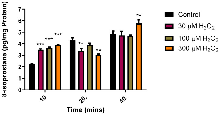
Isoprostanes are stable compounds whose production increases with exposure to OS, thus making them dependable markers of oxidative damage in both in vivo and in vitro animal models [46,47]. In a series of experiments, we studied the effects of time and different concentrations of H2O2 on the production of 8-isoprostane in an isolated bovine neural retina. As illustrated in Figure 1, 10–40 min insults with H2O2 (30–300 µM) elicited a concentration- and time-dependent increase in the 8-isoprostane levels (Figure 1). There was a time-dependent increase in the basal 8-isoprostane concentrations, reaching a maximum at 40 min. The effects elicited by H2O2 (100 µM) also showed a time-dependent increase reaching a maximum at 40 min. As a result of the data obtained in the studies described in Figure 1, all subsequent experiments assessing the pharmacological actions of H2S-producing compounds against H2O2-induced damage on isolated bovine retinae were performed in the presence of H2O2 (100 µM) for 30 min.
Figure 1.
Time- and concentration-dependent effect of H2O2 on 8-isoprostane production in the bovine retinae. Each value represents the mean ± SEM for n = 12; *** p < 0.001 is significantly different from the control and ** p < 0.01 is significantly different from the control.
2.2. Effects of H2S-Releasing Compounds on Lipid Peroxidation in the Bovine Retina
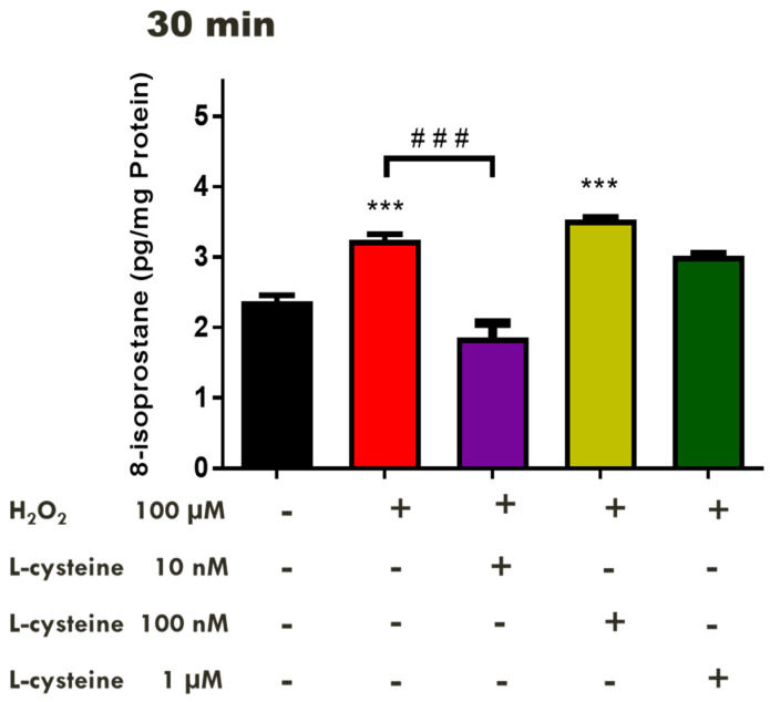
Although evidence in the literature supports a protective role for H2S against OS in neurons in vivo [46,48], the neuroprotective action of this gas in ocular tissues under conditions of OS induced by H2O2 in vitro warrants investigation. To assess the pharmacological role of endogenously produced H2S in the presence of the substrate, L-cysteine, isolated retinal tissues were pretreated with L-cysteine (10 nM–1 µM) for 30 min before an insult with H2O2 (100 µM). L-cysteine (10 nM) significantly (p < 0.001; n = 12) reversed the H2O2-induced increase in the 8-isoprostane levels, producing a 43% reduction in 8-isoprostane generation compared to the control tissues in the presence of H2O2. (Figure 2). In contrast, higher concentrations of L-cysteine (100 nM–1 µM) do not affect H2O2-induced damage.
Figure 2.
Concentration-dependent effect of L-cysteine on H2O2-induced oxidative stress in the bovine retinae. Each value represents the mean ± SEM for n = 12; *** p < 0.001 is significantly different from the control and ### p < 0.001 is significantly different from the H2O2 treated tissues.
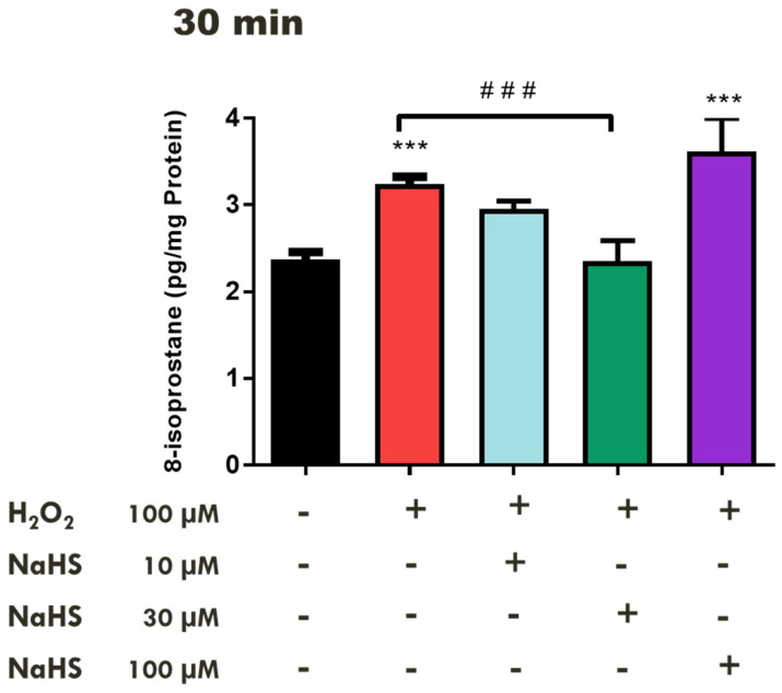
We then examined the effects of a fast-releasing H2S compound, NaHS, on 8-isoprostane production in the retina under conditions of H2O2-induced OS. Treatment of the tissues with NaHS (10–30 µM) for 10 min before the administration of H2O2 (100 µM) for 30 min significantly (p < 0.001; n = 12) attenuated 8-isoprostane production, whereas a high concentration of NaHS (100 µM) significantly (p < 0.001; n = 12) augmented the retinal 8-isoprostane levels, approximately 12% over the control (H2O2 treated tissues) (Figure 3).
Figure 3.
Concentration-dependent Effect of NaHS on H2O2–induced oxidative stress in the bovine retinae. Each value represents the mean ± SEM for n = 12; *** p < 0.001 is significantly different from the control and ### p < 0.001 is significantly different from the H2O2 treated tissues.
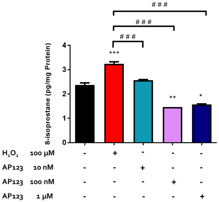
To examine the pharmacological action of the novel mitochondrial-targeting H2S-releasing compound, AP123, retinal tissues were exposed to varying concentrations of AP123 (10 nM–1 µM) before exposure to H2O2 (100 µM). After 30 min of treatment, AP123 (10 nM–1 µM) attenuated the H2O2-induced increase in 8-isoprostane production and even reduced the 8-isoprostane levels in the bovine retinae by 60% at higher concentrations (100 nM–1 µM) compared to the control tissues in the presence of H2O2. A maximal inhibitory effect on isoprostane production was observed with 100 nM of AP123 (Figure 4).
Figure 4.
Concentration-dependent effect of AP123 on H2O2-induced oxidative stress in the bovine retinae. Each value represents the mean ± SEM for n = 12; *** p < 0.001 is significantly different from the control, ** p < 0.01 is significantly different from the control, * p < 0.05 is significantly different from the control, and ### p < 0.001 is significantly different from the H2O2 treated tissues.
2.3. Effect of Inhibitors of CBS/CSE and 3-MST on the Antioxidant Actions of H2S-Producing Compounds
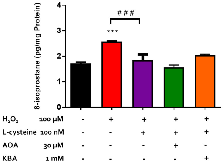
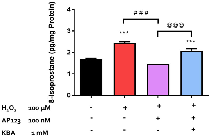
Endogenously produced H2S has been shown to protect retinal photoreceptors against light-induced oxidative damage [22]. In the present study, we investigated the role of endogenously produced H2S in the neuroprotective action of L-cysteine against H2O2-induced lipid peroxidation in isolated bovine neural retina. In a series of experiments, we tested the effect of an inhibitor of both CBS and CSE, amino-oxyacetic acid [AOAA, (30 µM)], and an inhibitor of 3-MST, ketobutyric acid [49] (KBA, 1 mM), on the pharmacological effect elicited by L-cysteine. Both AOAA (30 µM) and KBA (1 mM) did not affect the basal 8-isoprostane levels and had no significant effect on the reversal of H2O2-induced 8-isoprostane production caused by L-cysteine (100 µM) (Figure 5). The evidence in the literature suggests that AP123, the mitochondrial H2S donor, can exert antioxidant effects in the microvascular endothelial cells [27]. In the present study, we evaluated the role of endogenous H2S production through the 3-MST/H2S pathway in the antioxidant activity of AP123 in an isolated bovine retina under the conditions of H2O2-induced OS. On its own, KBA (1 mM) did not affect basal 8-isoprostane production. In the presence of KBA (1 mM), the inhibitory actions of AP123 on 8-isoprostane generation were significantly (p < 0.001; n = 12) reversed (Figure 6).
Figure 5.
Role of the 3-MST/CAT pathway in AP123-mediated neuroprotection. Each value represents the mean ± SEM for n = 12; *** p < 0.001 is significantly different from the control and ### p < 0.001 is significantly different from the H2O2 treated tissues.
Figure 6.
Role of CBS/CSE and the 3-MST/CAT pathway in L-cysteine-mediated neuroprotection. Each value represents the mean ± SEM for n = 12; *** p < 0.001 is significantly different from the control, ### p < 0.001 is significantly different from the H2O2 treated tissues, and @@@ p < 0.001 is significantly different from the AP123 + H2O2 treated tissues.
3. Discussion
ROS are formed during normal cellular metabolism, and there is evidence that they play an important, yet complex role in biological systems [1,2]. In excess, ROS can react with DNA, proteins, and lipids to form pharmacologically active metabolites that can perpetuate pathophysiological conditions [1,2]. Isoprostanes are chemically stable, prostaglandin-like lipid peroxidation products that are endogenously formed from oxidative damage to LCPUFAs [50]. These stable molecules are utilized as markers of lipid peroxidation in mammalian tissues, as their levels increase with oxidative damage [51,52]. While the evidence available supports the use of several markers to validate oxidative stress in biological samples [53,54], we selected 8-isoprostanes because of their direct relevance to the measurement of oxidative stress in retinal tissues [55,56,57]. In the present study, we found that various concentrations of H2O2 elicited a time-dependent increase in 8-isoprostane production in isolated bovine retinal tissues. The ability of exogenously administered H2O2 to cause a measurable increase in 8-isoprostane production supports the fact that lipid peroxidation occurs in response to this peroxide. The exposure of the tissues to increasing concentrations of H2O2 (30–300 µM) caused a time- and concentration-related increase in H2O2-induced lipid peroxidation, reaching a maximum at 300 µM. For this reason, an insult of 30 min duration with a submaximal concentration of H2O2 (100 µM) was selected as oxidative stimuli for all the experiments involving OS. It is pertinent to note that the concentration of H2O2 employed for inducing oxidative stress in retinal cells in the present study (100 µM) is much lower than that reported by other investigators such as Cui et al. [58] (200–800 µM), Hu et al. [59] (300–400 µM), and Wang et al. [60] (300 µM).
The biological actions of H2S have been documented in various mammalian tissues and systems [61]. This gaseous neurotransmitter is involved in a variety of pathophysiological processes such as inflammation, memory and learning, and in the regulation of blood pressure [16,62]. In the cardiovascular system, H2S has been shown to play a vital role in the maintenance of vascular smooth muscle tone, and in the central nervous system, this gas has been found to act as a neurotransmitter at synapses [63,64,65]. Cytoprotection is one of the many roles of this gas, as H2S has been reported to exert a neuroprotective action on neurons [48]. In 2011, Biermann and co-workers demonstrated that the exposure of animals to inhalational H2S decreased the apoptosis of retinal ganglion cells in a rat model of ischemia/reperfusion injury, suggesting a role for this gas as a neuroprotectant [66]. Because of the inherent challenges involved in the use of H2S gas in biological studies, the kinetic profiles of the release of this gas from some organic and inorganic compounds were extensively studied [28,67]. For instance, NaHS was reported to release H2S gas rapidly in biological media, whereas GYY4137 is a slow-releasing H2S compound [28]. There is evidence that H2S-releasing compounds such as NaHS can inhibit both light-induced degeneration and NMDA-induced oxidative damage in the retina, supporting a neuroprotective role for H2S [22,41]. H2S-releasing compounds have also been reported to inhibit excitatory amino acid neurotransmission (a marker of excitotoxicity) in an isolated bovine retina [38,68,69] and in H2O2-induced OS and cataract formation in cultured bovine lenses [70].
In the present study, the amino acid substrate for H2S production, L-cysteine, the fast-releasing H2S compound, NaHS, and the mitochondrial-targeted H2S compound, AP123, all protected the neural retina against H2O2-induced lipid peroxidation. Pretreatment with low concentrations of L-cysteine for 30 min attenuated lipid peroxidation whereas higher concentrations of this amino acid had no such action. The amino acid L-cysteine acts as a substrate for the enzymatic production of H2S and has been found to play a developmental and cytoprotective role in the CNS [71,72]. L-cysteine has been reported to promote the proliferation and differentiation of neural stem cells via the H2S/CBS pathway [72] and can protect against cellular damage in brain tissue [71]. Indeed, L-cysteine was found to upregulate the expression and activity of CBS and reduce brain edema in rats following a subarachnoid hemorrhage [71]. Taken together, our data suggest that the endogenous production of H2S from the substrate L-cysteine can protect the neural retina against H2O2-induced oxidative damage.
As observed with L-cysteine, pretreatment of tissues with low concentrations of NaHS for 30 min prevented H2O2-induced lipid peroxidation whereas a higher concentration of this fast-releasing H2S compound had no effect on oxidative damage. Interestingly, the pretreatment of tissues with a higher concentration of NaHS significantly increased (p < 0.001; n = 12) the levels of this lipid metabolite by 44% when compared with the control tissues. Both NaHS and N-acetyl-cysteine (NAC), a derivative of L-cysteine, have both been shown to exert concentration-related pharmacological actions that can be either protective or deleterious in biological systems [62,73,74]. Depending on the timing and duration of the H2S exposure prior to an oxidative insult as well as the concentration of the H2S-releasing compound utilized, the pharmacological actions of this gas can vary from cytoprotective and anti-inflammatory [62,71,74] to apoptotic and pro-inflammatory [62,73,74]. NaHS, an inorganic sulfide salt that instantaneously releases H2S in an aqueous solution, has been found to protect tissues from oxidative damage [45,66,75]. In a mouse model for diabetes, NaHS (100 µmol/kg) attenuated the production of ROS via the nicotine adenine dinucleotide phosphate (NADPH) oxidase and restored endothelial function in the aorta of diabetic mice [45]. The cortical neurons were protected against glutamate-induced excitotoxicity by NaHS (100 µM) via an observed increase in the activity of glutamate cysteine ligase, an enzyme involved in the production of the antioxidant GSH, and the augmentation of the intracellular GSH levels [75]. It is tempting to speculate that the observed protective effects of NaHS in the present study could be attributed to the stimulatory effect of NaHS on glutamate cysteine ligase and GSH production; however, further studies are needed to confirm this theory.
In the present study, the treatment of tissues with L-cysteine (10 nM) elicited a 43% reduction in the 8-isoprostane levels in the presence of H2O2, making it the most potent compound tested. The high potency of L-cysteine compared to the other H2S-releasing compounds has been observed in its pharmacological actions in ocular tissues. In a 2017 study, L-cysteine (100 nM) was found to increase the aqueous humor outflow facility by 150%, whereas a much higher dose of NaHS (10 µM) was required to exert a similar effect [76]. Similarly, Ohia and colleagues observed that L-cysteine elicited the most potent relaxation of carbachol-induced tone in porcine isolated irides when compared to the H2S-releasing compounds such as NaHS and Na2S [77].
AP123, a novel mitochondrial-targeting H2S-releasing compound, has been found to play a cytoprotective role in the cardiovascular system [27]. Indeed, AP123 (10 nM to 3 µM) was reported to stabilize mitochondrial membrane potential and significantly reduce the generation of ROS in hyperglycemic vascular endothelial cells [27]. In the present study, we found that the neuroprotective actions of AP123 in bovine retinae were observed in a similar concentration range as those demonstrated by Gero et al. [27]. Interestingly, AP123 not only prevented the H2O2-induced lipid peroxidation but it also elicited a further decrease in the basal isoprostane levels at higher concentrations. Through its actions as an electron donor in the electron transport chain, H2S has been found to stabilize membrane potential, thereby inhibiting the production of mitochondrial ROS in the vascular endothelium [78]. It is tempting to speculate that the observed marked effects of AP123 on lipid peroxidation in the present study may be related to its actions in H2S biosynthesis at the mitochondrial level.
In another series of experiments, we studied the role of endogenously produced H2S in the pharmacological action of L-cysteine against lipid peroxidation. The inhibitors of the enzymes responsible for the biosynthesis of H2S, (CBS, CSE, and 3-MST) were ineffective at diminishing the inhibitory effect elicited by L-cysteine on isoprostane production. Taken together, our data suggest that the protective actions of this amino acid in an isolated bovine retina are independent of its de novo enzymatic conversion to H2S. It is important to note that L-cysteine is also the rate-limiting agent in the production of glutathione (GSH), which possesses a cytoprotective function in various cell types due to its ROS-scavenging capabilities [8,79]. It may well be that the high potency displayed by L-cysteine in our studies could be related to its additional ability to regulate the production of GSH, a potent antioxidant.
3-MST is an enzyme that is constitutively expressed in mitochondria, and the production of H2S through this pathway is neuroprotective in the brain and retina [22,24]. In the present study, inhibition of 3-MST with KBA abolished the pharmacological actions of AP123, suggesting that the endogenous biosynthesis of H2S via the 3-MST pathway is involved in the pharmacological action of this H2S-releasing compound in an isolated bovine retina. Since AP123 is a mitochondrial-targeting H2S-releasing compound and 3-MST is localized to the mitochondria, it appears that the direct delivery of H2S to the mitochondria may account, at least in part, for the ability of KBA to effectively prevent the H2O2-induced lipid peroxidation. In summary, the data obtained using AP123 highlight a role for the mitochondria in serving as an important source of H2S in the observed protective action exhibited by some H2S-releasing compounds.
H2S-releasing compounds have been shown to reduce intraocular pressure in normotensive rabbits [80] and to increase the outflow facility in porcine ocular anterior segments, ex vivo [76], suggesting a potential role for this gas in glaucoma pharmacotherapy [81]. The current observation that H2S-releasing compounds can prevent oxidative damage in the retina supports a dual role for this gas in altering aqueous humor dynamics and serving as a neuroprotectant for the eye.
4. Materials and Methods
4.1. Chemicals
The H2O2, L-cysteine, sodium hydrosulfide (NaHS), amino-oxyacetic acid (AOAA), and α-ketobutyric acid (KBA) were purchased from Sigma Chemical (St. Louis, MO, USA). GYY4137, the 8-Isoprostane enzyme-linked immunoassay kit, and the protein determination assay kit were purchased from Cayman Chemical (Ann Harbor, MI, USA). AP123 was a gift from Dr. Matthew Whiteman’s laboratory. All test agents were freshly prepared immediately before use in the series of experiments. Stock solutions of AOAA were prepared in 70% ethanol. All other stock solutions were prepared in deionized water.
4.2. Tissue Preparation
Studies were performed using bovine eyes procured from a local slaughterhouse and transported to the laboratory in an ice bath. A cut was made along the equator of each eye, and the vitreous humor and lens were delicately dissected out. The neural retina was isolated by a gentle dislocation from the posterior segment of the eye and immediately immersed in a warm, oxygenated Krebs buffer solution (pH 7.4). After the isolation of the neural retina, the tissues were cut into four pieces and randomly assigned to treatment groups. The control groups received neither H2O2 nor H2S-releasing compounds/enzyme inhibitors. The time elapsed between the animal sacrifice and tissue preparation in the present study was less than 24 h; a protocol consistent with observations made by Murali et al. [82] who reported that human retinal tissues remained resilient to cell death, with a substantial number of cells remaining viable for up to five days postmortem.
4.3. 8-Isoprostane ELISA Assay
The methodology used for the extraction of 8-isoprostane was essentially the same as described by our laboratory and other investigators [83,84,85,86] with some modifications. The isolated bovine retinae were equilibrated in an oxygenated Krebs solution at 37 °C for 20 min. Tissues were then transferred and incubated in a Krebs solution in the presence and absence of H2O2 for 10 to 40 min. To determine the pharmacological effect of H2S-releasing compounds on H2O2-induced oxidative stress, the retinal tissues were treated with test compounds for 10–30 min before being subjected to a 30 min exposure to H2O2. To determine the role of enzymes in the biosynthesis of H2S, the retinae were treated with the CBS and CSE inhibitor (AOAA (30 µM)) and the noncompetitive 3-MST inhibitor, α-ketobutyric acid, (KBA (1 mM) to determine the involvement of the enzymes responsible for H2S biosynthesis in AP123- and L-cysteine-mediated actions. After incubation, tissues were homogenized in a 0.1 M phosphate buffer (pH 7.4) containing 1 mM of EDTA and 0.005% BHT (1 mL/100 mg tissue) and centrifuged at 3000 rpm for ten minutes at 5 °C. The supernatant was collected and purified using potassium hydroxide (15% w/v). The 8-isoprotane was extracted from purified samples using solid phase extraction cartridges and an ethyl acetate–methanol (99:1) mixture [87]. The 8-isoprotane was concentrated into a pellet by evaporating the ethyl acetate–methanol solution under N2 gas. The enzyme immunoassay buffer was used to re-suspend the 8-isoprotane prior to plating. Protein content was determined from the unpurified supernatant using a Cayman protein determination kit.
4.4. Data Analysis
The results were expressed as 8-isoprotane concentrations per milligram of soluble protein (pg/mg protein). Except where indicated, the values given are means ± S.E.M. The significance of differences between the values obtained in the control and drug-treated preparations were evaluated using one-way ANOVA followed by Tukey’s post hoc analysis. The time- and concentration-dependent effects were determined using two-way ANOVA. The differences in p values < 0.05 were accepted as statistically significant.
5. Conclusions
In conclusion, the substrate for the biosynthesis of H2S, L-cysteine, and H2S-releasing compounds, NaHS and AP123, can prevent H2O2-induced OS in an isolated bovine neural retina in a concentration-dependent manner. The pharmacological effect of L-cysteine was independent of the de novo intramural biosynthesis of H2S in the retina. On the other hand, the pharmacological action of the mitochondrial-targeted H2S-releasing compound, AP123, was mediated by the mechanisms that involve the 3-MST pathway for the biosynthesis of this gas in an isolated bovine retina. Taken together, our data support a neuroprotective role for H2S in the retina, a gas that has been reported to play a role in the regulation of aqueous humor dynamics in mammalian eyes.
Acknowledgments
The authors would like to thank Matthew Whiteman for providing the H2S-releasing compound AP123 for use in these studies.
Author Contributions
Conceptualization, L.B., S.E.O., and Y.F.N.M.; data curation, L.B., S.E.O., and Y.F.N.M.; formal analysis, L.B., S.E.O., and Y.F.N.M.; funding acquisition, C.A.O. and Y.F.N.M.; investigation, L.B., J.R., S.E.O., and Y.F.N.M.; methodology, L.B., S.E.O., and Y.F.N.M.; project administration, S.E.O. and Y.F.N.M.; resources, M.W. and Y.F.N.M.; supervision, C.A.O., M.W., S.E.O., and Y.F.N.M.; validation, L.B. and S.E.O.; visualization, L.B. and A.O.; writing—original draft, L.B.; writing—review & editing, L.B., J.R., A.O., F.M., C.A.O., S.E.O., and Y.F.N.M. All authors have read and agreed to the published version of the manuscript.
Institutional Review Board Statement
Not applicable.
Informed Consent Statement
Not applicable.
Data Availability Statement
The data are presented in the main text.
Conflicts of Interest
The authors declare that they have no known competing financial interests.
Funding Statement
This project was supported in part by Grant Number 1R15EY022215-01 from the National Institute of Health at the National Eye Institute.
Footnotes
Disclaimer/Publisher’s Note: The statements, opinions and data contained in all publications are solely those of the individual author(s) and contributor(s) and not of MDPI and/or the editor(s). MDPI and/or the editor(s) disclaim responsibility for any injury to people or property resulting from any ideas, methods, instructions or products referred to in the content.
References
- 1.Kim G.H., Kim J.E., Rhie S.J., Yoon S. The Role of Oxidative Stress in Neurodegenerative Diseases. Exp. Neurobiol. 2015;24:325–340. doi: 10.5607/en.2015.24.4.325. [DOI] [PMC free article] [PubMed] [Google Scholar]
- 2.Njie-Mbye Y.F., Kulkarni-Chitnis M., Opere C.A., Barrett A., Ohia S.E. Lipid peroxidation: Pathophysiological and pharmacological implications in the eye. Front. Physiol. 2013;4:366. doi: 10.3389/fphys.2013.00366. [DOI] [PMC free article] [PubMed] [Google Scholar]
- 3.Al-Shabrawey M., Smith S. Prediction of diabetic retinopathy: Role of oxidative stress and relevance of apoptotic biomarkers. EPMA J. 2010;1:56–72. doi: 10.1007/s13167-010-0002-9. [DOI] [PMC free article] [PubMed] [Google Scholar]
- 4.Berthoud V.M., Beyer E.C. Oxidative stress, lens gap junctions, and cataracts. Antioxid. Redox Signal. 2009;11:339–353. doi: 10.1089/ars.2008.2119. [DOI] [PMC free article] [PubMed] [Google Scholar]
- 5.Sacca S., Bolognesi C., Battistella A., Bagnis A., Izzotti A. Gene–environment interactions in ocular diseases. Mutat. Res. Mol. Mech. Mutagen. 2009;667:98–117. doi: 10.1016/j.mrfmmm.2008.11.002. [DOI] [PubMed] [Google Scholar]
- 6.Hollyfield J.G. Age-related macular degeneration: The molecular link between oxidative damage, tissue-specific inflammation and outer retinal disease: The Proctor lecture. Investig. Opthalmology Vis. Sci. 2010;51:1276–1281. doi: 10.1167/iovs.09-4478. [DOI] [PubMed] [Google Scholar]
- 7.De La Paz M.A., Zhang J., Fridovich I. Antioxidant enzymes of the human retina: Effect of age on enzyme activity of macula and periphery. Curr. Eye Res. 1996;15:273–278. doi: 10.3109/02713689609007621. [DOI] [PubMed] [Google Scholar]
- 8.Shih A.Y., Erb H., Sun X., Toda S., Kalivas P.W., Murphy T.H. Cystine/glutamate exchange modulates glutathione supply for neuroprotection from oxidative stress and cell proliferation. J. Neurosci. 2006;26:10514–10523. doi: 10.1523/JNEUROSCI.3178-06.2006. [DOI] [PMC free article] [PubMed] [Google Scholar]
- 9.Wielgus A.R., Sarna T. Ascorbate enhances photogeneration of hydrogen peroxide mediated by the iris melanin. Photochem. Photobiol. 2008;84:683–691. doi: 10.1111/j.1751-1097.2008.00341.x. [DOI] [PubMed] [Google Scholar]
- 10.Nishimura Y., Hara H., Kondo M., Hong S., Matsugi T. Oxidative Stress in Retinal Diseases. Oxidative Med. Cell. Longev. 2017;2017:4076518. doi: 10.1155/2017/4076518. [DOI] [PMC free article] [PubMed] [Google Scholar]
- 11.Greene E.L., Paller M.S. Xanthine oxidase produces O2−. in posthypoxic injury of renal epithelial cells. Pt 2Am. J. Physiol. Physiol. 1992;263:F251–F255. doi: 10.1152/ajprenal.1992.263.2.F251. [DOI] [PubMed] [Google Scholar]
- 12.SanGiovanni J.P., Chew E.Y. The role of omega-3 long-chain polyunsaturated fatty acids in health and disease of the retina. Prog. Retin. Eye Res. 2005;24:87–138. doi: 10.1016/j.preteyeres.2004.06.002. [DOI] [PubMed] [Google Scholar]
- 13.Shichi H. Cataract formation and prevention. Expert Opin. Investig. Drugs. 2004;13:691–701. doi: 10.1517/13543784.13.6.691. [DOI] [PubMed] [Google Scholar]
- 14.van Reyk D.M., Gillies M.C., Davies M.J. The retina: Oxidative stress and diabetes. Redox Rep. 2003;8:187–192. doi: 10.1179/135100003225002673. [DOI] [PubMed] [Google Scholar]
- 15.Ohia S.E., Opere C.A., LeDay A.M. Pharmacological consequences of oxidative stress in ocular tissues. Mutat. Res. Mol. Mech. Mutagen. 2005;579:22–36. doi: 10.1016/j.mrfmmm.2005.03.025. [DOI] [PubMed] [Google Scholar]
- 16.Łowicka E., Bełtowski J. Hydrogen sulfide (H2S)—The third gas of interest for pharmacologists. Pharmacol. Rep. 2007;59:4–24. [PubMed] [Google Scholar]
- 17.Goodwin L.R., Francom D., Dieken F.P., Taylor J.D., Warenycia M.W., Reiffenstein R.J., Dowling G. Determination of sulfide in brain tissue by gas dialysis/ion chromatography: Postmortem studies and two case reports. J. Anal. Toxicol. 1989;13:105–109. doi: 10.1093/jat/13.2.105. [DOI] [PubMed] [Google Scholar]
- 18.Lefer D.J. A new gaseous signaling molecule emerges: Cardioprotective role of hydrogen sulfide. Proc. Natl. Acad. Sci. USA. 2007;104:17907–17908. doi: 10.1073/pnas.0709010104. [DOI] [PMC free article] [PubMed] [Google Scholar]
- 19.Savage J., Gould D. Determination of sulfide in brain tissue and rumen fluid by ion-interaction reversed-phase high-performance liquid chromatography. J. Chromatogr. 1990;526:540–545. doi: 10.1016/S0378-4347(00)82537-2. [DOI] [PubMed] [Google Scholar]
- 20.Szabó C. Hydrogen sulphide and its therapeutic potential. Nat. Rev. Drug Discov. 2007;6:917–935. doi: 10.1038/nrd2425. [DOI] [PubMed] [Google Scholar]
- 21.Tan B.H., Wong P.T.-H., Bian J.-S. Hydrogen sulfide: A novel signaling molecule in the central nervous system. Neurochem. Int. 2010;56:3–10. doi: 10.1016/j.neuint.2009.08.008. [DOI] [PubMed] [Google Scholar]
- 22.Mikami Y., Shibuya N., Kimura Y., Nagahara N., Yamada M., Kimura H. Hydrogen sulfide protects the retina from light-induced degeneration by the modulation of Ca2+ influx. J. Biol. Chem. 2011;286:39379–39386. doi: 10.1074/jbc.M111.298208. [DOI] [PMC free article] [PubMed] [Google Scholar]
- 23.Shibuya N., Mikami Y., Kimura Y., Nagahara N., Kimura H. Vascular endothelium expresses 3-mercaptopyruvate sulfurtransferase and produces hydrogen sulfide. J. Biochem. 2009;146:623–626. doi: 10.1093/jb/mvp111. [DOI] [PubMed] [Google Scholar]
- 24.Shibuya N., Tanaka M., Yoshida M., Ogasawara Y., Togawa T., Ishii K., Kimura H. 3-Mercaptopyruvate sulfurtransferase produces hydrogen sulfide and bound sulfane sulfur in the brain. Antioxid. Redox Signal. 2009;11:703–714. doi: 10.1089/ars.2008.2253. [DOI] [PubMed] [Google Scholar]
- 25.Shibuya N., Kimura H. Production of hydrogen sulfide from d-cysteine and its therapeutic potential. Front. Endocrinol. 2013;4:57041. doi: 10.3389/fendo.2013.00087. [DOI] [PMC free article] [PubMed] [Google Scholar]
- 26.Shibuya N., Koike S., Tanaka M., Ishigami-Yuasa M., Kimura Y., Ogasawara Y., Fukui K., Nagahara N., Kimura H. A novel pathway for the production of hydrogen sulfide from D-cysteine in mammalian cells. Nat. Commun. 2013;4:1366. doi: 10.1038/ncomms2371. [DOI] [PubMed] [Google Scholar]
- 27.Gerő D., Torregrossa R., Perry A., Waters A., Le-Trionnaire S., Whatmore J.L., Wood M., Whiteman M. The novel mitochondria-targeted hydrogen sulfide (H2S) donors AP123 and AP39 protect against hyperglycemic injury in microvascular endothelial cells in vitro. Pt APharmacol. Res. 2016;113:186–198. doi: 10.1016/j.phrs.2016.08.019. [DOI] [PMC free article] [PubMed] [Google Scholar]
- 28.Li L., Whiteman M., Guan Y.Y., Neo K.L., Cheng Y., Lee S.W., Zhao Y., Baskar R., Tan C.H., Moore P.K. Characterization of a novel, water-soluble hydrogen sulfide-releasing molecule (GYY4137): New insights into the biology of hydrogen sulfide. Circulation. 2008;117:2351–2360. doi: 10.1161/CIRCULATIONAHA.107.753467. [DOI] [PubMed] [Google Scholar]
- 29.Zhao Y., Biggs T.D., Xian M. Hydrogen sulfide (H2S) releasing agents: Chemistry and biological applications. Chem. Commun. 2014;50:11788–11805. doi: 10.1039/C4CC00968A. [DOI] [PMC free article] [PubMed] [Google Scholar]
- 30.Abe K., Kimura H. The possible role of hydrogen sulfide as an endogenous neuromodulator. J. Neurosci. 1996;16:1066–1071. doi: 10.1523/JNEUROSCI.16-03-01066.1996. [DOI] [PMC free article] [PubMed] [Google Scholar]
- 31.Cheng Y., Ndisang J.F., Tang G., Cao K., Wang R. Hydrogen sulfide-induced relaxation of resistance mesenteric artery beds of rats. Am. J. Physiol. Heart Circ. Physiol. 2004;287:H2316–H2323. doi: 10.1152/ajpheart.00331.2004. [DOI] [PubMed] [Google Scholar]
- 32.Hosoki R., Matsuki N., Kimura H. The possible role of hydrogen sulfide as an endogenous smooth muscle relaxant in synergy with nitric oxide. Biochem. Biophys. Res. Commun. 1997;237:527–531. doi: 10.1006/bbrc.1997.6878. [DOI] [PubMed] [Google Scholar]
- 33.Kimura H. Hydrogen sulfide as a neuromodulator. Mol. Neurobiol. 2002;26:013–020. doi: 10.1385/MN:26:1:013. [DOI] [PubMed] [Google Scholar]
- 34.Qu K., Lee S.W., Bian J.S., Low C.-M., Wong P.T.-H. Hydrogen sulfide: Neurochemistry and neurobiology. Neurochem. Int. 2008;52:155–165. doi: 10.1016/j.neuint.2007.05.016. [DOI] [PubMed] [Google Scholar]
- 35.Kimura Y., Mikami Y., Osumi K., Tsugane M., Oka J., Kimura H. Polysulfides are possible H2S-derived signaling molecules in rat brain. FASEB J. 2013;27:2451–2457. doi: 10.1096/fj.12-226415. [DOI] [PubMed] [Google Scholar]
- 36.Zhang W., Xu C., Yang G., Wu L., Wang R. Interaction of H2S with Calcium Permeable Channels and Transporters. Oxid. Med. Cell Longev. 2015;2015:323269. doi: 10.1155/2015/323269. [DOI] [PMC free article] [PubMed] [Google Scholar]
- 37.Kulkarni M., Njie-Mbye Y.F., Okpobiri I., Zhao M., Opere C.A., Ohia S.E. Endogenous production of hydrogen sulfide in isolated bovine eye. Neurochem. Res. 2011;36:1540–1545. doi: 10.1007/s11064-011-0482-6. [DOI] [PubMed] [Google Scholar]
- 38.Bankhele P., Salvi A., Jamil J., Njie-Mbye F., Ohia S., Opere C.A. Comparative Effects of Hydrogen Sulfide-Releasing Compounds on [3H]D-Aspartate Release from Bovine Isolated Retinae. Neurochem. Res. 2018;43:692–701. doi: 10.1007/s11064-018-2471-5. [DOI] [PubMed] [Google Scholar]
- 39.Njie-Mbye Y.F., Bongmba O.Y.N., Onyema C.C., Chitnis A., Kulkarni M., Opere C.A., LeDay A.M., Ohia S.E. Effect of hydrogen sulfide on cyclic AMP production in isolated bovine and porcine neural retinae. Neurochem. Res. 2010;35:487–494. doi: 10.1007/s11064-009-0085-7. [DOI] [PubMed] [Google Scholar]
- 40.Njie-Mbye Y.F., Kulkarni M., Opere C.A., Ohia S.E. Mechanism of action of hydrogen sulfide on cyclic AMP formation in rat retinal pigment epithelial cells. Exp. Eye Res. 2012;98:16–22. doi: 10.1016/j.exer.2012.03.001. [DOI] [PubMed] [Google Scholar]
- 41.Cornwell A., Badiei A. The role of hydrogen sulfide in the retina. Exp. Eye Res. 2023;234:109568. doi: 10.1016/j.exer.2023.109568. [DOI] [PubMed] [Google Scholar]
- 42.Tribble J.R., Hui F., Quintero H., El Hajji S., Bell K., Di Polo A., Williams P.A. Neuroprotection in glaucoma: Mechanisms beyond intraocular pressure lowering. Mol. Asp. Med. 2023;92:101193. doi: 10.1016/j.mam.2023.101193. [DOI] [PubMed] [Google Scholar]
- 43.Tripathi S.J., Chakraborty S., Miller E., Pieper A.A., Paul B.D. Hydrogen sulfide signalling in neurodegenerative diseases. Br. J. Pharmacol. 2023 doi: 10.1111/bph.16170. [DOI] [PMC free article] [PubMed] [Google Scholar]
- 44.Liu H., Perumal N., Manicam C., Mercieca K., Prokosch V. Proteomics reveals the potential protective mechanism of hydrogen sulfide on retinal ganglion cells in an ischemia/reperfusion injury animal model. Pharmaceuticals. 2020;13:213. doi: 10.3390/ph13090213. [DOI] [PMC free article] [PubMed] [Google Scholar]
- 45.Sakamoto K., Suzuki Y., Kurauchi Y., Mori A., Nakahara T., Ishii K. Hydrogen sulfide attenuates NMDA-induced neuronal injury via its anti-oxidative activity in the rat retina. Exp. Eye Res. 2014;120:90–96. doi: 10.1016/j.exer.2014.01.008. [DOI] [PubMed] [Google Scholar]
- 46.Delanty N., Reilly M.P., Pratico D., Lawson J.A., McCarthy J.F., Wood A.E., Ohnishi S.T., Fitzgerald D.J., FitzGerald G.A. 8-epi PGF2 alpha generation during coronary reperfusion. A potential quantitative marker of oxidant stress in vivo. Circulation. 1997;95:2492–2499. doi: 10.1161/01.CIR.95.11.2492. [DOI] [PubMed] [Google Scholar]
- 47.Gopaul N.K., Anggard E.E., Mallet A.I., Betteridge D.J., Wolff S.P., Nourooz-Zadeh J. Plasma 8-epi-PGF2 alpha levels are elevated in individuals with non-insulin dependent diabetes mellitus. FEBS Lett. 1995;368:225–229. doi: 10.1016/0014-5793(95)00649-T. [DOI] [PubMed] [Google Scholar]
- 48.Kimura Y., Kimura H. Hydrogen sulfide protects neurons from oxidative stress. FASEB J. 2004;18:1165–1167. doi: 10.1096/fj.04-1815fje. [DOI] [PubMed] [Google Scholar]
- 49.Baskin S.I., Porter D.W., Rockwood G.A., Romano J.J.A., Patel H.C., Kiser R.C., Cook C.M., Ternay J.A.L. In vitro andin vivo comparison of sulfur donors as antidotes to acute cyanide intoxication. J. Appl. Toxicol. 1999;19:173–183. doi: 10.1002/(SICI)1099-1263(199905/06)19:3<173::AID-JAT556>3.0.CO;2-2. [DOI] [PubMed] [Google Scholar]
- 50.Milatovic D., Montine T.J., Aschner M. Measurement of isoprostanes as markers of oxidative stress. Methods Mol Biol. 2011;758:195–204. doi: 10.1007/978-1-61779-170-3_13. [DOI] [PMC free article] [PubMed] [Google Scholar]
- 51.Kadiiska M.B., Gladen B.C., Baird D., Germolec D., Graham L.B., Parker C.E., Nyska A., Wachsman J.T., Ames B.N., Basu S., et al. Biomarkers of oxidative stress study II: Are oxidation products of lipids, proteins, and DNA markers of CCl4 poisoning? Free Radic. Biol. Med. 2005;38:698–710. doi: 10.1016/j.freeradbiomed.2004.09.017. [DOI] [PubMed] [Google Scholar]
- 52.Labuschagne C.F., Broek N.J.F.v.D., Postma P., Berger R., Brenkman A.B. A protocol for quantifying lipid peroxidation in cellular systems by F2-isoprostane analysis. PLoS ONE. 2013;8:e80935. doi: 10.1371/journal.pone.0080935. [DOI] [PMC free article] [PubMed] [Google Scholar]
- 53.Frijhoff J., Winyard P.G., Zarkovic N., Davies S.S., Stocker R., Cheng D., Knight A.R., Taylor E.L., Oettrich J., Ruskovska T., et al. Clinical relevance of biomarkers of oxidative stress. Antioxid. Redox Signal. 2015;23:1144–1170. doi: 10.1089/ars.2015.6317. [DOI] [PMC free article] [PubMed] [Google Scholar]
- 54.Czerska M., Mikołajewska K., Zieliński M., Gromadzińska J., Wąsowicz W. Today’s oxidative stress markers. Med. Pr. 2015;66:393–405. doi: 10.13075/mp.5893.00137. [DOI] [PubMed] [Google Scholar]
- 55.Nourooz-Zadeh J., Pereira P. F2 isoprostanes, potential specific markers of oxidative damage in human retina. Ophthalmic Res. 2000;32:133–137. doi: 10.1159/000055603. [DOI] [PubMed] [Google Scholar]
- 56.Cervellati F., Cervellati C., Romani A., Cremonini E., Sticozzi C., Belmonte G., Pessina F., Valacchi G. Hypoxia induces cell damage via oxidative stress in retinal epithelial cells. Free Radic. Res. 2014;48:303–312. doi: 10.3109/10715762.2013.867484. [DOI] [PubMed] [Google Scholar]
- 57.Wong L.L., Pye Q.N., Chen L., Seal S., McGinnis J.F. Defining the catalytic activity of nanoceria in the P23H-1 rat, a photoreceptor degeneration model. PLoS ONE. 2015;10:e0121977. doi: 10.1371/journal.pone.0121977. [DOI] [PMC free article] [PubMed] [Google Scholar]
- 58.Cui Y., Xu N., Xu W., Xu G. Mesenchymal stem cells attenuate hydrogen peroxide-induced oxidative stress and enhance neuroprotective effects in retinal ganglion cells. Vitr. Cell. Dev. Biol.-Anim. 2017;53:328–335. doi: 10.1007/s11626-016-0115-0. [DOI] [PubMed] [Google Scholar]
- 59.Hu L., Guo J., Zhou L., Zhu S., Wang C., Liu J., Hu S., Yang M., Lin C. Hydrogen sulfide protects retinal pigment epithelial cells from oxidative stress-induced apoptosis and affects autophagy. Oxid. Med. Cell. Longev. 2020;2020:8868564. doi: 10.1155/2020/8868564. [DOI] [PMC free article] [PubMed] [Google Scholar]
- 60.Wang Y., Wang J., Zhang X., Feng Y., Yuan Y. Neuroprotective effects of idebenone on hydrogen peroxide-induced oxidative damage in retinal ganglion cells-5. Int. Ophthalmol. 2023;43:3831–3839. doi: 10.1007/s10792-023-02831-x. [DOI] [PubMed] [Google Scholar]
- 61.Wallace J.L., Wang R. Hydrogen sulfide-based therapeutics: Exploiting a unique but ubiquitous gasotransmitter. Nat. Rev. Drug Discov. 2015;14:329–345. doi: 10.1038/nrd4433. [DOI] [PubMed] [Google Scholar]
- 62.Wu D., Hu Q., Zhu Y. Therapeutic application of hydrogen sulfide donors: The potential and challenges. Front. Med. 2015;10:18–27. doi: 10.1007/s11684-015-0427-6. [DOI] [PubMed] [Google Scholar]
- 63.Van Goor H., van den Born J.C., Hillebrands J.-L., Joles J.A. Hydrogen sulfide in hypertension. Curr. Opin. Nephrol. Hypertens. 2016;25:107–113. doi: 10.1097/MNH.0000000000000206. [DOI] [PubMed] [Google Scholar]
- 64.Sattar M., Ahmad A., Rathore H., Khan S., Lazhari M., Afzal S., Hashmi F., Abdullah N., Johns E. A critical review of pharmacological significance of Hydrogen Sulfide in hypertension. Indian J. Pharmacol. 2015;47:243–247. doi: 10.4103/0253-7613.157106. [DOI] [PMC free article] [PubMed] [Google Scholar]
- 65.Kida K., Ichinose F. Hydrogen Sulfide and Neuroinflammation. Handb. Exp. Pharmacol. 2015;230:181–189. doi: 10.1007/978-3-319-18144-8_9. [DOI] [PubMed] [Google Scholar]
- 66.Biermann J., Lagrèze W.A., Schallner N., Schwer C.I., Goebel U. Inhalative preconditioning with hydrogen sulfide attenuated apoptosis after retinal ischemia/reperfusion injury. Mol. Vis. 2011;17:1275–1286. [PMC free article] [PubMed] [Google Scholar]
- 67.Powell C.R., Dillon K.M., Matson J.B. A review of hydrogen sulfide (H2S) donors: Chemistry and potential therapeutic applications. Biochem. Pharmacol. 2018;149:110–123. doi: 10.1016/j.bcp.2017.11.014. [DOI] [PMC free article] [PubMed] [Google Scholar]
- 68.Ohia S.E., Opere C.A., Awe S.O., Adams L., Sharif N.A. Human, bovine, and rabbit retinal glutamate-induced [3H]D-aspartate release: Role in excitotoxicity. Neurochem. Res. 2000;25:853–860. doi: 10.1023/A:1007525725996. [DOI] [PubMed] [Google Scholar]
- 69.Opere C.A., Heruye S., Njie-Mbye Y.-F., Ohia S.E., Sharif N.A. Regulation of excitatory amino acid transmission in the retina: Studies on neuroprotection. J. Ocul. Pharmacol. Ther. 2018;34:107–118. doi: 10.1089/jop.2017.0085. [DOI] [PubMed] [Google Scholar]
- 70.Heruye S.H., Mbye Y.F., Ohia S.E., Opere C.A. Protective action of hydrogen sulfide-releasing compounds against oxidative stress-induced cataract formation in cultured bovine lenses. Curr. Eye Res. 2022;47:239–245. doi: 10.1080/02713683.2021.1975764. [DOI] [PubMed] [Google Scholar]
- 71.Li T., Wang L., Hu Q., Liu S., Bai X., Xie Y., Zhang T., Bo S., Gao X., Wu S., et al. Neuroprotective Roles of l-Cysteine in Attenuating Early Brain Injury and Improving Synaptic Density via the CBS/H2S Pathway Following Subarachnoid Hemorrhage in Rats. Front. Neurol. 2017;8:176. doi: 10.3389/fneur.2017.00176. [DOI] [PMC free article] [PubMed] [Google Scholar]
- 72.Wang Z., Liu D.-X., Wang F.-W., Zhang Q., Du Z.-X., Zhan J.-M., Yuan Q.-H., Ling E.-A., Hao A.-J. L-Cysteine promotes the proliferation and differentiation of neural stem cells via the CBS/H(2)S pathway. Neuroscience. 2013;237:106–117. doi: 10.1016/j.neuroscience.2012.12.057. [DOI] [PubMed] [Google Scholar]
- 73.Qanungo S., Wang M., Nieminen A.L. N-Acetyl-L-cysteine enhances apoptosis through inhibition of nuclear factor-kappaB in hypoxic murine embryonic fibroblasts. J. Biol. Chem. 2004;279:50455–50464. doi: 10.1074/jbc.M406749200. [DOI] [PubMed] [Google Scholar]
- 74.Whiteman M., Li L., Rose P., Tan C.-H., Parkinson D.B., Moore P.K. The effect of hydrogen sulfide donors on lipopolysaccharide-induced formation of inflammatory mediators in macrophages. Antioxid. Redox Signal. 2010;12:1147–1154. doi: 10.1089/ars.2009.2899. [DOI] [PMC free article] [PubMed] [Google Scholar]
- 75.Kimura Y., Goto Y.-I., Kimura H. Hydrogen sulfide increases glutathione production and suppresses oxidative stress in mitochondria. Antioxid. Redox Signal. 2010;12:1–13. doi: 10.1089/ars.2008.2282. [DOI] [PubMed] [Google Scholar]
- 76.Robinson J., Okoro E., Ezuedu C., Bush L., Opere C.A., Ohia S.E., Njie-Mbye Y.F. Effects of Hydrogen Sulfide-Releasing Compounds on Aqueous Humor Outflow Facility in Porcine Ocular Anterior Segments, Ex Vivo. J. Ocul. Pharmacol. Ther. 2017;33:91–97. doi: 10.1089/jop.2016.0037. [DOI] [PMC free article] [PubMed] [Google Scholar]
- 77.Ohia S.E., Opere C.A., Monjok E.M., Kouamou G., Leday A.M., Njie-Mbye Y.F. Role of hydrogen sulfide production in inhibitory action of L-cysteine on isolated porcine irides. Curr. Eye Res. 2010;35:402–407. doi: 10.3109/02713680903576716. [DOI] [PubMed] [Google Scholar]
- 78.Suzuki K., Olah G., Modis K., Coletta C., Kulp G., Gerö D., Szoleczky P., Chang T., Zhou Z., Wu L., et al. Hydrogen sulfide replacement therapy protects the vascular endothelium in hyperglycemia by preserving mitochondrial function. Proc. Natl. Acad. Sci. USA. 2011;108:13829–13834. doi: 10.1073/pnas.1105121108. [DOI] [PMC free article] [PubMed] [Google Scholar]
- 79.Franklin C.C., Backos D.S., Mohar I., White C.C., Forman H.J., Kavanagh T.J. Structure, function, and post-translational regulation of the catalytic and modifier subunits of glutamate cysteine ligase. Mol. Asp. Med. 2008;30:86–98. doi: 10.1016/j.mam.2008.08.009. [DOI] [PMC free article] [PubMed] [Google Scholar]
- 80.Salvi A., Bankhele P., Jamil J., Chitnis M.K., Njie-Mbye Y.F., Ohia S.E., Opere C.A. Effect of hydrogen sulfide donors on intraocular pressure in rabbits. J. Ocul. Pharmacol. Ther. 2016;32:371–375. doi: 10.1089/jop.2015.0144. [DOI] [PMC free article] [PubMed] [Google Scholar]
- 81.Ohia S.E., Robinson J., Mitchell L., Ngele K.K., Heruye S., Opere C.A., Njie-Mbye Y.F. Regulation of aqueous humor dynamics by hydrogen sulfide: Potential role in glaucoma pharmacotherapy. J. Ocul. Pharmacol. Ther. 2018;34:61–69. doi: 10.1089/jop.2017.0077. [DOI] [PMC free article] [PubMed] [Google Scholar]
- 82.Murali A., Ramlogan-Steel C.A., Steel J.C., Layton C.J. Characterisation and validation of the 8-fold quadrant dissected human retinal explant culture model for pre-clinical toxicology investigation. Toxicol. Vitr. 2020;63:104716. doi: 10.1016/j.tiv.2019.104716. [DOI] [PubMed] [Google Scholar]
- 83.Kulkarni P., Payne S. Eicosanoids in bovine retinal microcirculation. J. Ocul. Pharmacol. Ther. 1997;13:139–149. doi: 10.1089/jop.1997.13.139. [DOI] [PubMed] [Google Scholar]
- 84.LeDay A.M., Kulkarni K.H., Opere C.A., Ohia S.E. Arachidonic acid metabolites and peroxide-induced inhibition of [3H]D-aspartate release from bovine isolated retinae. Curr. Eye Res. 2004;28:367–372. doi: 10.1076/ceyr.28.5.367.28675. [DOI] [PubMed] [Google Scholar]
- 85.Matsuda K., Ohnishi K., Misaka E., Yamazaki M. Decrease of urinary prostaglandin E2 and prostaglandin F2 alpha excretion by nonsteroidal anti-inflammatory drugs in rats. Relationship to anti-inflammatory activity. Biochem. Pharmacol. 1983;32:1347–1352. doi: 10.1016/0006-2952(83)90445-8. [DOI] [PubMed] [Google Scholar]
- 86.Zhan G.-L., Ohia S., Camras C., Ohia E., Wang Y. Superior cervical ganglionectomy-induced lowering of intraocular pressure in rabbits: Role of prostaglandins and neuropeptide Y. Gen. Pharmacol. Vasc. Syst. 1999;32:189–194. doi: 10.1016/S0306-3623(98)00187-6. [DOI] [PubMed] [Google Scholar]
- 87.Nucci C., Gasperi V., Tartaglione R., Cerulli A., Terrinoni A., Bari M., De Simone C., Agro A.F., Morrone L.A., Corasaniti M.T., et al. Involvement of the endocannabinoid system in retinal damage after high intraocular pressure-induced ischemia in rats. Investig. Opthalmology Vis. Sci. 2007;48:2997–3004. doi: 10.1167/iovs.06-1355. [DOI] [PubMed] [Google Scholar]
Associated Data
This section collects any data citations, data availability statements, or supplementary materials included in this article.
Data Availability Statement
The data are presented in the main text.


