Error in Figure 5
In the original publication [1], Figure 5c showed an image that was edited to eliminate an electron-dense artifact (precipitate stain generated using the contrast technique in electron microscopy), which in no way modified the essence of what the authors wanted to highlight in the publication. However, the authors now consider it more appropriate to present the image highlighting only the area of interest.
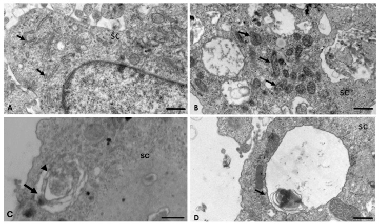
The corrected Figure 5c appears as follows:
Figure 5.
Interaction of A. culbertsoni and Schwann cells (SC) after 2 h of interaction. (A,B) Ultrastructural alterations in organelles of SC were frequently observed (arrows) in the rough endoplasmic reticulum (A) and mitochondria (B) Bars = 2 µm. (C,D) Multilamellar bodies with cytoplasmic material (arrows) persisted. In electron micrograph (C) Bar = 500 nm, a double membrane (arrow head) is evident, characteristic of these structures. Bar = 2 µm.
Addition of Supplementary File
The authors would like to include the following supporting information in the main text, under Section 2.4, “Analysis of the Interaction of A. culbertsoni Trophozoites with SC through TEM,” in the third paragraph.
The reference to the supplementary materials should be added to the third paragraph of Section 2.4. The sentence, “Multilamellar bodies with the characteristic double membrane persisted (Figure 5C),” should be revised to, “Multilamellar bodies with the characteristic double membrane persisted (Figure 5C and Supplementary Figure S1).”
In the Supplementary Materials section at the end of the paper, the authors included the following:
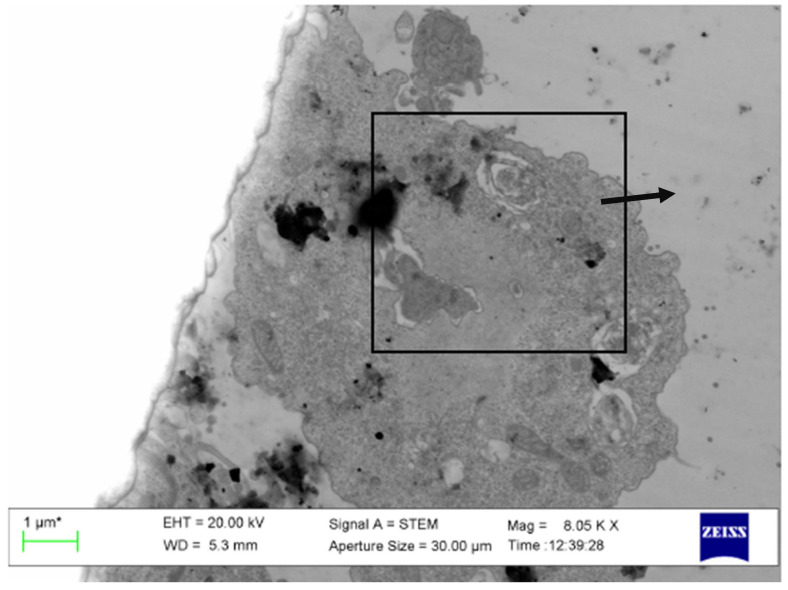
Supplementary Materials: The following supporting information can be downloaded at: http://www.mdpi.com/2076-0817/9/6/458/s1, Figure S1: Original image corresponding to Figure 5C in which Multilamellar body with cytoplasmic material (arrow) persisted are described.
The Figure S1 appears as follows:
Figure S1.
Original image corresponding to Figure 5C in which Multilamellar body with cytoplasmic material (arrow) persisted are described.
The authors state that the scientific conclusions are unaffected. These corrections were approved by the Academic Editor. The original publication has also been updated.
Footnotes
Disclaimer/Publisher’s Note: The statements, opinions and data contained in all publications are solely those of the individual author(s) and contributor(s) and not of MDPI and/or the editor(s). MDPI and/or the editor(s) disclaim responsibility for any injury to people or property resulting from any ideas, methods, instructions or products referred to in the content.
Reference
- 1.Castelan-Ramírez I., Salazar-Villatoro L., Chávez-Munguía B., Salinas-Lara C., Sánchez-Garibay C., Flores-Maldonado C., Hernández-Martínez D., Anaya-Martínez V., Ávila-Costa M.R., Méndez-Cruz A.R., et al. Schwann Cell Autophagy and Necrosis as Mechanisms of Cell Death by Acanthamoeba. Pathogens. 2020;9:458. doi: 10.3390/pathogens9060458. [DOI] [PMC free article] [PubMed] [Google Scholar]


