A 66-year-old male patient presented with incidentally detected lymphocytosis. On examination, no lymphadenopathy/hepatosplenomegaly/pallor/icterus was found. CBC showed a leukocyte count of 21.1 × 10⁹/L with normal hemoglobin (13.6 g/dL) and platelets (193X109/L). Smear examination showed an absolute lymphocyte count of 15.4 × 10⁹/L, predominantly composed of small lymphoid cells.
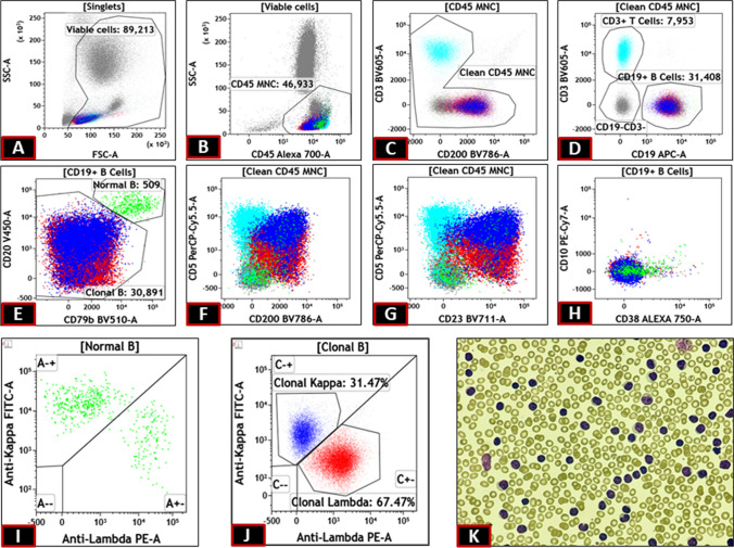
On flow-cytometric analysis, B cells constituted 34% of all leukocytes, showing multiple immunophenotypic abnormalities (Fig. 1) consistent with the diagnosis of chronic lymphocytic leukemia (CLL). The abnormalities included down-regulation of CD20 and CD79b, along with the expression of CD5, CD23, and CD200. CD10 and CD38 were negative. However, on light chain analysis, one of these populations was positive for kappa (blue population), and the other was positive for lambda (red population). On back-gating, both of these populations had the same immunophenotype, except CD5, which was brighter in the kappa-positive population. A final diagnosis of bi-clonal CLL was made.
Fig. 1.
Mononuclear cell were gated on SSC Vs CD45 plot (B) after removal of debris on SSC Vs FSC (A). CD19 positive cells were gated (D) after removal of junk on CD3 Vs CD200 plot(C). Normal B cells were identified as bright CD20 and CD79b positive cells (E). The rest abnormal B cells showed immunophenotype consistent with CLL (E to H) but with split on kappa lambda plot (J) but with dimmer intensity than the normal B cells (I). Peripheral smear showing small cell lymphocytosis (K)
Bi-clonal CLL is an uncommon disease, with a reported incidence of 3.4% of CLL cases. [1]Their clinical behavior is akin to their normal counterpart; however, they show significantly lower WBC counts and a greater incidence of splenomegaly [1]. The bi-clonal nature of the disease may pose a difficulty in diagnosis on tissue biopsy, as clonality assessment is often done using immunohistochemistry for light chains. The patient is currently on observation as his counts are normal and is not requiring treatment.
Declarations
Disclaimers
The identity of the patient is not disclosed here in this case. The authors state that there is no conflict of interest present.
Conflict of Interest
1. Author 1 declares that he has no conflict of interest.
2. Author 2 declares that he has no conflict of interest.
3. Author 3 declares that he has no conflict of interest.
4. Author 4 declares that he has no conflict of interest.
Footnotes
Publisher's Note
Springer Nature remains neutral with regard to jurisdictional claims in published maps and institutional affiliations.
Reference
- 1.Sanchez ML, Almeida J, Gonzalez M et al (2003) Incidence and clinicobiologic characteristics of leukemic B-cell chronic lymphoproliferative disorders with more than one B-cell clone. Blood 102:2994–3002 [DOI] [PubMed] [Google Scholar]


