Abstract
BACKGROUND
According to current statistics, renal cancer accounts for 3% of all cancers worldwide. Renal cell carcinoma (RCC) is the most common solid lesion in the kidney and accounts for approximately 90% of all renal malignancies. Increasing evidence has shown an association between immune infiltration in RCC and clinical outcomes. To discover possible targets for the immune system, we investigated the link between tumor-infiltrating immune cells (TIICs) and the prognosis of RCC.
AIM
To investigate the effects of 22 TIICs on the prognosis of RCC patients and identify potential therapeutic targets for RCC immunotherapy.
METHODS
The CIBERSORT algorithm partitioned the 22 TIICs from the Cancer Genome Atlas cohort into proportions. Cox regression analysis was employed to evaluate the impact of 22 TIICs on the probability of developing RCC. A predictive model for immunological risk was developed by analyzing the statistical relationship between the subpopulations of TIICs and survival outcomes. Furthermore, multivariate Cox regression analysis was used to investigate independent factors for the prognostic prediction of RCC. A value of P < 0.05 was regarded as statistically significant.
RESULTS
Compared to normal tissues, RCC tissues exhibited a distinct infiltration of immune cells. An immune risk score model was established and univariate Cox regression analysis revealed a significant association between four immune cell types and the survival risk connected to RCC. High-risk individuals were correlated to poorer outcomes according to the Kaplan-Meier survival curve (P = 1E−05). The immunological risk score model was demonstrated to be a dependable predictor of survival risk (area under the curve = 0.747) via the receiver operating characteristic curve. According to multivariate Cox regression analysis, the immune risk score model independently predicted RCC patients' prognosis (hazard ratio = 1.550, 95%CI: 1.342–1.791; P < 0.001). Finally, we established a nomogram that accurately and comprehensively forecast the survival of patients with RCC.
CONCLUSION
TIICs play various roles in RCC prognosis. The immunological risk score is an independent predictor of poor survival in kidney cancer cases.
Keywords: Renal cell carcinoma, Tumor-infiltrating immune cells, Prognosis, Immune risk score model, Nomogram
Core Tip: Renal cell carcinoma (RCC) is a prevalent form of renal cancer that typically arises subtly and is distinguished by its propensity for easy metastasis and poor prognosis. Patients with advanced RCC frequently exhibit low overall survival rates owing to the extensive spread of cancer. However, the recent advent of immunotherapy has resulted in an improvement in the median overall survival for all risk groups of patients, making it imperative to identify potential immune targets. Our research has led to the development of an immune risk assessment model that underscores the critical role of tumor-infiltrating immune cells-based immune models in the prognostic prediction and clinical management of RCC.
INTRODUCTION
Renal cancer is a highly prevalent cancer that causes malignant tumors worldwide. Its prevalence has been increasing over the past decade, accounting for up to 2%–3% of all newly diagnosed tumor cases[1,2]. Histologically, renal cell carcinoma (RCC) is a prominent subtype of renal cancer, comprising approximately 85% of the total renal cancer cases[3-5]. Despite great developments in conception, diagnosis, surgery, and various tumor drug treatments, the clinical results of RCC are still insufficient. Therefore, the accurate assessment of patient survival risk and prognostic prediction is critical.
The human immune system effectively protects the body from both exogenous and endogenous diseases. When uncontrolled proliferation of mutated or abnormal cells occurs in an organism, invading and spreading throughout the body, the immune system effectively recognizes the differences between tumors and healthy tissues, thereby secreting relevant signals to inhibit tumor growth. However, this process may co-opted by the cancer cells. Research has shown that during tumor progression, cancer cells take advantage of oncogenic mutations in the immune system to help escape immune system attack and promote growth[6,7]. These findings indicate that there are diverse immune escape mechanisms by which tumor cells reduce killing by the immune system during progression. In addition, during tumor progression, changes in immune phenotype and composition cause secondary changes in immune responses within and outside the tumor microenvironment (TME)[8]. Reconstruction of tumor immunity occurs in a wide range of tumor types. Among them, RCC has a high metastatic rate and poor prognosis, with 30% of patients already having metastases at the first diagnosis, and nearly 30% of the remaining patients having metastatic foci detected during the course of follow-up[9]. This characteristic is closely related to the interactions among the stroma, immune cells, and tumor cells in the RCC immune microenvironment, which leads to reconstruction of the microenvironment[10]. Research on the complexity of the immune heterogeneity of RCC with respect to this characteristic will help us clinically assess tumor heterogeneity and thus facilitate the development of more effective and personalized therapies.
The TME is highly heterogeneous. Previous research has revealed that dynamic interactions between tumor cells and their microenvironment promote the formation, progression, metastasis, and development of drug resistance in solid tumors[11]. There is substantial evidence that the composition of multiple immune cells within the microenvironment affects antitumor immunity and immunotherapeutic responsiveness. An in-depth understanding of the TME, particularly the characteristics of tumor-infiltrating immune cells (TIICs), is essential for identifying critical regulatory molecules for tumor development and immunotherapy.
Most previous studies on RCC prognostic modeling have included only traditional clinical information, and the model has limitations such as poor sensitivity and lag, which makes it impossible to comprehensively assess the clinical application value of the model[12-14]. CIBERSORT is an algorithm that employs gene expression data to analyze barcode gene values of expression and estimate the components of immune cells. The CIBERSORT algorithm is more efficient and precise than traditional immunohistochemical and flow cytometry approaches for calculating the relative proportions of 22 types of invasive TIICs[15]. Based on the effectiveness of this methodology, numerous studies have recently applied it to examine whether patient prognosis is affected by the 22 TIICs subtypes[16-18].
Hence, our study aimed to explore the relationship between TIICs and RCC. To investigate potential relationships between immunotherapy and RCC patients, we constructed an immune risk score methodology to prognostic prediction and thoroughly examined the impact of 22 TIICs subgroups on the prognosis of RCC patients by utilizing the CIBERSORT algorithm. The result provided a new avenue for the development of prognostic prediction models for RCC patients as well as the identification of therapeutic targets for immunotherapy.
MATERIALS AND METHODS
Data acquisition
The entire transcriptome data of RCC utilized in this investigation were obtained from The Cancer Genome Atlas (TCGA) database. The research project encompassed a cohort of 957 patients diagnosed with RCC. The transcriptome data was obtained from the TCGA database using the R package "TCGA-Assembler". This dataset consisted of 128 cases of normal tissue and 829 cases of malignant tissue. Furthermore, we collected essential clinical information, including age, sex, TNM stage, grade, survival status, and survival duration, from a total of 829 patients diagnosed with RCC. The R software's "limma" package was executed to rectify the transcriptome data.
Assessment of immune infiltration
The deconvolution algorithm CIBERSORT divides 547 tag gene expression levels to describe the immune cell composition in tissues. In this research, the relative proportions of 22 TIICs in the transcriptome data of corrected RCC patients were estimated via this algorithm. We uploaded the transcriptome to the CIBERSORT website (http://cibersort.stanford.edu/) and configured the algorithm to process 1000 rows. The criterion to calculate statistical significance was set at a P value < 0.05.
Statistical analysis
The analyses were conducted via Statistical Package for the Social Sciences 23.0 and R 3.5.3. Statistical tests were performed as two-sided tests. The criterion to calculate statistical significance was set at a P value < 0.05. The Kaplan-Meier curve was assessed via the log-rank test to examine the correlation between immune cell infiltration and overall survival. The sensitivity and specificity of the recurrence prediction model were analyzed using time-dependent receiver operating characteristic (ROC) curves. Independent impact factors linked to survival were examined using a multivariate Cox regression model, whereas the impacts of individual variables on survival were examined using a one-factor Cox regression model. A nomogram was generated to represent the regression coefficients in the Cox analysis.
RESULTS
Distribution of TIICs
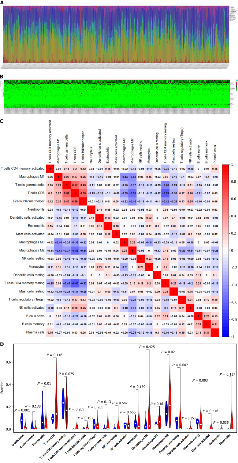
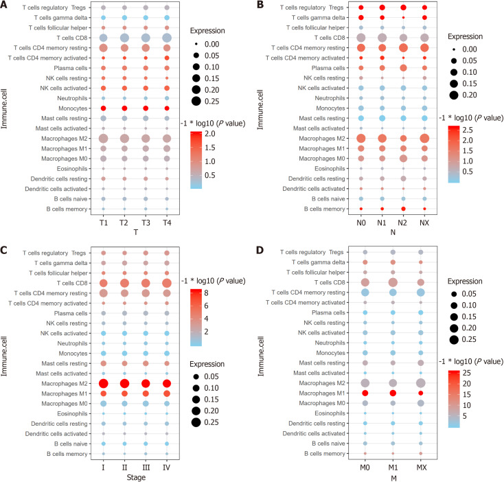
With the CIBERSORT algorithm, we conducted immune cell infiltration analysis to facilitate the identification of relevant immune cell levels in all RCC patients in the TCGA database. The dataset included 829 patients with RCC and 128 healthy tissues. The clinicopathological characteristics of the samples are shown in Table 1. Figure 1A-C display the arrangement of TIICs in RCC samples and the relationship with various immune cell subtypes. The violin diagram demonstrates the main advantages in composition between normal and RCC tissues in 22 TIICs (Figure 1D). The invasion of immune cells, such as native B cells, plasma cells, the alternating-activated subset (M2) macrophages, dendritic cells, and eosinophils, did not change between normal tissues and RCC tissues. Further analysis revealed that the normal activated subset (M1) macrophages, M2 macrophages, monocytes, and T cells exhibited the strongest correlation with tumor advancement (Figure 2). On the basis of the above analysis, we explored the types and distributions of TIICs subpopulations that are closely related to RCC, which provides potential research directions for clinical immunotherapy.
Table 1.
Clinical characteristics of patients
|
Variables
|
Clinical characters
|
Numbers
|
| Age in years | ≤ 65 | 343 |
| > 65 | 181 | |
| Sex | Female | 185 |
| Male | 339 | |
| T stage | T1 | 269 |
| T2 | 66 | |
| T3 | 178 | |
| T4 | 11 | |
| M stage | M0 | 418 |
| M1 | 78 | |
| MX | 28 | |
| N stage | N0 | 236 |
| N1 | 15 | |
| N2 | 273 | |
| Stage | Stage I | 263 |
| Stage II | 54 | |
| Stage III | 124 | |
| Stage IV | 83 | |
| Grade | G1 | 14 |
| G2 | 227 | |
| G3 | 206 | |
| G4 | 77 |
The 305 cases of kidney cancer patients lack clinical data.
Figure 1.
Analysis of the expression levels of 22 types of tumor-infiltrating immune cells and their correlation in 829 renal cell carcinoma patients and 128 normal controls. A and B: Expression of 22 tumor-infiltrating immune cells (TIICs) in renal cell carcinoma (RCC) and normal samples; C: Correlation analysis of 22 TIICs and immune cells between 829 RCC samples; D: Differences in the composition of TIICs between normal tissue and RCC tissue in 829 RCC samples.
Figure 2.
Correlation analysis between TNM stage and 22 tumor-infiltrating immune cells in 829 renal cell carcinoma patients. A: Tumor stage and the expression of 22 tumor-infiltrating immune cells (TIICs) in 829 renal cell carcinoma (RCC) patients; B: Node stage and the expression of 22 TIICs in 829 RCC patients; C: Metastasis stage and the expression of 22 TIICs in 829 RCC patients; D: Pathologic stage and the expression of 22 TIICs in 829 RCC patients. NK: Natural killer; M1: Normal activated subset; M2: Alternating-activated subset.
Univariate and multivariate Cox regression analyses to construct an immune risk score
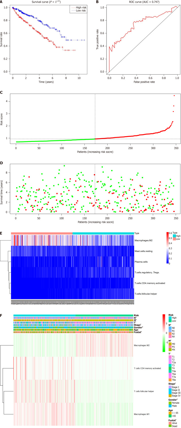
We analyzed 22 immune cell types using a univariate Cox regression technique to identify a subset that was significantly related to RCC survival (Table 2). P < 0.01 was implemented as the screening threshold. Results suggested that the risk of RCC survival was related to macrophages M1 and M2, CD4+ memory activated T cells, and follicular helper T cells. Based on the assumption that these four immune cells significantly influence the survival risk of RCC patients, an immune risk scoring model for these four immune cells was generated via the multivariate Cox regression approach (Table 3). A risk score based on the model was assigned to each patient. The patients were separated into high-risk and low-risk groups based on their median risk score. Based on the Kaplan-Meier survival curve analysis, individuals classified in the high-risk category exhibited a significantly unfavorable prognosis (Figure 3A). According to the ROC curve, the risk model was useful in forecasting survival risk and had excellent specificity and sensitivity (Figure 3B). Figures 3C-E present the distributions of the immune risk score, patient survival status, and four immune cell types in patients with RCC. The connections between the risk assessments and clinical indicators are presented in Figure 3F.
Table 2.
Results of univariate cox regression analysis for 22 immune cells in renal cell carcinoma
|
Immune cells
|
HR
|
HR 95L
|
HR 95H
|
P value
|
| B cells naive | 23260.95811 | 7.327755285 | 73838733.88 | < 0.001 |
| B cells memory | 367.0654108 | 1.02E-14 | 1.32528E+19 | 0.761 |
| Plasma cells | 7.884077616 | 0.471946257 | 131.7071149 | 0.151 |
| T cells CD8+ | 2.305757787 | 0.842062146 | 6.313689546 | 0.104 |
| T cells CD4+ memory resting | 0.350135006 | 0.061633114 | 1.989101538 | 0.236 |
| T cells CD4+ memory activated | 1100224.032 | 1157.449769 | 1045827605 | < 0.001 |
| T cells follicular helper | 14026508.28 | 1930.466435 | 1.01915E+11 | < 0.001 |
| T cells regulatory | 477.9895258 | 1.97885441 | 115457.704 | 0.028 |
| T cells gamma delta | 1.185772305 | 0.027650702 | 50.85064158 | 0.929 |
| NK cells resting | 14.09528528 | 0.052612618 | 3776.224703 | 0.354 |
| NK cells activated | 3.21804095 | 0.010005648 | 1034.994197 | 0.692 |
| Monocytes | 4.094791848 | 0.200859787 | 83.47773603 | 0.359 |
| Macrophages M0 | 0.386593443 | 0.064142169 | 2.330050464 | 0.300 |
| Macrophages M1 | 1864.763728 | 50.85250386 | 68380.97431 | < 0.001 |
| Macrophages M2 | 0.04122243 | 0.009170583 | 0.18529779 | < 0.001 |
| Dendritic cells resting | 0.893700613 | 0.002682052 | 297.7946697 | 0.970 |
| Dendritic cells activated | 3.99E-09 | 2.39E-24 | 6648051.131 | 0.279 |
| Mast cells resting | 0.006154535 | 4.70E-05 | 0.806155892 | 0.051 |
| Mast cells activated | 2.46E-06 | 4.49E-18 | 1344373.927 | 0.349 |
| Eosinophils | 725332583.3 | 3.12E-14 | 1.69E+31 | 0.437 |
| Neutrophils | 18.26220316 | 0.311769833 | 1069.725255 | 0.162 |
| T cells naive | \ | \ | \ | \ |
HR 95L: Lower limit of the 95% confidence interval; HR 95H: Upper limit of the 95% confidence interval; NK: Natural killer; HR: Hazard ratio; M1: Normal activated subset; M2: Alternating-activated subset.
Table 3.
Results of multivariate cox regression analysis for four immune cells in renal cell carcinoma patients
|
Immune cells
|
HR
|
HR 95L
|
HR 95H
|
P value
|
| T cells CD4+ memory activated | 72976.763 | 55.302 | 96300449.399 | < 0.05 |
| T cells follicular helper | 3049.111 | 0.102 | 90834209.409 | < 0.05 |
| Macrophages M1 | 296.374 | 4.843 | 18138.002 | < 0.05 |
| Macrophages M2 | 0.230 | 0.036 | 1.452 | < 0.05 |
HR 95L: Lower limit of the 95% confidence interval; HR 95H: Upper limit of the 95% confidence interval; HR: Hazard ratio; M1: Normal activated subset; M2: Alternating-activated subset.
Figure 3.
Univariate Cox regression analysis for factors affecting overall survival. A: Kaplan-Meier survival curve of overall survival between the high-risk and low-risk groups; B: Receiver operating characteristic (ROC) curve area under the curve (AUC) statistics assess the predictive capability of the immune risk score model; C: Distribution of the immune risk score in renal cell carcinoma (RCC) patients; D: Distribution of the survival status of RCC patients; E: Distribution of five immune cells in the high-risk and low-risk groups; F: Correlation between the immune risk score and clinical indicators.
An impartial predictor of the prognosis for RCC patients was the immunological risk score model
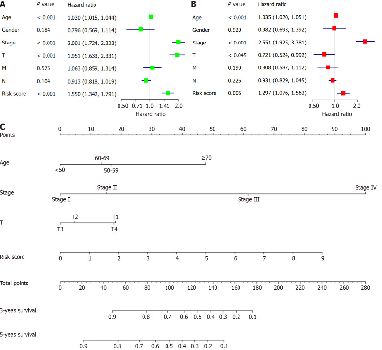
Age, sex, stage, TNM, and risk score were all analyzed using univariate and multivariate Cox regression algorithms (Figure 4A and B) to observe whether the established immune risk scoring model was independent of patient age, sex, stage, and other clinicopathological statistics. Univariate analysis suggested that age, stage, T stage, and the risk score were related to prognosis (P < 0.05). Multifactor Cox proportional risk regression was used to construct predictive models. The results of multivariate regression analysis revealed that the risk score, age, stage, and T stage were all independent predictive factors for RCC. To predict patient survival, we constructed a nomogram utilizing the multivariate Cox regression analysis coefficients (Figure 4C).
Figure 4.
Cox proportional hazard model of correlative factors in renal cell carcinoma patients. A: Univariate Cox regression analysis of seven clinicopathological parameters affecting overall survival; B: Multivariate Cox regression analysis of seven clinicopathological parameters affecting overall survival; C: An established nomogram to predict survival based on the Cox model.
DISCUSSION
Cancer tissues contain fibroblasts, immune cells, endothelial cells, and a variety of chemokines, growth factors, and cytokines in malignant tumor cells. The TME consists of these components and their interactions, which usually have an inhibitory effect on malignant cells[19,20]. Nevertheless, tumor cells have the ability to evade these inhibitory signals as they spread, utilizing immune cells and other favorable conditions encountered in their own surroundings to facilitate their expansion, infiltration, and metastasis. Research has shown that cancer prognosis is closely associated with the TME, especially TIICs. TIICs can promote tumor growth by providing signals, and this field of research has attracted much attention in recent years[21,22]. Consequently, investigating immune-infiltrating cell subsets to evaluate risk and tumor prognosis is crucial.
By deconvoluting data derived from an extensive array of samples, we performed an exhaustive and thorough evaluation of immune cell infiltration in RCC. This study illustrated the diversity of immune cells in RCC patients as well as the identification of particular TIICs. It is well recognized that different immune cell subgroups locally penetrate RCC[23]. These distinct immune cells provide a unique "immune signature map" for each patient and provide novel proposals for specialized immunotherapy for RCC in the future. First, we explored immune cell infiltration in all RCC patients via the TCGA database. The difference analysis results indicated that native B cells, plasma cells, M2 macrophages, dendritic cells, and eosinophils differentially infiltrated healthy and RCC tissues. (P < 0.05). These outcomes correspond with conclusions from earlier research. The number of eosinophils and dendritic cells is greater in the blood of RCC patients than in that of normal individuals[24]. The proportions of tumor-infiltrating lymphocytes, native B cells, and plasma cells are increased[25]. In addition, the proportion of M2 macrophages is increased in RCC tissues, and the increased proportion of this subpopulation significantly promotes the production of inflammatory cytokines and facilitates tumor growth[26].
A thorough investigation was conducted into the relationship between the prognosis of RCC patients and the subpopulations of TIICs. Additional analysis revealed that CD4+ memory-activated T cells, follicular helper T cells, M1 macrophages, and M2 macrophages were associated with RCC survival risk. Tumor-associated immune cells are considered important factors for predicting the prognosis of patients with tumors[27]. Previous studies revealed that infiltrating CD4+ T cells could promote RCC cell proliferation through the TGFβ1/YBX1/HIF2α axis both ex vivo and in vivo[28]. Thompson et al[29] observed that the interaction between programmed cell death 1 (PD-1) and B7-H1 expressed by activated T-cell PD-1 receptor immune cells may promote tumor progression by promoting immune dysfunction in patients with RCC, which is associated with the survival risk of the disease. In addition, Menard et al[30] demonstrated that an increased proportion of CD4+ T cells in RCC tissues was significantly associated with RCC death according to flow analysis (P = 0.004). The results of our analysis further confirmed that an increased proportion of CD4+ T cells was negatively associated with the prognosis of patients with RCC. Furthermore, studies have suggested that helper T cells, which are highly immunosuppressive, play important roles in organismal immunomodulation and the promotion of RCC immune escape[31]. This finding is consistent with our conclusion of a low overall survival rate in RCC patients with high infiltration of helper T cells.
Multiple studies have demonstrated that macrophages have significant functions in the inflammatory response throughout the process of tumor formation and growth[32,33]. In cancer and inflammation, the primary macrophage subsets are categorized into two distinct types: M1 and M2. M1 macrophages have anticancer properties by facilitating the host's immune response against microbes and cancer cells. In contrast, M2 macrophages possess anti-inflammatory characteristics and promote tumor growth, which could contribute to tumor growth and migration. There is plenty of evidence that tumor-associated macrophages (TAMs) are related to tumor progression and metastasis. Tumors are able to utilize the remodeling ability of the macrophage matrix, allowing them to enter the surrounding matrix and migrate through it. However, TAMs are more highly expressed in highly malignant tumors. These findings reveal that high numbers of TAMs in the TME are more likely to predict adverse outcomes of the disease. Recent research has demonstrated that macrophages have a vital role in the progression of RCC, and specifically, M1 macrophages are strongly linked to the stage of RCC and the grade of tumor histology. Geissler et al[27] reported the phenotypic characteristics of macrophages in RCC. They reported that the number of TAMs slightly increased with the degree of dedifferentiation. The ratio of macrophages to T cells was highest in G1-stage tumors. In addition, Yang et al[34] found that macrophages were more easily absorbed into RCC tissues than into nontumor tissues. The results revealed a new mechanism by which macrophages in the RCC TME increase RCC metastasis by activating AKT/mTOR signaling. These results further suggest that immune monitoring of RCC patients could be an effective tool for predicting the prognosis of RCC patients.
Additionally, a multivariate Cox regression method was employed to develop an immunological risk score model for the four categories of immune cells. The Kaplan-Meier survival curve demonstrated a significant association between high-risk individuals and unfavorable outcomes (P = 1E−05). With an area under the curve value of 0.747, the ROC curve demonstrated the validity of the immunological risk evaluation model in forecasting the risk of survival. A multivariate Cox regression analysis was performed on the risk score, age, sex, stage, and TNM stage. Research studies demonstrated that the immunological risk score model was a significant and separate factor in predicting the prognosis of RCC patients (hazard ratio = 1.550, 95%CI: 1.342–1.791; P < 0.001). A nomogram was developed to accurately forecast the overall survival of patients with RCC by utilizing the outcomes of a multivariate Cox regression assessment. Currently, the prognostic judgment of patients after RCC is mainly based on TNM staging[35]. As a visually and clinically valuable medical prediction model, a nomogram can objectively evaluate new predictive variables and provide a more accurate and personalized prognostic assessment.
Due to the rapid evolution of high-throughput sequencing technologies, many tumor markers associated with tumor prognosis have been identified, but the number of these markers is still limited, and their value for clinical application still needs to be considered. Hence, it is imperative to investigate additional therapeutic targets to accurately forecast the prognosis of patients with RCC and to guide the development of clinically individualized treatment regimens. Compared to other solid tumors, RCC is a highly heterogeneous tumor with wide variations in therapeutic efficacy, and relevant specific therapeutic targets are still lacking. In the context of precision tumor therapy and multiline therapy, rational individualized selection of diverse treatments has become an important clinical issue for maximizing the efficacy of comprehensive treatment for advanced RCC. Traditional cancer treatments have many drawbacks, such as pathological tissues that cannot be completely resected by surgery, leading to recurrence, drug resistance, or related serious adverse reactions in patients during radiotherapy and chemotherapy. In addition, the poor prognosis and easy metastasis of RCC make it difficult to effectively control the disease. Currently, immunotherapy is recognized as one of the most promising therapeutic tools for RCC treatment. Since 2005, with the introduction of a variety of antitumor neovascularization tyrosine kinase inhibitors, targeted therapy has become the main systemic treatment for RCC[36]. Immune combinations (pabrolizumab + axitinib, avelumab + axitinib, nabulizumab + cabozantinib, and pabrolizumab + lenvatinib) and dual immune combinations (nabulizumab + ipilizumab) have been approved for the treatment of RCC. Macrophages, CD4+ T cells and helper T cells play crucial roles in cancer immunotherapy. Therefore, we explored the role of four critical TIICs immune subpopulations, M1 macrophages, M2 macrophages, CD4+ T cells and helper T cells in a model for RCC immune risk assessment. This model was confirmed as an independent predictor of RCC. In other words, the immune risk assessment model is a reliable indicator for determining RCC prognosis before immunotherapy. These findings suggest that patients with RCC with high levels of M1 macrophages, M2 macrophages, CD4+ T cells and helper T cells may benefit from immunotherapy. At present, only a few studies have modeled and predicted the prognosis of RCC patients via immune risk scores. However, in other fields, an increasing number of researchers have realized the importance of establishing immune risk score models. Research has shown that the prognostic immune risk scoring model established by researchers on the basis of systematic assessment of the immune status of cancer patients is not only an independent prognostic factor for the recurrence-free survival of cancer patients but also has better prognostic value than TNM staging. This conclusion was also verified in our research, and the established immune risk assessment model has greater risk stratification ability, accuracy of prognostic judgment, and value in guiding clinical decision-making than the current traditional single prognostic model for RCC. The model can be used to assess the prognosis of patients with RCC and the therapeutic benefits of related drugs, which can help clinicians develop personalized treatment plans and improve the survival rate of patients with RCC.
There were specific constraints in our investigation. Initially, it is crucial to note that the public dataset contains a restricted amount of data. Consequently, the clinical pathology characteristics that were specifically chosen for examination in this study are not exhaustive and may result in possible errors or biases. In subsequent studies, the sample size should be further expanded to reduce bias and confirm the results. Furthermore, we did not consider the variability of the immunological microenvironment in relation to the specific site of immune infiltration. Lastly, the outcomes of the study may not be applicable to patients in Asian countries because all of the data sets that were downloaded for developing an immunological risk scoring model were from Western countries. More investigation is needed to verify this.
CONCLUSION
In this study, we explored all immune cells that are closely associated with RCC. We identified and validated the types and distributions of key TIIC subpopulations closely associated with patients with RCC. Additionally, our research incorporated the major factors that influence the prognosis of patients and constructed a prognostic prediction-related model. The predictive model for immune risk assessment on the basis of key subgroups has good predictive value for patients in the RCC cohort. Considering the rapid growth of high-throughput technology, we are optimistic that our immunological risk score model provides significant promise for application in clinical practice. These results would potentially be extremely valuable for investigating novel immunodiagnosis and therapeutic approaches for cancer.
Footnotes
Conflict-of-interest statement: The authors declare no conflicts of interest.
Provenance and peer review: Unsolicited article; Externally peer reviewed.
Peer-review model: Single blind
Specialty type: Oncology
Country of origin: China
Peer-review report’s classification
Scientific Quality: Grade B, Grade C
Novelty: Grade B, Grade B
Creativity or Innovation: Grade B, Grade B
Scientific Significance: Grade A, Grade B
P-Reviewer: Zhou ZL S-Editor: Luo ML L-Editor: Filipodia P-Editor: Wang WB
Contributor Information
Guo-Hao Wei, Department of Oncology, The Second Hospital of Nanjing, Affiliated to Nanjing University of Chinese Medicine, Nanjing 210003, Jiangsu Province, China.
Xi-Yi Wei, The State Key Laboratory of Reproductive, Department of Urology, The First Affiliated Hospital of Nanjing Medical University, Nanjing 210003, Jiangsu Province, China.
Ling-Yao Fan, Department of Oncology, The Second Hospital of Nanjing, Affiliated to Nanjing University of Chinese Medicine, Nanjing 210003, Jiangsu Province, China.
Wen-Zheng Zhou, Department of Oncology, The Second Hospital of Nanjing, Affiliated to Nanjing University of Chinese Medicine, Nanjing 210003, Jiangsu Province, China.
Ming Sun, Department of Oncology, The Second Hospital of Nanjing, Affiliated to Nanjing University of Chinese Medicine, Nanjing 210003, Jiangsu Province, China.
Chuan-Dong Zhu, Department of Oncology, The Second Hospital of Nanjing, Affiliated to Nanjing University of Chinese Medicine, Nanjing 210003, Jiangsu Province, China. zhucd@njucm.edu.cn.
References
- 1.Siegel RL, Wagle NS, Cercek A, Smith RA, Jemal A. Colorectal cancer statistics, 2023. CA Cancer J Clin. 2023;73:233–254. doi: 10.3322/caac.21772. [DOI] [PubMed] [Google Scholar]
- 2.Brown JE, Symeonides SN. Treatment Strategies in Metastatic Renal Cancer: Dose Titration in Clear Cell Renal Cell Carcinoma. Eur Urol. 2022;82:293–294. doi: 10.1016/j.eururo.2022.04.018. [DOI] [PubMed] [Google Scholar]
- 3.Lv J, Liu Y, Mo S, Zhou Y, Chen F, Cheng F, Li C, Saimi D, Liu M, Zhang H, Tang K, Ma J, Wang Z, Zhu Q, Tong WM, Huang B. Gasdermin E mediates resistance of pancreatic adenocarcinoma to enzymatic digestion through a YBX1-mucin pathway. Nat Cell Biol. 2022;24:364–372. doi: 10.1038/s41556-022-00857-4. [DOI] [PMC free article] [PubMed] [Google Scholar]
- 4.Chen YW, Wang L, Panian J, Dhanji S, Derweesh I, Rose B, Bagrodia A, McKay RR. Treatment Landscape of Renal Cell Carcinoma. Curr Treat Options Oncol. 2023;24:1889–1916. doi: 10.1007/s11864-023-01161-5. [DOI] [PMC free article] [PubMed] [Google Scholar]
- 5.Yang JC. The ongoing mystery of renal cell cancer. Cell Rep Med. 2021;2:100445. doi: 10.1016/j.xcrm.2021.100445. [DOI] [PMC free article] [PubMed] [Google Scholar]
- 6.Martin TD, Patel RS, Cook DR, Choi MY, Patil A, Liang AC, Li MZ, Haigis KM, Elledge SJ. The adaptive immune system is a major driver of selection for tumor suppressor gene inactivation. Science. 2021;373:1327–1335. doi: 10.1126/science.abg5784. [DOI] [PubMed] [Google Scholar]
- 7.Del Poggetto E, Ho IL, Balestrieri C, Yen EY, Zhang S, Citron F, Shah R, Corti D, Diaferia GR, Li CY, Loponte S, Carbone F, Hayakawa Y, Valenti G, Jiang S, Sapio L, Jiang H, Dey P, Gao S, Deem AK, Rose-John S, Yao W, Ying H, Rhim AD, Genovese G, Heffernan TP, Maitra A, Wang TC, Wang L, Draetta GF, Carugo A, Natoli G, Viale A. Epithelial memory of inflammation limits tissue damage while promoting pancreatic tumorigenesis. Science. 2021;373:eabj0486. doi: 10.1126/science.abj0486. [DOI] [PMC free article] [PubMed] [Google Scholar]
- 8.Iglesias-Escudero M, Arias-González N, Martínez-Cáceres E. Regulatory cells and the effect of cancer immunotherapy. Mol Cancer. 2023;22:26. doi: 10.1186/s12943-023-01714-0. [DOI] [PMC free article] [PubMed] [Google Scholar]
- 9.Bukavina L, Bensalah K, Bray F, Carlo M, Challacombe B, Karam JA, Kassouf W, Mitchell T, Montironi R, O'Brien T, Panebianco V, Scelo G, Shuch B, van Poppel H, Blosser CD, Psutka SP. Epidemiology of Renal Cell Carcinoma: 2022 Update. Eur Urol. 2022;82:529–542. doi: 10.1016/j.eururo.2022.08.019. [DOI] [PubMed] [Google Scholar]
- 10.Navani V, Heng DYC. Immunotherapy in renal cell carcinoma. Lancet Oncol. 2023;24:1164–1166. doi: 10.1016/S1470-2045(23)00473-4. [DOI] [PubMed] [Google Scholar]
- 11.Dai S, Zeng H, Liu Z, Jin K, Jiang W, Wang Z, Lin Z, Xiong Y, Wang J, Chang Y, Bai Q, Xia Y, Liu L, Zhu Y, Xu L, Qu Y, Guo J, Xu J. Intratumoral CXCL13(+)CD8(+)T cell infiltration determines poor clinical outcomes and immunoevasive contexture in patients with clear cell renal cell carcinoma. J Immunother Cancer. 2021;9:e001823. doi: 10.1136/jitc-2020-001823. [DOI] [PMC free article] [PubMed] [Google Scholar]
- 12.Eskelinen T, Veitonmäki T, Kotsar A, Tammela TLJ, Pöyhönen A, Murtola TJ. Improved renal cancer prognosis among users of drugs targeting renin-angiotensin system. Cancer Causes Control. 2022;33:313–320. doi: 10.1007/s10552-021-01527-w. [DOI] [PMC free article] [PubMed] [Google Scholar]
- 13.Xu C, Zeng H, Fan J, Huang W, Yu X, Li S, Wang F, Long X. A novel nine-microRNA-based model to improve prognosis prediction of renal cell carcinoma. BMC Cancer. 2022;22:264. doi: 10.1186/s12885-022-09322-9. [DOI] [PMC free article] [PubMed] [Google Scholar]
- 14.Wang Z, Xu C, Liu W, Zhang M, Zou J, Shao M, Feng X, Yang Q, Li W, Shi X, Zang G, Yin C. A clinical prediction model for predicting the risk of liver metastasis from renal cell carcinoma based on machine learning. Front Endocrinol (Lausanne) 2022;13:1083569. doi: 10.3389/fendo.2022.1083569. [DOI] [PMC free article] [PubMed] [Google Scholar]
- 15.Yang Y, Cao Y, Han X, Ma X, Li R, Wang R, Xiao L, Xie L. Revealing EXPH5 as a potential diagnostic gene biomarker of the late stage of COPD based on machine learning analysis. Comput Biol Med. 2023;154:106621. doi: 10.1016/j.compbiomed.2023.106621. [DOI] [PubMed] [Google Scholar]
- 16.Li J, Zhang Y, Lu T, Liang R, Wu Z, Liu M, Qin L, Chen H, Yan X, Deng S, Zheng J, Liu Q. Identification of diagnostic genes for both Alzheimer's disease and Metabolic syndrome by the machine learning algorithm. Front Immunol. 2022;13:1037318. doi: 10.3389/fimmu.2022.1037318. [DOI] [PMC free article] [PubMed] [Google Scholar]
- 17.Jiang H, Zhang X, Wu Y, Zhang B, Wei J, Li J, Huang Y, Chen L, He X. Bioinformatics identification and validation of biomarkers and infiltrating immune cells in endometriosis. Front Immunol. 2022;13:944683. doi: 10.3389/fimmu.2022.944683. [DOI] [PMC free article] [PubMed] [Google Scholar]
- 18.Zhang Y, Xia R, Lv M, Li Z, Jin L, Chen X, Han Y, Shi C, Jiang Y, Jin S. Machine-Learning Algorithm-Based Prediction of Diagnostic Gene Biomarkers Related to Immune Infiltration in Patients With Chronic Obstructive Pulmonary Disease. Front Immunol. 2022;13:740513. doi: 10.3389/fimmu.2022.740513. [DOI] [PMC free article] [PubMed] [Google Scholar]
- 19.Bejarano L, Jordāo MJC, Joyce JA. Therapeutic Targeting of the Tumor Microenvironment. Cancer Discov. 2021;11:933–959. doi: 10.1158/2159-8290.CD-20-1808. [DOI] [PubMed] [Google Scholar]
- 20.Peng C, Xu Y, Wu J, Wu D, Zhou L, Xia X. TME-Related Biomimetic Strategies Against Cancer. Int J Nanomedicine. 2024;19:109–135. doi: 10.2147/IJN.S441135. [DOI] [PMC free article] [PubMed] [Google Scholar]
- 21.Tay C, Tanaka A, Sakaguchi S. Tumor-infiltrating regulatory T cells as targets of cancer immunotherapy. Cancer Cell. 2023;41:450–465. doi: 10.1016/j.ccell.2023.02.014. [DOI] [PubMed] [Google Scholar]
- 22.Dai Q, Wu W, Amei A, Yan X, Lu L, Wang Z. Regulation and characterization of tumor-infiltrating immune cells in breast cancer. Int Immunopharmacol. 2021;90:107167. doi: 10.1016/j.intimp.2020.107167. [DOI] [PMC free article] [PubMed] [Google Scholar]
- 23.Díaz-Montero CM, Rini BI, Finke JH. The immunology of renal cell carcinoma. Nat Rev Nephrol. 2020;16:721–735. doi: 10.1038/s41581-020-0316-3. [DOI] [PubMed] [Google Scholar]
- 24.Minárik I, Lašťovička J, Budinský V, Kayserová J, Spíšek R, Jarolím L, Fialová A, Babjuk M, Bartůňková J. Regulatory T cells, dendritic cells and neutrophils in patients with renal cell carcinoma. Immunol Lett. 2013;152:144–150. doi: 10.1016/j.imlet.2013.05.010. [DOI] [PubMed] [Google Scholar]
- 25.Meylan M, Petitprez F, Becht E, Bougoüin A, Pupier G, Calvez A, Giglioli I, Verkarre V, Lacroix G, Verneau J, Sun CM, Laurent-Puig P, Vano YA, Elaïdi R, Méjean A, Sanchez-Salas R, Barret E, Cathelineau X, Oudard S, Reynaud CA, de Reyniès A, Sautès-Fridman C, Fridman WH. Tertiary lymphoid structures generate and propagate anti-tumor antibody-producing plasma cells in renal cell cancer. Immunity. 2022;55:527–541. doi: 10.1016/j.immuni.2022.02.001. [DOI] [PubMed] [Google Scholar]
- 26.Zhang X, Sun Y, Ma Y, Gao C, Zhang Y, Yang X, Zhao X, Wang W, Wang L. Tumor-associated M2 macrophages in the immune microenvironment influence the progression of renal clear cell carcinoma by regulating M2 macrophage-associated genes. Front Oncol. 2023;13:1157861. doi: 10.3389/fonc.2023.1157861. [DOI] [PMC free article] [PubMed] [Google Scholar]
- 27.Geissler K, Fornara P, Lautenschläger C, Holzhausen HJ, Seliger B, Riemann D. Immune signature of tumor infiltrating immune cells in renal cancer. Oncoimmunology. 2015;4:e985082. doi: 10.4161/2162402X.2014.985082. [DOI] [PMC free article] [PubMed] [Google Scholar]
- 28.Wang Y, Wang Y, Xu L, Lu X, Fu D, Su J, Geng H, Qin G, Chen R, Quan C, Niu Y, Yue D. CD4 + T cells promote renal cell carcinoma proliferation via modulating YBX1. Exp Cell Res. 2018;363:95–101. doi: 10.1016/j.yexcr.2017.12.026. [DOI] [PubMed] [Google Scholar]
- 29.Thompson RH, Dong H, Lohse CM, Leibovich BC, Blute ML, Cheville JC, Kwon ED. PD-1 is expressed by tumor-infiltrating immune cells and is associated with poor outcome for patients with renal cell carcinoma. Clin Cancer Res. 2007;13:1757–1761. doi: 10.1158/1078-0432.CCR-06-2599. [DOI] [PubMed] [Google Scholar]
- 30.Menard LC, Fischer P, Kakrecha B, Linsley PS, Wambre E, Liu MC, Rust BJ, Lee D, Penhallow B, Manjarrez Orduno N, Nadler SG. Renal Cell Carcinoma (RCC) Tumors Display Large Expansion of Double Positive (DP) CD4+CD8+ T Cells With Expression of Exhaustion Markers. Front Immunol. 2018;9:2728. doi: 10.3389/fimmu.2018.02728. [DOI] [PMC free article] [PubMed] [Google Scholar]
- 31.Gutiérrez-Melo N, Baumjohann D. T follicular helper cells in cancer. Trends Cancer. 2023;9:309–325. doi: 10.1016/j.trecan.2022.12.007. [DOI] [PubMed] [Google Scholar]
- 32.Li C, Xu X, Wei S, Jiang P, Xue L, Wang J Senior Correspondence. Tumor-associated macrophages: potential therapeutic strategies and future prospects in cancer. J Immunother Cancer. 2021;9:e001341. doi: 10.1136/jitc-2020-001341. [DOI] [PMC free article] [PubMed] [Google Scholar]
- 33.Li M, Yang Y, Xiong L, Jiang P, Wang J, Li C. Metabolism, metabolites, and macrophages in cancer. J Hematol Oncol. 2023;16:80. doi: 10.1186/s13045-023-01478-6. [DOI] [PMC free article] [PubMed] [Google Scholar]
- 34.Yang Z, Xie H, He D, Li L. Infiltrating macrophages increase RCC epithelial mesenchymal transition (EMT) and stem cell-like populations via AKT and mTOR signaling. Oncotarget. 2016;7:44478–44491. doi: 10.18632/oncotarget.9873. [DOI] [PMC free article] [PubMed] [Google Scholar]
- 35.Delahunt B, Eble JN, Samaratunga H, Thunders M, Yaxley JW, Egevad L. Staging of renal cell carcinoma: current progress and potential advances. Pathology. 2021;53:120–128. doi: 10.1016/j.pathol.2020.08.007. [DOI] [PubMed] [Google Scholar]
- 36.Rathmell WK, Rumble RB, Van Veldhuizen PJ, Al-Ahmadie H, Emamekhoo H, Hauke RJ, Louie AV, Milowsky MI, Molina AM, Rose TL, Siva S, Zaorsky NG, Zhang T, Qamar R, Kungel TM, Lewis B, Singer EA. Management of Metastatic Clear Cell Renal Cell Carcinoma: ASCO Guideline. J Clin Oncol. 2022;40:2957–2995. doi: 10.1200/JCO.22.00868. [DOI] [PubMed] [Google Scholar]


