



Abstract
Background
Early laparoscopic cholecystectomy (ELC) in the setting of acute calculous cholecystitis (ACC) requires to be performed by highly-skilled surgeons to avoid complications. The purpose of this study is to identify preoperative factors that would predict difficult ELC among patients with ACC prior to proceeding with surgery.
Methods
We retrospectively reviewed all patients who received ELC within 10 days from the onset of symptoms of ACC between August 1, 2018, and December 31, 2022. They were divided into 2 groups according to the difficulty of surgery.
Results
149 patients with ACC received ELC during the study period. ELC was considered difficult in 52 patients (35%). Five preoperative factors were identified as significant predictors of difficult ELC (DELC) on multivariate analysis: duration of acute attack ≥ 4 days from the onset of symptoms till surgery (OR 34.4, P < 0.001), ultrasound showing largest gallstone size > 20 mm (OR 20.2, P = 0.029), ultrasound showing gallstone impaction in Hartmann’s pouch (OR 7.2, P = 0.017), history of prior episode(s) of acute attack (OR 6.8, P = 0.048), and diabetes mellitus (OR 5.8, P = 0.046).
Conclusion
Careful preoperative assessment of patients with ACC is crucial among junior surgeons with limited surgical expertise prior to proceeding with ELC to identify those at risk of DELC to potentially reduce postoperative morbidity and mortality. If encountered, a management plan should be made, and surgery should proceed only upon confirming the availability of experienced surgeons in the field of biliary and laparoscopic surgery to supervise or assist in the procedure. Alternatively, such group of patients should rather be transferred to more advanced surgical centers which offer higher level of care to maintain patient safety and optimal surgical outcomes. More importantly, bail-out procedures should always be resorted to whenever DELC is encountered intraoperatively to prevent further surgical damage.
Keywords: Acute calculous cholecystitis, Difficult early laparoscopic cholecystectomy, Predictors, Risk factors
Introduction
40% of patients with gallstones will experience complications including acute calculous cholecystitis (ACC) which could be the primary presentation among 10–15% [1–3]. Early laparoscopic cholecystectomy (ELC) performed within 7 days from hospital admission and 10 days from the onset symptoms of acute attack is considered safe, feasible and preferred over intermediate or delayed laparoscopic cholecystectomy (LC) whenever possible for it offers shorter hospital stay, fewer re-admissions from the repeated attacks, and lower financial burden [4–6].
Nonetheless, it requires to be performed by skilled surgeons with adequate experience and profound training in the field of hepatobiliary and minimally invasive surgery to be able to deal with the complex nature of such highly demanding surgery. It is also mandatory for the surgery to be performed in advanced surgical centers with intensive care professionals, interventional radiologists, and importantly having operative theaters well-equipped with advanced laparoscopic equipment. The most dreaded complication of LC is iatrogenic bile duct injury (BDI) which is usually attributed to the disturbed anatomy of the region caused by the florid inflammatory process of the gallbladder especially at the infundibular region. Acute cholecystitis has been reported as a strong risk factor for BDI among patients undergoing emergency LC [7]. Advocates of patient safety and well-being recommend exercising great care and extreme caution during dissection while performing ELC in the setting of ACC to prevent complications that could potentially be life-threatening and they recommend opting to bailout procedures such as fundus-first LC, subtotal cholecystectomy, or conversion to open surgery to avoid such complications [8, 9].
Accordingly, our aim was to identify preoperative factors which could predict challenging or difficult ELC among patients with ACC before proceeding with surgery to guard against intra- and/or postoperative complications so as to mitigate surgical outcomes, and to give the chance for surgeons to place a more sound surgical plan ahead of time.
Materials and methods
Definitions and study design
Since August 2018, our policy for the optimal timing of ELC for patients with ACC was modified in compliance with the recommendations endorsed by the World Society of Emergency Surgery guidelines (2016 WSES) [10]. ELC was planned and performed within 10 days from the onset of symptoms of ACC provided that the patients were fit for surgery according to the Charlson comorbidity index (CCI) and the American Society of Anesthesiologists physical status classification (ASA-PS) [11, 12]. The duration of acute attack was calculated from the time of onset of symptoms correlating with ACC until the time of ELC. The diagnosis of ACC was achieved by a combination of clinical (fever, right upper quadrant pain, and positive Murphy’s sign), laboratory (elevated total leukocytic count [TLC] and C-reactive protein [CRP]), and imaging criteria (transabdominal ultrasound showing increased gallbladder wall thickness and pericholecystic fluid collection) as recommended by 2016 WSES and Tokyo Guidelines (TG18) [5, 10]. Whenever ultrasound was incapable of achieving a definitive diagnosis of ACC, a more dedicated imaging modality was resorted to as abdomino-pelvic contrast-enhanced computed tomography (CECT). Magnetic resonance imaging (MRI) / magnetic resonance cholangiopancreatography (MRCP) was required if intravenous contrast injection was contraindicated or hyperbilirubinemia and/or elevated liver and cholestatic enzymes were encountered to exclude concomitant common bile duct stones (CBDS). The grading of severity and treatment algorithms of ACC were implemented according to the recommendations of TG18 guidelines [5].
We retrospectively reviewed the data of all patients who received LC from a prospectively maintained database in the period from August 1, 2018, to December 31, 2022. We excluded patients who received delayed LC after resolution of the acute attack or those who received LC for chronic calculous cholecystitis. The remaining patients who received ELC within 10 days from the onset of symptoms of the acute attack were included and constituted our study cohort. They were divided into 2 groups according to difficulty of LC: simple early LC (SELC) and difficult early LC (DELC).
DELC was considered whenever one or more of the following criteria was documented:
Resorting to bail-out procedures (fundus-first cholecystectomy, subtotal cholecystectomy, or conversion to open surgery).
Net operative time exceeded 90 min attributed only to the difficulty and the technical challenging nature of surgery related to the established acute inflammatory process of the gallbladder as well as the surrounding tissues.
The net operative time was calculated by eliminating from the overall operative time any time-intervals of undue operative lengthening that did not directly correlate with the complexity of the surgery. Overall operative represented the time-lapse between skin incision done for the first port-site and skin closure of the last wound. Examples of time intervals that were eliminated form overall operative time included intraoperative delays due to accidental instrumental failure, device malfunction, or time wasted during switching of the nursing staff or scrubbing in of more assisting surgeons during which the flow of the surgery was suspended until the whole set up became ready again for resumption of the procedure.
All of the surgical procedures were video-recorded starting from time of the first skin incision until closure of the last skin wound. Recorded media were saved on portable external hard drive that was connected to the camera control and processing unit via universal serial bus (USB) port. The recording was kept running throughout the whole procedure without any interruption and a free-handed surgery resident was provided with a paper checklist to record every instance of surgical delay or interruption not related to the complexity of surgical dissection or severity of ACC. All insignificant delays were recorded as time intervals with proper explanation for the reason of interruption. After conclusion of the procedure, overall operative time was identified from the length of the recorded video file and the intervals of insignificant operative delays were subtracted from it to achieve the net operative time which would reflect the actual operative time explaining the surgical complexity pertaining to the duration of extensive dissection, manipulation, and completion of removal of the acutely inflamed gallbladder.
The study was approved by the Ethics Committee and the Institutional Review Board (IRB) of Alexandria University, Faculty of Medicine (IRB No. 00012098, FWA No. 00018699) under serial number 0305608.
Preoperative management and surgical technique
Upon admission, all patients were restricted from oral intake and placed on intravenous fluids, parenteral analgesic/anti-inflammatory drugs, and proton pump inhibitors until the conclusion of laboratory and imaging investigations. Parenteral antibiotics were required for patients with grade II or grade III ACC using a combination of fluoroquinolones (ciprofloxacin) and metronidazole. Patients with grade III ACC who presented with organ failure (cardiac, respiratory, renal, or neurologic), elevated serum bilirubin, and/or markedly elevated liver enzymes were admitted to the intensive care unit where they were placed on aggressive supportive care to ameliorate their general condition and improve organ functions after which they were reassessed for surgical candidacy to undergo ELC.
We followed the standard 4-port technique for ELC (two 10 mm ports, and two 5 mm ports) after creation of closed pneumoperitoneum using Veress needle. Open pneumoperitoneum was performed alternatively whenever upper abdominal scars were present to avoid any inadvertent visceral/vascular injury. The use of 10 mm, 30o telescope was mandatory in all of the procedures to guarantee optimal vision of the operative field in such complex and technically demanding surgery. Once identified, the fundus of the gallbladder was grasped and retracted upwards, and the Hartmann’s pouch was grasped using non-traumatic Endo-Clinch laparoscopic forceps. Dissection and hemostasis were primarily achieved using monopolar electrocautery connected to either laparoscopic hook, Maryland forceps, or laparoscopic spatula. Bipolar electrocautery was occasionally used in certain circumstances to achieve proper hemostasis of the gallbladder liver bed. Meticulous dissection alternating between sharp and blunt dissection with jet saline flow from the suction/irrigation nozzle (hydro-dissection) was used to deal with tough omental/visceral adhesions concealing gallbladder fundus or at the region of Calot’s triangle. Achieving the critical view of safety (CVS) was mandatory prior to clip-controlling any structure to confirm proper identification of both, the cystic duct and artery. In cases where the cystic duct was deemed to be very short and wide preventing proper clip control, the stump was suture-closed using 3/0 absorbable suture material (polydioxanone suture [PDS] or vicryl). If the CVS was difficult to achieve related to the severe inflammatory process, fibrosis, or presence of dense adhesions, fundus-first cholecystectomy was the most initial option to resort to prior to opting for other bail-out alternatives as subtotal cholecystectomy or conversion to open surgery. When ELC was successfully concluded via laparoscopy, the gallbladder was routinely placed in specimen endo-bag and then extracted through the epigastric port. Abdominal drain insertion was not routinely practiced and was kept at the discretion of the operating surgeon which were mostly used in certain circumstances as extensive adhesolysis, subtotal cholecystectomy, or conversion to open surgery.
Data collection
Preoperative data included patients’ demographic characteristics, associated comorbidities, history of prior abdominal surgery, preoperative laboratory work-up [serum total and direct bilirubin, alanine aminotransferase (ALT), aspartate aminotransferase (AST), alkaline phosphatase (ALP), CRP, TLC, polymorphonuclear leukocytic (PMNL) count], ultrasound documenting stone impaction in Hartmann’s pouch, the size of the largest gallstone, severity grade of ACC (mild or grade I, moderate or grade II, and severe or grade III), and the duration of acute attack from the onset of symptoms. Operative data included the net operative time, and records of challenging intraoperative encounters as gallbladder concealed by dense omental (Fig. 1A) or visceral (Fig. 1B) adhesions, markedly distended gallbladder that is difficult to grasp (Fig. 1C), marked gallbladder wall thickness and edema (Fig. 1D), gangrenous gallbladder wall (Fig. 2A), fibrosis or dense adhesions obscuring a frozen Calot’s triangle (Fig. 2B), large stone impacted in Hartmann’s pouch impairing proper manipulation (Fig. 2C), challenging short and wide cystic duct requiring suture-ligation for proper control (Fig. 2D), and friability of gallbladder wall with perforation and/or stone spillage.
Fig. 1.

Intraoperative findings during ELC showing GB concealed by dense omental adhesions (A); dense visceral adhesions (duodenum) with the GB (B); GB difficult to grasp due to marked distension necessitating percutaneous needle aspiration (C); and acute cholecystitis difficult to grasp due to marked wall thickness and edema (D)
Fig. 2.

Intraoperative findings during ELC showing GB wall gangrene (A); frozen Calot’s triangle by dense adhesions (B); large stone impaction in Hartmann’s pouch (C); and large stone impacted in gallbladder neck with very short and wide cystic duct requiring suture-ligation (D)
Postoperative data included the length of postoperative hospital stay (days) and record of any encountered postoperative morbidity as BDI, visceral injury, bleeding, or wound infection graded according to the Clavien-Dindo classification for surgical complications [13].
Statistical analysis
Descriptive statistics were reported for the entire study cohort; categorical variables as number and percentage, and continuous variables as median (50th percentile) and interquartile range (IQR, the range between 25th and 75th percentile). Univariate comparison of the 2 groups was performed using Pearson’s Chi-square/Fisher’s exact test for categorical variables and Mann-Whitney U test for continuous variables. Statistical significance was considered at P < 0.05. Preoperative independent variables significant at P < 0.1 and those expected to be of significant contribution to the analysis on clinical grounds were entered into a multivariate logistic regression analysis using the backward elimination stepwise approach to identify preoperative risk factors that would significantly predict DELC. The goodness-of-fit of the resultant model was assessed by the area under the receiver operating characteristic curve (AUROC), [14] Hosmer-Lemeshow test, [15] and the coefficient of discrimination (R2) [16]. All the analyses were performed using IBM® SPSS® Statistics version 26 (IBM Corporation, Armonk, NY).
Results
The flowchart of the study population is displayed in Fig. 3. Over 53-month-period, 151 patients with ACC received ELC within 10 days from the onset of symptoms of the acute attack during the index hospital admission.
Fig. 3.

Flow chart showing study design and the population of patients included in the analysis over 53-month-period
The majority of the patients were females (n = 88, 59.16%), the median age was 46 years (IQR 39–53), and the median BMI was 32 kg/m2 (IQR = 30–35). Thirty-three patients (22%) were diabetic, 27 were hypertensive (18%), 6 patients had bilharzial hepatic fibrosis (4%), and 3 had chronic HCV infection (2%). Serum hyperbilirubinemia was encountered in 2 patients which required further preoperative biliary assessment via MRCP which excluded concomitant CBDS, however, external biliary compression by the inflamed gallbladder infundibulum was identified as the cause. The median duration of acute attack from onset of symptoms till ELC was 3 days (IQR = 2–5). While the majority of the ELCs were concluded laparoscopically (n = 140, 92.7%), conversion to open was required in 11 patients (7.3%) due to frozen Calot’s triangle with dense adhesions rendering CVS difficult to achieve (n = 4, 2.6%), intractable bleeding that was difficult to control laparoscopically (n = 2, 1.3%), dense visceral adhesions between the gallbladder and duodenum/colon precluding safe dissection (n = 2, 1.3%), failure to grasp or manipulate a severely fibrotic gallbladder contracted over large stone (n = 1, 0.7%), and accidentaly discovered advanced grade of Mirizzi Syndrome (n = 2, 1.3%).
Upon conversion to open surgery, cholecysto-choledochal fistula was identified in both patients that was not clearly documented on preoperative MRCP. It was type II in the first patient and type III in the second one, and both were managed by cholecystectomy and Roux-en-Y hepaticojejunostomy. Owing to the presence of ACC with concomitant biliary complication that was not discovered on imaging among those 2 patients, they were excluded from further analysis to ensure uniformity of the compared groups. Statistical comparisons were thus performed on the remaining 149 patients who presented with ACC only without any concomitant biliary complication.
Fifty-two out of 149 patients (35%) fulfilled the criteria of DELC. They were compared to the remaining 97 patients who constituted the SELC group. Patient demographic characteristics, associated comorbidities, preoperative laboratory and imaging findings, and severity of ACC are summarized in Table 1.
Table 1.
Patient demographic data, associated comorbidities, and preoperative investigations among DELC and SELC groups
| Variables | DELC (N = 52) |
SELC (N = 97) |
P |
|---|---|---|---|
| Demographic data | |||
| Age (y) | 48 (42–55) | 44 (39–53) | 0.07 |
| Gender, male | 27 (51.9%) | 34 (35.1%) | 0.05 |
| BMI (Kg/m2) | 33 (30–36) | 32 (30–34) | 0.2 |
| Comorbidities | |||
| Diabetes | 25 (48.1%) | 8 (8.2%) | < 0.001* |
| Hypertension | 12 (23.1%) | 15 (15.5%) | 0.3 |
| HCV infection | 1 (1.9%) | 1 (1%) | 1 |
| Bilharzial hepatic fibrosis | 4 (7.7%) | 2 (2.1%) | 0.2 |
| Cardiovascular disease | 2 (3.8%) | 1 (1%) | 0.3 |
| Prior upper abdominal surgery | 5 (9.6%) | 6 (6.2%) | 0.5 |
| History of prior episode(s) of ACC | 29 (55.8%) | 3 (3.1%) | < 0.001* |
| Duration of acute attack (from onset till ELC) | 6 (4–8) | 2 (2–3) | < 0.001* |
| Duration of acute attack ≥ 4 days | 41 (78.8%) | 17 (17.5%) | < 0.001* |
| Preoperative laboratory work-up | |||
| Total bilirubin (mg/dL) | 0.8 (0.6–1) | 0.8 (0.6–1) | 0.97 |
| Direct bilirubin (mg/dL) | 0.2 (0.1–0.3) | 0.2 (0.2–0.3) | 0.93 |
| AST (U/L) | 27 (21–29) | 24 (20–29) | 0.19 |
| ALT (U/L) | 29 (23–37) | 26 (22–32) | 0.14 |
| ALP (U/L) | 114 (99–128) | 108 (97–119) | 0.046* |
| Hb (g/dL) | 12.8 (11.8–14.1) | 12.8 (11.9–14.1) | 0.87 |
| TLC (x103/µL) | 12.1 (11–14) | 9.9 (8.6–10.9) | < 0.001* |
| PMNL count (x103/µL) | 9.5 (8.7–11.2) | 7.3 (5.9–8.2) | < 0.001* |
| CRP (mg/L) | 57.5 (34.3–73.5) | 15 (9.9–24) | < 0.001* |
| Ultrasound findings | |||
| Largest GB stone size (mm) | 20 (17–25) | 10 (9–13) | < 0.001* |
| Largest GB stone > 20 mm | 23 (44.2%) | 1 (1%) | < 0.001* |
| Largest GB stone > 25 mm | 9 (17.3%) | 1 (1%) | < 0.001* |
| Stone impacted in Hartmann’s pouch | 37 (71.2%) | 11 (11.3%) | < 0.001* |
| Severity of acute cholecystitis | |||
| Grade I | 10 (19.2%) | 75 (77.3%) | < 0.001* |
| Grade II | 40 (76.9%) | 22 (22.7%) | < 0.001* |
| Grade III | 2 (3.8%) | 0 | 0.012* |
* Statistical significance at P < 0.05
Continuous variables are reported as median (interquartile range)
Categorical variables are reported as n (%)
Intraoperative data and postoperative outcomes are illustrated in Table 2. No mortalities were recorded throughout the study period, and none of the patients required further surgical reintervention. None of the patients experienced postoperative bleeding or iatrogenic biliary injury according to Strasburg-Bismuth classification for BDI [17]. Nonetheless, postoperative bile leak was encountered in 2 patients (1.3%) who presented during the first 24 h postoperatively with bilious output through the abdominal drain. Urgent MRCP was performed which excluded any sort of iatrogenic BDI or missed CBDS. The source of leak in both patients, however, was identified to come from the residual gallbladder stump following subtotal cholecystectomy. Both patients were managed conservatively with close follow-up, serial laboratory and imaging investigations while keeping the abdominal drain in place for as long as it was draining bile. Upon cessation of bilious drainage, confirmatory abdomino-pelvic CECT scan was performed which excluded free or loculated intraperitoneal fluid collection. The drains were then removed, and the patients were allowed to resume their regular lives thereafter.
Table 2.
Operative data and postoperative outcomes among DELC and SELC groups
| Variables | DELC (N = 52) |
SELC (N = 97) |
P |
|---|---|---|---|
| Net operative time (min) | 111 (100–131) | 70 (65–78) | < 0.001* |
| Gallbladder concealed by dense adhesions | 39 (75%) | 3 (3.1%) | < 0.001* |
| Friable gallbladder wall | 22 (42.3%) | 8 (8.2%) | < 0.001* |
| Gangrenous gallbladder wall | 4 (7.7%) | 0 | 0.014* |
| Gallbladder perforation / stone spillage | 28 (53.8%) | 8 (8.2%) | < 0.001* |
| Frozen Calot’s triangle | 37 (71.2%) | 12 (12.4%) | < 0.001* |
| Very short and wide cystic duct requiring suturing | 43 (82.7%) | 4 (4.1%) | < 0.001* |
| Absent gallbladder / liver plane of dissection | 33 (63.5%) | 9 (9.3%) | < 0.001* |
| Abdominal drain insertion | 25 (48.1%) | 5 (5.2%) | < 0.001* |
| Bail-out techniques | |||
| Subtotal cholecystectomy | 8 (15.4%) | 0 | < 0.001* |
| Conversion to open surgery | 9 (17.3%) | 0 | < 0.001* |
| Length of postoperative hospital stay (days) | 1 (1–2) | 1 (0.5–1) | < 0.001* |
| Clavien-Dindo Classification | |||
| Grade I | 17 (32.7%) | 4 (4.1%) | < 0.001* |
| Grade II | 4 (7.7%) | 0 | |
| Postoperative morbidity | |||
| Bile leak | 2 (3.8%) | 0 | 0.1 |
| Superficial wound infection | 6 (11.5%) | 3 (3.1%) | 0.07 |
* Statistical significance at P < 0.05
Continuous variables are reported as median (interquartile range)
Categorical variables are reported as n (%)
All patients who developed surgical site infection were successfully managed conservatively by frequent sterile dressing under strict antibiotic coverage based on culture and sensitivity.
Multivariate analysis for predictors of DELC in ACC
The following preoperative independent variables were entered into multivariate logistic regression using the backward stepwise elimination approach to identify risk factors that would significantly predict DELC: duration of acute attack ≥ 4 days, history of prior episode(s) of acute attack, patient’s age, gender, BMI, diabetes mellitus, scar of prior upper abdominal surgery, serum ALT, serum ALP, TLC, PMNL count, CRP, ultrasound evidence of stone impaction in Hartmann’s pouch, largest gallstone size on ultrasound (> 20 mm and > 25 mm), and severity grade of ACC.
Five preoperative factors were identified as significant predictors of DELC among patients with ACC as shown in Table 3.
Table 3.
Multivariate analysis of preoperative predictors of DELC in ACC
| Variables | OR | 95% CI | P |
|---|---|---|---|
| Duration of acute attack ≥ 4 days | 34.4 | 7.3–160.9 | < 0.001* |
| Ultrasound with GB stone size > 20 mm | 20.2 | 1.37–298.2 | 0.029* |
| Ultrasound showing stone impaction in Hartmann’s pouch | 7.2 | 1.4–36.1 | 0.017* |
| History of previous episode(s) of ACC | 6.8 | 1.01–46.03 | 0.048* |
| Diabetes mellitus | 5.8 | 1.03–32.9 | 0.046* |
* Statistical significance at P < 0.05
The model with the 5 preoperative predictors showed statistical significance over the constant-only-model with X2(5, n = 149) = 128.8, at P < 0.001. The model was able to correctly classify 91% of the cases with sensitivity of 83%, specificity of 95%, positive predictive value of 90%, and negative predictive value of 91%. The goodness-of-fit of the model was assessed by the AUROC as shown in Fig. 4. Hosmer-Lemeshow test indicated good fitness of our model with X2 = 6.17 at P = 0.19. The 5 variables statistically significantly predicted DELC with F (5, 143) = 59.36, at P < 0.001, and coefficient of discrimination (R2) = 0.675.
Fig. 4.
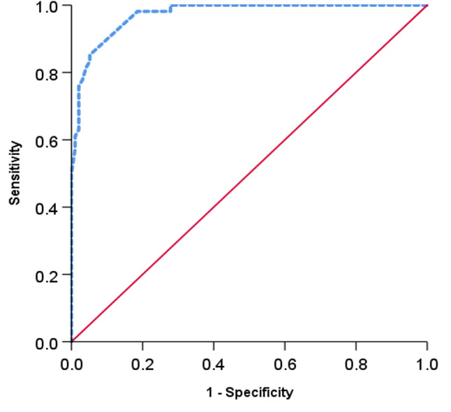
ROC curve showing goodness-of-fit of the model with AUROC = 0.97 (95% CI 0.95–0.99, P < 0.001)
Discussion
ELC performed as soon as possible after the acute attack is established to be safe, feasible, and more preferred over delayed LC for the management of ACC provided the availability of surgeons with adequate expertise in the fields of hepatobiliary and minimally invasive surgery to achieve favorable postoperative outcomes [4, 5, 18–20].
Despite the limited availability of data regarding the incidence of BDI following emergency LC for ACC in Egypt, hepatobiliary surgeons have been observing steep uprising trends evidenced by the increased frequency of patient referrals with BDI to tertiary care hospitals following ELC. The majority were performed by surgeons with limited experience in the field of hepatobiliary surgery or by junior surgeons in the prime of their career who insist on concluding the surgery laparoscopically regardless the degree of difficulty carrying the false perception that conversion to open represents a stigma and a sign of technical incapability among the community of laparoscopic surgeons.
This study was performed to address these surgeons with limited experience in biliary and laparoscopic surgery, regardless of their age, to further urge and promote their vigilance during the preoperative assessment of patients with ACC to guide them to properly select those who lack the risk factors for DELC with expected straightforward ELC. Exercising great caution and wisdom are required at all times whenever patients expected to have DELC are encountered.
Several studies analyzed clinical, radiological, and pathological factors that could predict difficulty of LC among patients with gallstone disease, [21–27] however, data remain limited regarding the situation of difficult ELC specifically among patients with acute cholecystitis [28–31].
Over the past 2 decades, literature became studded with many studies which developed several scoring systems to predict difficult LC, [26, 32–36] however, none of them remains to be universally accepted and surgeons keep researching in this area either to validate the already present models or to propose new ones. The main aim that intrigued us to perform this study was to shed a focused light on the population of patients with ACC who are specifically undergoing ELC very soon within 10 days from the onset of symptoms since they have not been intensively sought in literature. While every proposed scoring system had its own unique risk factors displayed to predict difficult LC, several of these factors were common among them as male gender, high BMI, palpable gallbladder, gallbladder wall thickness ≥ 4 mm, pericholecystic collection, dense adhesions, acute cholecystitis, marked gallbladder distension, preoperative endoscopic retrograde cholangiopancreatography (ERCP), history of acute cholecystitis, and leukocytosis [26, 32–35, 37].
We believe that a main reason behind the multiplicity of proposed models remains in the heterogeneity of the criteria and lack of a standardized definition for difficult LC. Every study proposed its own unique definition of difficult LC based on the experience and volume of practice of its practicing surgeons which has finally urged a group of Spanish experts to perform a study to develop a consensus towards standardizing the definition for difficult LC. They enumerated few criteria which received the highest votes from experts to be considered as markers of difficulty of the surgery such as encountering bile duct injury, non-evident anatomy, Mirizzi syndrome, severe inflammation of Calot’s triangle, conversion to laparotomy, time since last acute cholecystitis, scleroatrophic gallbladder, and pericholecystic abscess [38]. Other studies included significant bleeding, vascular injuries, and subtotal cholecystectomy as markers of difficulty [25, 27, 29, 30]. This is in accordance with the criteria we encountered to define DELC in our patient population with acute cholecystitis. However, we added fundus-first cholecystectomy as a marker of difficulty as it represents a deviation from the normal flow of a straightforward LC and the most initial bail-out option to resort to whenever the surgery deems challenging to continue. Several reports also suggested the length of operation as a valid indicator for difficult LC whenever it exceeded a specific cut-off point which varied widely among the studies ranging from 60 to 180 min [21, 28, 29, 39].
To avoid any potential bias while using operative time as a surrogate for DELC, we thought to introduce the more accurate “net operative time” which, we argue, was not clearly presented in literature the way it was demonstrated in this study. It has contributed well to the accuracy of our analysis by correctly classifying patients with longer operative time exceeding 90 min to the difficult group for the length of operation was strictly reflecting the difficulty and challenging nature of the ELC surgery. The incidence of DELC in our study was comparable to the rate reported in literature (20–61%) [28–31, 39]. The wide interval of incidence obviously depicts the degree of heterogeneity of the criteria used to define difficult LC across the studies added to discrepancy in the efficiency, skills, and amount of surgical experience between the operating surgeons which creates a sort of subjectivity at which the procedure of LC would be considered.
Conversion to open was relatively low in our study and remains be comparable to the rates reported in literature (5–23%) [28, 30, 31, 40]. The reasons for conversion in our experience were in accordance with those reported by Terho et al., attributing the main reason of conversion to the severity of gallbladder inflammation and disturbed anatomy at the region of Calot’s triangle [31]. Similarly, Hayama et al. in their study of risk factors predicting difficult ELC in patients with ACC reported inflammation and adhesions as the most common reasons for conversion, followed by bleeding, and encountered bile leak [28].
While we identified five significant risk factors to strongly predict DELC among patients with ACC, several factors failed to show statistical significance on multivariate analysis as male gender, TLC, PMNL count, CRP, and ALP despite being significant on univariate analysis. The grade of severity of ACC was not also a significant predictor of DELC among the other factors. Similarly, Maehira et al. in their study of predictors of difficult LC in acute cholecystitis did not find CRP, ALP, or male gender to be significant on multivariate analysis [39]. Di Buono et al. found male gender not a significant predictor of difficult LC [29]. Their conclusive analyses, however, identified elevated TLC and fibrinogen level as significant predictors for difficult LC. In the prospective study performed by Nidoni et al. on 180 patients, they found that > 2 previous attacks of cholecystitis, TLC > 11,000/cmm, GB wall thickness > 3 mm, and pericholecystic collection to significantly predict difficult LC and conversion to open laparotomy among patients with ACC [27]. In contrast, Terho et al. reported that high CRP and gallbladder gangrene or abscess were strong determinants for difficult LC with increasing risk for conversion [31]. However, it is worth mentioning that gallbladder abscess or gangrene were only detected intraoperatively as explained in their study and not on preoperative assessment.
We are quite aware with the limiations of our study. The retrospective nature of the study and the relatively small cohort of patients included have potentially affected the degree of precision as observed in the relatively wide range of confidence intervals of few factors displayed on multivariate analysis. Additionally, this is a single center experience which will require external validation of the reported results on larger scale of patients from other centers to confirm their reproducibility. However, we remain very confident about the accuracy and completeness of the collected data, which augments the reliability and competence of the reported results. Furthermore, we reckon that performing all the ELC surgeries by only 2 surgeons with high surgical expertise and almost equivalent technical skills would contribute to the robustness of the data reported by potentially reducing any form of bias in management which may happen if several surgeons with varying degrees of surgical experience and hand skills were involved.
The proposed model with the five preoperative risk factors to predict difficult LC that are simple and easy to identify preoperatively among patients with ACC prior to proceeding with ELC. Our major aim is to encourage junior surgeons and those with limited surgical experience in the field of hepatobiliary and/or laparoscopic surgery, regardless of their age, to exercise more vigilance and look thoroughly for the risk factors and possibly proceed with surgery only for patients lacking them where surgery is expected to be uneventful and straightforward. The degree of experience of a biliary surgeon is usually measured by the number of surgeries he performs both, laparoscopically and open, under elective or emergency settings, while maintaining proper knowledge for all the possible biliary tract and vascular anatomy of the region. In contrast, whenever risk factors are identified, DELC should be considered and thus greater caution should be exercised in dealing with them and a rather alternative plan should be constructed to avoid exposing them to undesired intraoperative events and/or postoperative complications which can be life-threatening. Surgeons should actively disregard several misconceptions and express more readiness to take responsibility through offering this high-risk group of patients the best management possible. This can be achieved by proper consultation of surgeons who are well-experienced in the field and seeking their supervision and/or assistance during such technically demanding surgery to be able to properly guide them and deal with any potentially challenging events encountered intraoperatively. Otherwise, those patients should not proceed with surgery and must be transferred to advanced surgical centers provided with experienced surgeons, anesthesiologists, intensivists, and interventional radiologists, and equipped with advanced laparoscopic equipment to offer the patients the highest level of care they deserve to ensure optimal surgical outcomes. The experience of a surgeon in cholecystectomy could be measured by the number of surgeries performed in both, elective and emergency settings, the ability of the surgeon to perform the cholecystectomy both, laparoscopically and open, and the wide experience with variant biliary tract anatomy. Under all circumstances, the most important message to be delivered is that whenever DELC is already encountered intraoperatively, patient safety and well-being should remain the highest priority to the operating surgeon and they should always opt for bail-out procedures which help to conclude the procedure safely and limit further surgical damage.
Conclusion
Understanding the complexity of ELC in the setting of ACC is mandatory to avoid complications. Identification of preoperative predictors of DELC will help proper selection of patients and better decision making towards either proceeding with the surgery among those expected to have SELC at minimal to no surgical risk, or optimize the surrounding circumstances for those expected to have DELC. Placing a proper management plan ahead of the surgery for those expected to have DELC is very crucial and it is the responsibility of the operating surgeon to ensure the availability of any required experienced surgeons on site, experienced anesthesiologists, nursing staff, as well as the availability of proper surgical and laparoscopic equipment to guarantee smooth flow of the surgery and utmost patient safety. Whenever LC seems difficult and challenging to conclude laparoscopically, surgeon should be responsible and wise enough to opt for bail-out procedures as the lifebuoy to save the patient and prevent further damage.
Author contributions
IMK constructed the design and concept of the study, performed data acquisition, analyzed and interpreted the data, prepared the tables and figures of the manuscript, and drafted the manuscript.IMK revised the manuscript and responded point-by-point to the editors’ and reviewers’ comments.IMK and SSB performed the surgeries described in this manuscript, and they critically revised the manuscript.
Funding
The authors did not receive any form of funding to declare for this study.
Data availability
While the research data and/or materials are not available online, we -the authors- confirm that they are available with us and can be presented upon request by contacting the corresponding author, Islam M. Korayem, MD, PhD, at his email address islam.korayem@alexmed.edu.eg.
Declarations
Ethics approval and consent to participate
The study was approved by the Ethics Committee and the Institutional Review Board of Alexandria University Faculty of Medicine (IRB No. 00012098, FWA No. 00018699) under study protocol serial number 0305608. A formal informed consent was obtained from all the patients included in the study prior to proceeding with surgery explaining the details of surgery, its expected outcomes, and all the potential complications in compliance with the Declaration of Helsinki.
Consent for publication
Not applicable.
Competing interests
The authors declare no competing interests.
Study serial number
0305608
Footnotes
Publisher’s note
Springer Nature remains neutral with regard to jurisdictional claims in published maps and institutional affiliations.
References
- 1.Shaffer EA. Epidemiology and risk factors for gallstone disease: has the paradigm changed in the 21st century? Curr Gastroenterol Rep. 2005;7(2):132–40. [DOI] [PubMed] [Google Scholar]
- 2.Kimura Y, Takada T, Strasberg SM, Pitt HA, Gouma DJ, Garden OJ, et al. TG13 current terminology, etiology, and epidemiology of acute cholangitis and cholecystitis. J Hepatobiliary Pancreat Sci. 2013;20(1):8–23. [DOI] [PubMed] [Google Scholar]
- 3.Stinton LM, Myers RP, Shaffer EA. Epidemiology of gallstones. Gastroenterol Clin North Am. 2010;39(2):157–69. vii. [DOI] [PubMed] [Google Scholar]
- 4.Pisano M, Allievi N, Gurusamy K, Borzellino G, Cimbanassi S, Boerna D, et al. 2020 World Society of Emergency Surgery updated guidelines for the diagnosis and treatment of acute calculus cholecystitis. World J Emerg Surg. 2020;15(1):61. [DOI] [PMC free article] [PubMed] [Google Scholar]
- 5.Yokoe M, Hata J, Takada T, Strasberg SM, Asbun HJ, Wakabayashi G, et al. Tokyo guidelines 2018: diagnostic criteria and severity grading of acute cholecystitis (with videos). J Hepatobiliary Pancreat Sci. 2018;25(1):41–54. [DOI] [PubMed] [Google Scholar]
- 6.Lyu Y, Cheng Y, Wang B, Zhao S, Chen L. Early versus delayed laparoscopic cholecystectomy for acute cholecystitis: an up-to-date meta-analysis of randomized controlled trials. Surg Endosc. 2018;32(12):4728–41. [DOI] [PubMed] [Google Scholar]
- 7.Tornqvist B, Stromberg C, Akre O, Enochsson L, Nilsson M. Selective intraoperative cholangiography and risk of bile duct injury during cholecystectomy. Br J Surg. 2015;102(8):952–8. [DOI] [PubMed] [Google Scholar]
- 8.Flum DR, Dellinger EP, Cheadle A, Chan L, Koepsell T. Intraoperative cholangiography and risk of common bile duct injury during cholecystectomy. JAMA. 2003;289(13):1639–44. [DOI] [PubMed] [Google Scholar]
- 9.Toro A, Teodoro M, Khan M, Schembari E, Di Saverio S, Catena F, et al. Subtotal cholecystectomy for difficult acute cholecystitis: how to finalize safely by laparoscopy-a systematic review. World J Emerg Surg. 2021;16(1):45. [DOI] [PMC free article] [PubMed] [Google Scholar]
- 10.Ansaloni L, Pisano M, Coccolini F, Peitzmann AB, Fingerhut A, Catena F, et al. 2016 WSES guidelines on acute calculous cholecystitis. World J Emerg Surg. 2016;11:25. [DOI] [PMC free article] [PubMed] [Google Scholar]
- 11.Glasheen WP, Cordier T, Gumpina R, Haugh G, Davis J, Renda A. Charlson Comorbidity Index: ICD-9 update and ICD-10 translation. Am Health Drug Benefits. 2019;12(4):188–97. [PMC free article] [PubMed] [Google Scholar]
- 12.Daabiss M. American Society of Anaesthesiologists physical status classification. Indian J Anaesth. 2011;55(2):111–5. [DOI] [PMC free article] [PubMed] [Google Scholar]
- 13.Clavien PA, Barkun J, de Oliveira ML, Vauthey JN, Dindo D, Schulick RD, et al. The Clavien-Dindo classification of surgical complications: five-year experience. Ann Surg. 2009;250(2):187–96. [DOI] [PubMed] [Google Scholar]
- 14.Hanley JA, McNeil BJ. The meaning and use of the area under a receiver operating characteristic (ROC) curve. Radiology. 1982;143(1):29–36. [DOI] [PubMed] [Google Scholar]
- 15.Fagerland MW, Hosmer DW, Bofin AM. Multinomial goodness-of-fit tests for logistic regression models. Stat Med. 2008;27(21):4238–53. [DOI] [PubMed] [Google Scholar]
- 16.Tjur T. Coefficients of determination in logistic regression Models—A new proposal: the coefficient of discrimination. Am Stat. 2009;63(4):366–72. [Google Scholar]
- 17.Strasberg SM, Hertl M, Soper NJ. An analysis of the problem of biliary injury during laparoscopic cholecystectomy. J Am Coll Surg. 1995;180(1):101–25. [PubMed] [Google Scholar]
- 18.Wakabayashi G, Iwashita Y, Hibi T, Takada T, Strasberg SM, Asbun HJ, et al. Tokyo guidelines 2018: surgical management of acute cholecystitis: safe steps in laparoscopic cholecystectomy for acute cholecystitis (with videos). J Hepatobiliary Pancreat Sci. 2018;25(1):73–86. [DOI] [PubMed] [Google Scholar]
- 19.Hibi T, Iwashita Y, Ohyama T, Honda G, Yoshida M, Takada T, et al. The right way is not always popular: comparison of surgeons’ perceptions during laparoscopic cholecystectomy for acute cholecystitis among experts from Japan, Korea and Taiwan. J Hepatobiliary Pancreat Sci. 2017;24(1):24–32. [DOI] [PubMed] [Google Scholar]
- 20.Okamoto K, Suzuki K, Takada T, Strasberg SM, Asbun HJ, Endo I, et al. Tokyo guidelines 2018: flowchart for the management of acute cholecystitis. J Hepatobiliary Pancreat Sci. 2018;25(1):55–72. [DOI] [PubMed] [Google Scholar]
- 21.Veerank N, Togale MD. Validation of a Scoring System to predict difficult laparoscopic cholecystectomy: a one-year cross-sectional study. J West Afr Coll Surg. 2018;8(1):23–39. [PMC free article] [PubMed] [Google Scholar]
- 22.Nachnani J, Supe A. Pre-operative prediction of difficult laparoscopic cholecystectomy using clinical and ultrasonographic parameters. Indian J Gastroenterol. 2005;24(1):16–8. [PubMed] [Google Scholar]
- 23.Agrawal N, Singh S, Khichy S. Preoperative prediction of difficult laparoscopic cholecystectomy: a Scoring Method. Niger J Surg. 2015;21(2):130–3. [DOI] [PMC free article] [PubMed] [Google Scholar]
- 24.Vivek MA, Augustine AJ, Rao R. A comprehensive predictive scoring method for difficult laparoscopic cholecystectomy. J Minim Access Surg. 2014;10(2):62–7. [DOI] [PMC free article] [PubMed] [Google Scholar]
- 25.Bhandari TR, Khan SA, Jha JL. Prediction of difficult laparoscopic cholecystectomy: an observational study. Ann Med Surg (Lond). 2021;72:103060. [DOI] [PMC free article] [PubMed] [Google Scholar]
- 26.Randhawa JS, Pujahari AK. Preoperative prediction of difficult lap chole: a scoring method. Indian J Surg. 2009;71(4):198–201. [DOI] [PMC free article] [PubMed] [Google Scholar]
- 27.Nidoni R, Udachan TV, Sasnur P, Baloorkar R, Sindgikar V, Narasangi B. Predicting difficult laparoscopic cholecystectomy based on Clinicoradiological Assessment. J Clin Diagn Res. 2015;9(12):PC09–12. [DOI] [PMC free article] [PubMed] [Google Scholar]
- 28.Hayama S, Ohtaka K, Shoji Y, Ichimura T, Fujita M, Senmaru N et al. Risk factors for difficult laparoscopic cholecystectomy in Acute Cholecystitis. JSLS. 2016;20(4). [DOI] [PMC free article] [PubMed]
- 29.Di Buono G, Romano G, Galia M, Amato G, Maienza E, Vernuccio F, et al. Difficult laparoscopic cholecystectomy and preoperative predictive factors. Sci Rep. 2021;11(1):2559. [DOI] [PMC free article] [PubMed] [Google Scholar]
- 30.Wennmacker SZ, Bhimani N, van Dijk AH, Hugh TJ, de Reuver PR. Predicting operative difficulty of laparoscopic cholecystectomy in patients with acute biliary presentations. ANZ J Surg. 2019;89(11):1451–6. [DOI] [PMC free article] [PubMed] [Google Scholar]
- 31.Terho PM, Leppaniemi AK, Mentula PJ. Laparoscopic cholecystectomy for acute calculous cholecystitis: a retrospective study assessing risk factors for conversion and complications. World J Emerg Surg. 2016;11:54. [DOI] [PMC free article] [PubMed] [Google Scholar]
- 32.Nassar AHM, Hodson J, Ng HJ, Vohra RS, Katbeh T, Zino S, et al. Predicting the difficult laparoscopic cholecystectomy: development and validation of a pre-operative risk score using an objective operative difficulty grading system. Surg Endosc. 2020;34(10):4549–61. [DOI] [PubMed] [Google Scholar]
- 33.Tongyoo A, Liwattanakun A, Sriussadaporn E, Limpavitayaporn P, Mingmalairak C. The modification of a preoperative Scoring System to predict difficult elective laparoscopic cholecystectomy. J Laparoendosc Adv Surg Tech A. 2023;33(3):269–75. [DOI] [PMC free article] [PubMed] [Google Scholar]
- 34.Gupta N, Ranjan G, Arora MP, Goswami B, Chaudhary P, Kapur A, et al. Validation of a scoring system to predict difficult laparoscopic cholecystectomy. Int J Surg. 2013;11(9):1002–6. [DOI] [PubMed] [Google Scholar]
- 35.Ramirez-Giraldo C, Isaza-Restrepo A, Conde Monroy D, Castillo-Barbosa AC, Rubio-Avilez JJ, Van-Londono I. What is the best score for predicting difficult laparoscopic cholecystectomy? A diagnostic trial study. Int J Surg. 2023;109(7):1871–9. [DOI] [PMC free article] [PubMed] [Google Scholar]
- 36.Lipman JM, Claridge JA, Haridas M, Martin MD, Yao DC, Grimes KL, et al. Preoperative findings predict conversion from laparoscopic to open cholecystectomy. Surgery. 2007;142(4):556–63. discussion 63 – 5. [DOI] [PubMed] [Google Scholar]
- 37.Bulbuller N, Ilhan YS, Baktir A, Kirkil C, Dogru O. Implementation of a scoring system for assessing difficult cholecystectomies in a single center. Surg Today. 2006;36(1):37–40. [DOI] [PubMed] [Google Scholar]
- 38.Manuel-Vazquez A, Latorre-Fragua R, Alcazar C, Requena PM, de la Plaza R, Blanco Fernandez G, et al. Reaching a consensus on the definition of difficult cholecystectomy among Spanish experts. A Delphi project. A qualitative study. Int J Surg. 2022;102:106649. [DOI] [PubMed] [Google Scholar]
- 39.Maehira H, Kawasaki M, Itoh A, Ogawa M, Mizumura N, Toyoda S, et al. Prediction of difficult laparoscopic cholecystectomy for acute cholecystitis. J Surg Res. 2017;216:143–8. [DOI] [PubMed] [Google Scholar]
- 40.Morales-Maza J, Rodriguez-Quintero JH, Santes O, Aguilar-Frasco JL, Romero-Velez G, Garcia-Ramos ES, et al. Conversion from laparoscopic to open cholecystectomy: risk factor analysis based on clinical, laboratory, and ultrasound parameters. Rev Gastroenterol Mex (Engl Ed; 2020. [DOI] [PubMed]
Associated Data
This section collects any data citations, data availability statements, or supplementary materials included in this article.
Data Availability Statement
While the research data and/or materials are not available online, we -the authors- confirm that they are available with us and can be presented upon request by contacting the corresponding author, Islam M. Korayem, MD, PhD, at his email address islam.korayem@alexmed.edu.eg.