ABSTRACT
Despite being the foundation of, or supplement to, many skull base exposures, the retrolabyrinthine approach has not been adequately illustrated in the skull base literature. As an aid to skull base surgeons in training, this article provides a step-by-step description of the microsurgical anatomy and operative nuances of this important technique.
Keywords: Cerebellopontine angle, retrolabyrinthine approach, temporal bone
The retrolabyrinthine approach is a true skull base approach that preserves hearing by following a direct route through the temporal bone to expose the cerebellopontine angle without manipulation of neural structures. The most common indications for this approach are resection of cerebellopontine angle and posterior petrous ridge tumors, vestibular neuronectomy, partial section of the sensory root of the fifth cranial nerve, fenestration of symptomatic arachnoid cysts, and biopsy of brain stem lesions. This technique is also a primary component of many other more extensive skull base exposures, including the translabyrinthine, transcochlear, infratemporal, and combined transpetrosal approaches. The retrolabyrinthine bone removal also may supplement far-lateral and retrosigmoid craniotomies.
Despite its importance and ubiquity in treating skull base pathology, many skull base surgeons are uncomfortable performing this powerful approach. At many institutions the neuro-otologists are solely responsible for this component of the surgery. Furthermore, an adequate, concise description of this technique with anatomically correct illustrations has not been available to skull base surgeons in training, especially neurosurgeons. Mastering this approach will help surgeons to become a more vital component of their interdisciplinary skull base team. This article therefore details the microsurgical anatomy and operative nuances of the retrolabyrinthine approach.
SURGICAL PROCEDURE
Patient Preparation
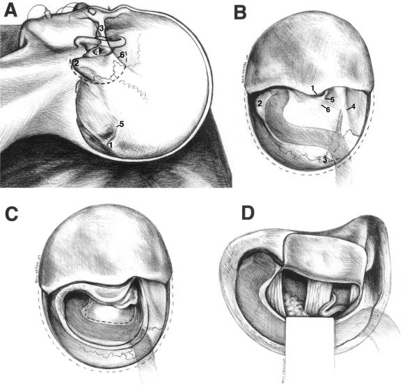
The operating table is reversed head-to-foot allowing access to the table controls and permitting the surgeon to work at desktop height while sitting comfortably. After general anesthesia is induced, the patient is positioned supine on the operating room table and the head is rotated 70 degrees lateral with the face away from the surgeon (Fig. 1A). For patients with poor cervical flexibility a small shoulder roll can be used. The head is secured to the operating room table with adhesive tape. Three-pin fixation is not used to avoid the risk of contralateral jugular vein occlusion from excessive neck rotation that can occur when using rigid head fixation. Electrodes for intraoperative neurophysiologic monitoring of the facial (VII) and cochlear (VIII) nerves are attached to the patient, away from the sterile field. A large craniotomy drape with a collecting bag is positioned posteriorly to receive the large amount of irrigation used during the exposure. Appropriate antibiotic prophylaxis is given before the skin incision is made.
Figure 1.
(A) Patient position for the retrolabyrinthine approach (left-sided approach). The inion (1), mastoid process (2), zygomatic arch (3), external auditory meatus (4), superior nuchal line (5), and linea temporalis (supramastoid crest) (6), all of which may be palpated before the skin incision, are illustrated. (B) The mastoid surface after elevation of the scalp. External landmarks, which define the extent of retrolabyrinthine bone removal, are visualized, including the posterior rim of the external auditory meatus (1), mastoid tip (2), asterion (3), linea temporalis (4), spine of Henle (5), and the cribrose area (6). (C) After the mastoidectomy and before the dura is opened, the sigmoid sinus is completely skeletonized, the lateral bony labyrinth and fallopian canal are visualized, and the dura of the presigmoid, middle fossa, and posterior fossa are exposed. (D) View of the cerebellopontine angle with the VII–VIII cranial nerve complex emerging from the lateral pontomedullary junction as seen through the retrolabyrinthine exposure. The anterior inferior cerebellar artery can be seen looping near this complex. The trigeminal nerve, choroid plexus, and IX–X cranial nerve complex are also visible with this approach.
Although not required, a preoperative temporal bone computed tomography (CT) scan may be obtained with 1 mm high-resolution cuts in both the axial and coronal planes for planning the surgical approach. Considering the high interpatient variability of anatomic structures in the temporal bone, this study provides useful information for the surgeon in training, especially the degree of mastoid aeration, size and orientation of the sigmoid sinus and jugular bulb, and limits of the semicircular canals.
External Cranial Landmarks
To obtain optimal exposure of the mastoid region for the retrolabyrinthine approach, the surgeon carefully determines the location of the skin incision by palpating external osseous landmarks (see Fig. 1A). The external acoustic meatus, zygomatic arch, external occipital protuberance (inion), and mastoid process are identified. An imaginary line extending from the zygoma to the inion passes just superior to the external acoustic meatus and overlies, in sequential order, the linea temporalis (supramastoid crest), asterion, and superior nuchal line. The linea temporalis, a subtle ridge of bone that originates at the root of the zygoma, runs almost parallel to the zygomatic arch toward the asterion. This crest demarcates the superior extent of the mastoid bone and thus the floor of the middle cranial fossa. The posterior extent of the linea temporalis corresponds to the most posterior aspect of the mastoid body. The asterion overlies the transverse-sigmoid junction and is the nexus of three sutures: the lambdoid, parietomastoid, and occipitomastoid. The superior nuchal line overlies the transverse sinus and is the site of attachment for the cervical musculature to the occiput.
Scalp Elevation
A C-shaped skin incision is made with a No. 15 blade or electrocauterization through the galea. The periosteum, which is continuous with the fascia of the temporalis muscle and the cervical musculature, is preserved. The incision begins slightly superior to the pinna, extends posteriorly one-third of the distance from the root of the zygoma to the inion, and ends 1 cm below and slightly anterior to the mastoid tip. Sharp dissection is used to release the galea from the underlying periosteum until the skin flap is reflected anteriorly and the external acoustic meatus can be palpated.
The incision line for the periosteal flap is created 1 cm under the initial skin incision using skin rakes to provide exposure. Once this incision is completed, the periosteum and muscular fascia are detached from the cranium with a periosteal dissector and also reflected forward. The scalp (superficial) and periosteal (deep) flap incisions do not override, thus providing a barrier against postoperative cerebrospinal fluid (CSF) leakage. During this dual-layer flap elevation, a mastoid emissary vein is usually encountered 1 cm inferior to the asterion along and slightly anterior to the occipitomastoid suture. This vein connects the sigmoid sinus to the cutaneous venous plexus and may be a source of bleeding and potential air embolus if torn. Bleeding from the emissary vein is controlled by firmly placing bone wax in the mastoid emissary foramen. The attachments of the sternocleidomastoid and splenius capitus muscles to the mastoid tip are freed using a periosteal dissector or electrocautery. The two-layered scalp flap is padded underneath with a surgical sponge and then retracted anteriorly with multiple fishhooks. Hemostasis is achieved with bipolar cauterization.
Retrolabyrinthine Bone Removal
The surgeon uses skull topography to visualize the location and relationships of deep temporal bone structures. Therefore, all bony landmarks are identified before the mastoidectomy is performed, including the root of the zygoma, external auditory meatus, linea temporalis, mastoid emissary foramen, asterion, and spine of Henle (Figs. 1B and 2A). The spine of Henle, or suprameatal spine, is a small horizontal ridge of bone located at the posterior–superior rim of the external auditory meatus. This landmark, along with the cribrose area, defines the path to the mastoid antrum, which is located about 15 mm deep to the spine of Henle.
Figure 2.
Serial intraoperative photomicrographs of a left-sided retrolabyrinthine approach. (A) Mastoid surface after scalp elevation. The mastoid tip (mt), posterior margin of the external acoustic meatus (eam), spine of Henle (*), and linea temporalis (lt) are visualized. (B) After the initial bone is removed, the mastoid air cells are apparent, as is the contour of the transverse-sigmoid junction (tsj). The dissector is over the sigmoid sinus covered with cortical bone. (C) Bone covering the midportion of the sigmoid sinus (ss) has been removed. The dissector tip touches the dense cortical bone of the horizontal semicircular canal (hc). (D) A magnified view of facial nerve (fn) in the fallopian canal. The horizontal semicircular canal (hc) is also visualized. (E) A magnified view of the retrofacial air cells being removed with a diamond bur. The facial nerve (fn) in the fallopian canal is seen. (F) After the bone removal is completed, the presigmoid dura, endolymphatic duct (ed), facial nerve (fn), horizontal (hc) and posterior (pc) semicircular canals, and sigmoid sinus are exposed.
The mastoid bone removal is performed using a high-speed cutting burr and continuous suction irrigation. Shallow troughs outlining the superior and anterior perimeters of the mastoidectomy are created. The superior trough extends from the zygomatic root to the asterion, just inferior to the linea temporalis. The anterior trough is perpendicular to the superior one and extends from the posterior–superior rim of the external auditory meatus down to the mastoid tip. Working both posterior and inferior to these margins, cortical bone is removed with brushlike strokes exposing a variable number of mastoid air cells. A uniform depth of dissection is maintained as more air cells are exposed and the cortical bone covering the sigmoid sinus becomes apparent (Fig. 2B). A thin, depressible layer of bone may be left on the surface of the sigmoid sinus for protection. Approximately 1 cm of posterior fossa dura is exposed posterior to the sinus. This additional bone removal behind the sigmoid sinus allows retraction of the sinus posteriorly, providing improved exposure medial to the bony labyrinth.
Once the sigmoid sinus is fully skeletonized, additional air cells anterior to the sigmoid sinus are removed to expose the reflection of the dura of the middle and posterior fossae. This bone removal is continued until the middle fossa dura is completely exposed below the linea temporalis. Successively smaller cutting burrs are used as the mastoidectomy proceeds. The sigmoid sinus is followed inferiorly and the air cells in the mastoid tip are removed exposing the inferior segment of the sigmoid sinus and digastric ridge. The mastoid antrum is opened, and the dense cortical bone overlying the horizontal semicircular canal (Koerner's septum) is identified and preserved (Fig. 2C). The short process of the incus is identified in the incudal fossa. Care is taken to avoid bur contact with the incus, which may conduct to the inner ear causing sensorineuronal hearing loss.
Higher magnification and a diamond bur are used to skeletonize the bony labyrinth, fallopian canal (containing the facial nerve), and presigmoid dura. The bone covering the presigmoid dura at the sinodural angle (of Citelli) is removed. Removal of this bone is paramount to achieve adequate dural exposure. The horizontal semicircular canal on the medial wall of the mastoid antrum and the posterior semicircular canal on the postero-medial aspect of the antrum are also skeletonized. The bony labyrinth must not be violated during this critical stage of the retrolabyrinthine approach or hearing loss may occur. Changing the angle of view by moving the microscope and depressing the dura away from the thinned bone covering the presigmoid dura facilitate safe bone removal and maximize the final exposure.
If the bony labyrinth is accidentally violated with the drill but the membranous labyrinth remains intact, a small amount of bone wax can be used to occlude the defect. However, if both the bony and membranous components of the labyrinth are transected, the surgeon must act quickly and cork each end of the defect with a small piece of muscle to prevent excessive leakage of the peri- and endolymph, which may cause significant vertigo after surgery.
The surgeon now removes the remaining aerated bone overhanging the antero-medial rim of the mastoidectomy, which contains the vertical segment of the facial nerve in the fallopian canal (Fig. 2D). A thin layer of cortical bone is left covering the fallopian canal. Neurophysiologic monitoring of the facial nerve helps prevent inadvertent injury of the facial nerve during this stage of the dissection. Osseous dissection of most temporal bones can also be carried medially and anteriorly from the posterior semicircular canal to the porus acousticus. The retrofacial air cells are also removed (Fig. 2E). These maneuvers provide further exposure of the presigmoid dura.
Intradural Dissection
To obtain a maximal dural exposure, the sigmoid sinus and jugular bulb are skeletonized completely, the lateral and posterior semicircular canals are defined precisely, the entire course of the facial nerve through the mastoid bone is visualized, and the dura of the presigmoid, middle fossa, and sinodural angle is exposed completely (Figs. 1C and 2F).
The dura is opened with an anteriorly based C-shaped flap using a No. 11 blade and microscissors. The dura is tacked up with 4-0 braided nylin sutures. The endolymphatic sac will be visible inferior to the posterior semicircular canal as a whitish, thickened area of dura emerging from the vestibular aqueduct. To maintain its integrity, the endolymphatic sac often defines the anterior limit of the dural opening. Routinely, the middle fossa dura is not opened and the superior petrosal sinus is not sacrificed.
The cerebellum resting against the petrous temporal bone is visualized after the dura is opened. Using microsurgical technique under high magnification, the arachnoid adhesions between the cerebellum and petrous ridge are placed under tension by gently retracting the cerebellum posteriorly with a cottonoid-padded microsuction. These adhesions are dissected sharply. The cerebellopontine angle and prepontine arachnoid cisterns are opened sharply and CSF is released. With transection of cerebellar adhesions and release of CSF from the subarachnoid space, the cerebellum falls away from the petrous ridge providing adequate exposure of the cerebellopontine angle and posterior face of the petrous ridge without static retraction on the cerebellum.
Through the retrolabyrinthine exposure, both the brain stem origin and cisternal segment of the VII–VIII cranial nerve complex are well visualized in the cerebellopontine angle (Fig. 1D). A loop of the anterior inferior cerebellar artery (AICA) is often found intimately associated with this complex. A microdissector and hand-held nerve stimulator are used to identify the components of the VII–VIII cranial nerve complex: the facial nerve, acoustic nerve, and vestibular nerve. In most patients the porus of the internal auditory canal is inaccessible for exposure through this approach because the sigmoid sinus limits a more obtuse viewing angle in relation to the posterior petrous ridge. The ninth and tenth cranial nerves are visible inferiorly, anterior to the level of the jugular bulb. The fifth cranial nerve and basilar artery can also be examined through this exposure.
Wound Closure
The dura is reapproximated with multiple 4-0 interrupted braided nylon sutures, but a watertight closure is seldom possible. In all cases, a 2 cm sphere of adipose tissue is harvested from the lower left abdomen via a 2 cm, periumbilical incision. The fat graft is placed in the mastoid craniectomy defect, with smaller pieces of fat corking the gaps in the dural closure. Fibrin sealant (Tisseal, 5 cc, Haemacure Inc., Sarasota, FL, USA) is used along the dural incision and again over the secured fat graft to provide a watertight seal. All facial recess and retrofacial temporal bone air cells are carefully sealed with bone wax and covered with additional fat graft.
The periosteal flap is closed with interrupted 3-0 vicryl sutures spaced 5 mm apart, which firmly cinches the fat graft in the mastoid defect. In a similar manner, the galeal-skin flap is closed with inverted 3-0 vicryl sutures. As already described, the two flap incisions do not overlap, providing an additional barrier to CSF leakage. The skin is closed with a running, locked 3-0 nylon suture. A mastoid compression dressing is placed and maintained 3 days after surgery to promote wound healing and to prevent pseudomeningocele formation by assisting the fresh tissue planes to fix.
ILLUSTRATIVE CASES
Case 1
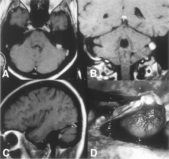
A 34-year-old woman had experienced violent bouts of vertigo for 6 months. Radiographic evaluation revealed a 1 cm meningioma on the left posterior petrous ridge adjacent to the endolymphatic sac (Fig. 3). Using a left-sided retrolabyrinthine approach, the meningioma was removed en bloc with a margin of surrounding dura. Postoperatively, her symptoms of vertigo resolved. Her hearing was maintained despite transection of the endolymphatic duct.
Figure 3.
Axial (A), coronal (B), and sagittal (C) T1-weighted magnetic resonance images after gadolinium administration show a 1cm left posterior petrous ridge meningioma near the endolymphatic duct in a patient with paroxysms of vertigo. (D) Intraoperative photomicrograph demonstrating en bloc removal of the meningioma via the retrolabyrinthine approach.
Case 2
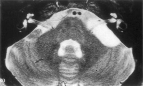
A 43-year-old woman developed progressive, left-sided hearing loss over a few months. An audiogram documented unilateral sensorineuronal hearing loss with speech discrimination of 52%. Radiographic evaluation revealed a left cerebellopontine angle arachnoid cyst (Fig. 4). The arachnoid cyst was fenestrated through a retrolabyrinthine exposure. Postoperatively, her speech discrimination improved to 92%.
Figure 4.
Axial T2-weighted magnetic resonance image shows a left cerebellopontine angle arachnoid cyst in a patient with progressive, unilateral sensorineuronal hearing loss.
DISCUSSION
Initially, the retrolabyrinthine approach was popularized for functional procedures of the cerebellopontine angle, including vestibular and sensory trigeminal neuronectomies.1,2,3,4 With popularization of the “keyhole” concept in neurosurgery, where adjustments in trajectory through a relatively small dural opening were used to create a significantly larger operative field while minimizing exposure of nearby eloquent structures, the retrolabyrinthine approach was subsequently reported useful for the management of other cerebellopontine angle pathologies.5,6,7,8 For example, Darrouzet et al6 used the retrolabyrinthine approach to resect 60 acoustic neuromas, with facial nerve and hearing preservation outcomes similar to those of previous series using more extensive retrosigmoid and translabyrinthine approaches. In their series, 63% of tumors were larger than 2 cm and 15% were larger than 3 cm. These authors concluded that the retrolabyrinthine approach may be used to resect acoustic neuromas of almost any size.
Although we believe the exposure provided by the retrolabyrinthine approach is too limited for large cerebellopontine angle tumors, it provides adequate exposure for smaller tumors, especially petrous ridge and jugular fossa meningiomas, epidermoids, and arachnoid cysts, and for biopsy of intrinsic brain stem lesions. Despite the risk of hearing loss, some authors advocate partial labyrinthectomies to widen the exposure.9 Our philosophy is that even a small chance of hearing loss is unacceptable when using the retrolabyrinthine approach. If a wider exposure is required, we would opt either to sacrifice hearing with a translabyrinthine approach, when indicated, or to perform a retrosigmoid craniotomy, thus preserving hearing and obtaining a wider surgical corridor.
A retrosigmoid craniotomy can be used to expose the same region and anatomical structures as the retrolabyrinthine approach. However, for small lesions located adjacent to the presigmoid dura, we believe the retrolabyrinthine approach has certain advantages. First, it provides a direct approach through the temporal bone. It preserves hearing, which may be compromised with cerebellar retraction during the retrosigmoid approach. It also requires no retraction or manipulation of neural elements. Finally, using a small craniectomy without transection of the musculature avoids postoperative cervicalgia.
The most frequent approach-related complication associated with the retrolabyrinthine exposure is CSF leakage. For all major clinical series, the incidence of CSF leakage after this approach has ranged from 3 to 7%.1,2,3,4,6,7 Although uncommon, injury to the facial nerve in the fallopian canal and damage to the labyrinth causing hearing loss can occur during bone removal.
The retrolabyrinthine craniectomy is an important technique for skull base surgeons to understand and master. It not only provides safe and direct exposure to select pathology of the cerebellopontine angle as a primary surgical approach, it also is the foundation for more common and extensive cranial base exposures of the posterior and middle fossae. It is the initial step when performing a translabyrinthine, transcochlear, infratemporal, or combined petrosal approach to both infra- and supratentorial compartments. When indicated, it can provide additional exposure for retrosigmoid and far-lateral craniotomies. Day et al7 used the retrolabyrinthine approach with transection of the sigmoid sinus “as a simple alternative to more involved strategies” to manage 11 aneurysms of the lower basilar trunk and AICA.
Despite frequent use of the retrolabyrinthine approach alone, with modification, or as the foundation of a more extensive cranial base approach, the skull base literature has lacked an adequate description of the procedure. We hope this communication also encourages renewed interest in this approach in the neurosurgical community.
ACKNOWLEDGMENTS
We thank Benjamin Kress for creating Figure 1 and Sigrid Hahn, M.D. for editorial assistance.
REFERENCES
- Kemink J L, Hoff J T. Retrolabyrinthine vestibular nerve section: analysis of results. Laryngoscope. 1986;96:33–36. doi: 10.1288/00005537-198601000-00006. [DOI] [PubMed] [Google Scholar]
- McElveen J T, Jr, Shelton C, Hitselberger W E, Brackmann D E. Retrolabyrinthine vestibular neurectomy: a reevaluation. Laryngoscope. 1988;98:502–506. doi: 10.1288/00005537-198805000-00005. [DOI] [PubMed] [Google Scholar]
- Nguyen C D, Brackmann D E, Crane R T, Linthicum F H, Hitselberger W E. Retrolabyrinthine vestibular nerve section: evaluation of technical modification in 143 cases. Am J Otol. 1992;13:328–332. [PubMed] [Google Scholar]
- Silverstein H, Norrell H, Rosenberg S. The resurrection of vestibular neurectomy: a 10-year experience with 115 cases. J Neurosurg. 1990;72:533–540. doi: 10.3171/jns.1990.72.4.0533. [DOI] [PubMed] [Google Scholar]
- Arriaga M, Shelton C, Nassif P, Brackmann D E. Selection of surgical approaches for meningiomas affecting the temporal bone. Otolaryngol Head Neck Surg. 1992;107:738–744. doi: 10.1177/019459988910700605.1. [DOI] [PubMed] [Google Scholar]
- Darrouzet V, Guerin J, Aouad N, Dutkiewics J, Blayney A W, Bebear J P. The widened retrolabyrinthe approach: a new concept in acoustic neuroma surgery. J Neurosurg. 1997;86:812–821. doi: 10.3171/jns.1997.86.5.0812. [DOI] [PubMed] [Google Scholar]
- Day J D, Fukushima T, Giannotta S L. Cranial base approaches to posterior circulation aneurysms. J Neurosurg. 1997;87:544–554. doi: 10.3171/jns.1997.87.4.0544. [DOI] [PubMed] [Google Scholar]
- Kinney S E, Hughes G B, Little J R. Retrolabyrinthine transtentorial approach to lesions of the anterior cerebellopontine angle. Am J Otol. 1992;13:426–430. [PubMed] [Google Scholar]
- Horgan M A, Delashaw J B, Schwartz M S, Kellogg J X, Spektor S, McMenomey S O. Transcrusal approach to the petroclival region with hearing preservation. Technical note and illustrative cases. J Neurosurg. 2001;94:660–666. doi: 10.3171/jns.2001.94.4.0660. [DOI] [PubMed] [Google Scholar]


