Abstract
A chemically defined medium in combination with an airlift fermentor system was used to study the growth and sporulation of Bacillus cereus ATCC 14579. The medium contained six amino acids and lactate as the main carbon sources. The amino acids were depleted during exponential growth, while lactate was metabolized mainly during stationary phase. Two concentrations of glutamate were used: high (20 mM; YLHG) and low (2.5 mM; YLLG). Under both conditions, sporulation was complete and synchronous. Sporulation started and was completed while significant amounts of carbon and nitrogen sources were still present in the medium, indicating that starvation was not the trigger for sporulation. Analysis of amino acids and NH4+ in the culture supernatant showed that most of the nitrogen assimilated by the bacteria was taken up during sporulation. The consumption of glutamate depended on the initial concentration; in YLLG, all of the glutamate was used early during exponential growth, while in YLHG, almost all of the glutamate was used during sporulation. In YLLG, but not in YLHG, NH4+ was taken up by the cells during sporulation. The total amount of nitrogen used by the bacteria in YLLG was less than that used by the bacteria in YLHG, although a significant amount of NH4+ was present in the medium throughout sporulation. Despite these differences, growth and temporal expression of key sigma factors involved in sporulation were parallel, indicating that the genetic time frames of sporulation were similar under both conditions. Nevertheless, in YLHG, dipicolinic acid production started later and the spores were released from the mother cells much later than in YLLG. Notably, spores had a higher heat resistance when obtained after growth in YLHG than when obtained after growth in YLLG, and the spores germinated more rapidly and completely in response to inosine, l-alanine, and a combination of these two germinants.
Bacillus cereus is a gram-positive, facultative anaerobic rod-shaped bacterium able to form spores. It is a ubiquitous bacterium found in soil and in many raw and processed foods, such as rice, milk and dairy products, spices, and vegetables (8, 12, 20, 44). Many strains of B. cereus are able to produce toxins and cause distinct types of food poisoning (19, 31). Concerns over B. cereus contamination have increased over the past few years because of the rapidly expanding market of chilled foods that may be pasteurized but still contain viable spores (8, 20, 34). Spores from B. cereus can germinate and outgrow during storage, even at low temperatures (8, 11, 20). To address this increasing problem, major efforts focus on determining the causes of spore resistance and the mechanisms of germination.
It has been well established that bacterial spore properties are affected by the conditions during sporulation (1, 17, 18, 33, 41). In most studies, spores are routinely produced from fortified agar or rich liquid media, which results in heterogeneous sporulation conditions for the individual cells. This prevents careful analysis of the metabolism during growth and sporulation. Recent studies describing the effect of sporulation conditions on spore properties involved modulation of sporulation temperature (1, 18, 33, 41, 42) or compared spores produced from different media (9, 32).
Studies employing defined conditions and media to link substrate use with sporogenesis are relatively rare; moreover, the effect of carbon sources on sporulation has not been studied systematically in recent decades. The most common carbon source used in sporulation media is glucose, but in natural conditions, B. cereus may encounter rather different substrates, such as lactate. We have previously shown that B. cereus is able to metabolize lactate (14), which is formed in the natural environment by the fermentation of a variety of naturally occurring polymers, such as lactose in dairy or plant sugars in silage. Indeed, silage is a known source of B. cereus contamination of milk (47). Furthermore, in contrast to glucose, lactate does not cause catabolite repression or repression of the tricarboxylic acid (TCA) cycle, which is required for activation of the Spo0A phosphorelay, which in turn leads to sporulation (25). Therefore, we investigated the growth and sporulation of B. cereus ATCC 14579, which has been characterized at the genome level (26), on a chemically defined medium with lactate instead of glucose as the main carbon source. Secondly, because glutamate has been reported to have a large impact on sporulation as well as spore properties of bacilli (7, 10, 29), we used two different concentrations of glutamate: low (2.5 mM; YLLG) and high (20 mM; YLHG).
MATERIALS AND METHODS
Strains, medium, and fermentation.
B. cereus strain ATCC 14579 was obtained from the American Type Culture Collection and used throughout this study. It was cultivated in a chemically defined medium described previously (Table 1) (14). Fermentation conditions and inoculation were as described before (14). The spores were harvested and washed as described previously and were stored in 10 mM KPO4, pH 7, with 0.1% Tween 80 to prevent clumping and attachment of spores to plastic laboratory equipment. The addition of Tween 80 had no effect on spore heat resistance.
TABLE 1.
Medium compositions of YLHG and YLLG
| Component | Concn
|
|
|---|---|---|
| YLHG | YLLG | |
| d/l-Lactic acid (mM) | 25 | 25 |
| l-Glutamic acid (mM) | 20 | 2.5 |
| l-Leucine (mM) | 6 | 6 |
| l-Valine (mM) | 2.6 | 2.6 |
| l-Threonine (mM) | 1.4 | 1.4 |
| l-Methionine (mM) | 0.47 | 0.47 |
| l-Histidine (mM) | 0.32 | 0.32 |
| Acetic acid (mM) | 1 | 1 |
| FeCl3 (μM) | 50 | 50 |
| CuCl2 (μM) | 2.5 | 2.5 |
| ZnCl2 (μM) | 12.5 | 12.5 |
| MnSO4 (μM) | 66 | 66 |
| MgCl2 (mM) | 1 | 1 |
| (NH4)2SO4 (mM) | 5 | 5 |
| Na2MoO4 (μM) | 2.5 | 2.5 |
| CoCl2 (μM) | 2.5 | 2.5 |
| Ca(NO3)2 (mM) | 1 | 1 |
Analytical procedures.
Concentrations of d- and l-lactic acid and glucose in culture supernatant were determined by the UV method with enzymatic bioanalysis kits from Boehringer Mannheim (Darmstadt, Germany). Phase-contrast microscopy was performed with an Axioskop microscope from Zeiss. Amino acids in culture supernatant, cells, and spores were determined with the PicoTaq high-performance liquid chromatography method (5). The amino acids and NH4+ from culture supernatant were derivatized with the AccQ-Fluor kit from Waters (Etten-Leur, The Netherlands) according to manufacturer's instructions. For cell and spore analyses, samples were autoclaved and acid hydrolyzed prior to derivatization.
Dipicolinic acid (DPA) was measured with a fluorescent assay modified from Hindle and Hall (23). Spores or cells were suspended in Tris-NaCl-Tw buffer (Tris-HCl [10 mM, pH 7.5] buffer with 10 mM NaCl and 0.1% Tween 80), destroyed by autoclaving (121°C for 15 min), a treatment known to release all DPA (27), and pelleted at maximum speed in an Eppendorf centrifuge for 2 min. The supernatant was assayed for DPA as follows: 80 μl of supernatant was used in a final volume of 200 μl Tris-NaCl-Tw buffer with freshly added TbCl3 at a final concentration of 100 μM, and the fluorescence was measured with the Safire microplate reader from Tecan (Salzburg, Austria) in combination with XFLUOR4 software, version 4.40. Standard curves of DPA with this method showed straight lines (R2 > 0.99) up to 150 μM of DPA with this Tb3+ concentration, with a detection limit of about 0.5 μM. The settings of the machine were as follows: the excitation wavelength was 272 nm, the emission wavelength was 547 nm, the bandwidth excitation and emission wavelengths were 7.5 nm, the lag time was 10 μs, and the integration time was 2,000 μs.
Spore properties.
For the heat resistance assay, 100 μl spores suspended at an A600 of 0.7 to 1.0 in Tris-NaCl-Tw buffer was sealed in the tip of a Pasteur pipette, placed in an oil bath calibrated to the designated temperature, and cooled after a set time in ice-cold water. Because of the small diameters of the Pasteur pipette tips and the small volume of spore suspension, we assume that the heating and cooling of the spore suspensions were instantaneous. Serial dilutions were made in Tris-NaCl-Tw buffer and plated onto diluted nutrient broth (2.6 g/liter; Difco) solidified with 1.5% agar. Colonies were counted after overnight incubation at 30°C.
The density of the whole spore was measured with a Percoll gradient according to the method described by Tisa et al. (48). Spores were concentrated in 0.15 M NaCl, added to 90% (vol/vol) Percoll (Amersham Pharmacia Biotech) solution with 0.15 M NaCl, and centrifuged for 20 min at 30,000 × g. Whole-spore densities were derived by comparison of the positions of the spores and of density marker beads (Amersham Pharmacia Biotech) in the self-established gradient. The spore core density was determined with Nycodenz (Nygaard, Oslo, Norway) density gradients (43) according to the method described by Lindsay et al. (30) as follows: spores were permeabilized with a decoating solution containing 0.1 M dithiothreitol, 0.1 M NaCl, and 0.5% sodium dodecyl sulfate according to the method described by Vary (51). Permeabilized spores were equilibrated in 30% Nycodenz (1.159 g/ml). Continuous gradients were made by letting discontinuous gradients diffuse overnight at 5°C (43) or for a few hours at room temperature. After centrifugation in a swing-out rotor for 45 min at 3,700 rpm in a Centaur 2 centrifuge, the spores formed a band at a certain position. The refractive index, which was linearly correlated with the density, from samples taken just above and below the band was measured with a precision refractometer, and the average yielded the spore core density.
Spore germination was measured by the drop in A600 of spore suspensions at 30°C by using a Safire microplate reader from Tecan (Salzburg, Austria) in combination with XFLUOR4 software, version 4.40. Spores were suspended at an A600 of 0.7 to 1.0 in Tris-NaCl-Tw buffer, and after the addition of the germinants, the A600 was followed automatically with intermittent shaking to prevent settling of the spores.
RT-PCR.
Real-time reverse transcription (RT)-PCR was used to monitor the expression of the genes for key sigma factors (sigA, sigB, sigF, and sigG) and performed as described earlier (14). The level of expression was calibrated by using the positive control, sigA expression, as a reference. All the samples in this communication were handled in exactly the same way to enable a good comparison of the gene expressions in the two conditions we tested. Real-time RT-PCRs were carried out in duplicate, and the duplicates differed less than 5% in all cases.
RESULTS AND DISCUSSION
Carbon and nitrogen use during growth and sporulation.
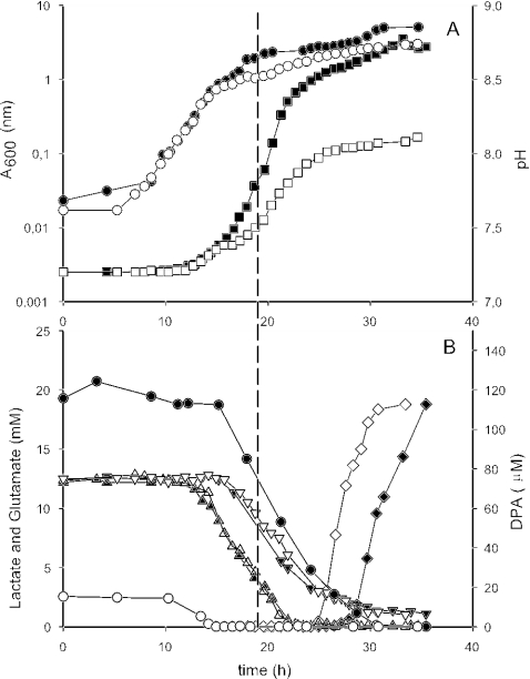
B. cereus was able to grow and sporulate in the chemically defined medium devoid of glucose. With lactate as the main carbon source, the exponential growth phases of B. cereus with low and high glutamate, YLLG and YLHG, respectively, were identical (Fig. 1A). The A600 curves and pH curves ran exactly parallel up to the point where the glutamate in YLLG is used up (Fig. 1B), while during growth in YLHG, glutamate was barely consumed at this time. From the moment of glutamate depletion in YLLG, the absorbance and pH differed between cultures grown in YLLG or YLHG, although lactic acid was metabolized in a parallel manner for both media (Fig. 1B). During the growth of culture in YLHG, the extra rise in pH coincided with glutamate consumption, indicating that it was a result of glutamate metabolism.
FIG. 1.
Growth and sporulation of B. cereus cultures in YLHG (closed symbols) and YLLG (open symbols). (A) Circles, A600; squares, pHs. (B) Triangles, l-lactate; inverted triangles, d-lactate; diamonds, DPA; circles, glutamate. The dashed line indicates the moment of upregulation of sigG transcription.
The higher A600 that was reached during the growth of culture in YLHG suggests that many more cells are present in this culture than in the culture grown in YLLG. However, neither plate counts nor direct microscopic observations confirmed that this was the case, although plate counts were difficult because the cells formed large clusters at the end of exponential growth, as we observed before (14). It is possible that the A600 is influenced by the pH, because we noticed that in the medium that we used a high pH (above 8.5) results in some precipitation, presumably of phosphate with calcium or magnesium. Furthermore, a dark brown color developed in the supernatant during sporulation in YLHG but not in YLLG, further raising the A600. This makes it plausible that although the final A600 reached with the culture grown in YLHG was higher, the actual cell density was quite similar to that of the culture grown in YLLG. Indeed, the total biomasses, as measured by nitrogen contents, were similar for the two cultures at the end of sporulation, both cultures produced similar amounts of DPA, sporulation frequency was >99% in both cases, and the cleaned spores had similar DPA contents (Table 2). This confirmed that, indeed, equal amounts of cells were present in the cultures in YLLG and YLHG.
TABLE 2.
Properties of spores from YLLG and YLHGa
| Spores from indicated medium | Cell-bound N at 35 h ± SD (mM) | DPA content ± SD (μM/OD600 unit) | Density (g/ml)
|
D95 (± SD) (min) | |
|---|---|---|---|---|---|
| Whole spore | Spore core (± SD) | ||||
| YLLG | 15.10 ± 3.20 | 112 ± 1.35 | 1.13 | 1.345 ± 0.008 | 1.52 ± 0.07 |
| YLHG | 16.03 ± 1.05 | 115 ± 2.56 | 1.13 | 1.309 ± 0.012 | 2.64 ± 0.16 |
OD600, optical density at 600 nm; D95, decimal reduction time at 95°C.
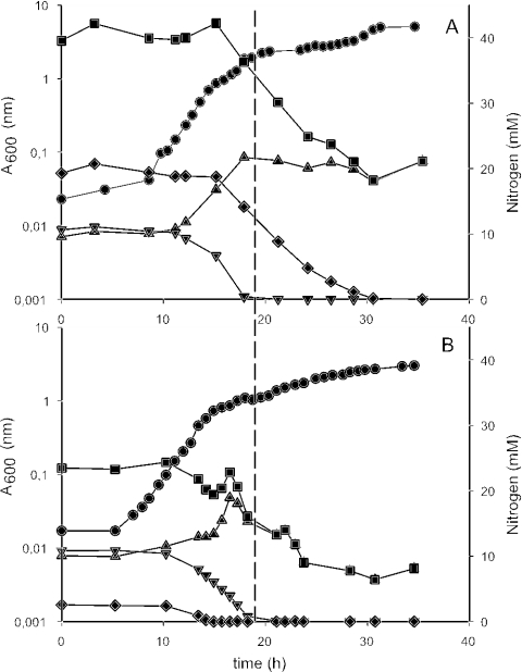
We initially expected the maximum growth rate of B. cereus in the medium with lactate to be lower than that of B. cereus in the same medium with the much more favorable substrate, glucose, but the rates were identical (about 0.45 h−1). However, the lag phase was much longer without glucose. This indicates that the exponential growth rate is primarily determined by the amino acids in the medium. Indeed, during exponential growth, the bacteria metabolized the amino acids present in the medium, while lactate was used upon entry into stationary phase (Fig. 1 and 2). The metabolism of a 10 mM concentration of amino acids during exponential growth was accompanied by a 10-mmol rise in NH4+ in the supernatant of both cultures, indicating that B. cereus used the amino acids as a carbon source and fuel, while most of the lactic acid was metabolized after the cells had entered stationary phase. These findings confirm the anticipated preference of B. cereus for amino acids, as derived from genome analysis studies (26). In the presence of either glucose or lactate, the amino acids were depleted in the same order: threonine was first, then valine, histidine, and leucine, and methionine was the last (data not shown). The moment of glutamate consumption depended on the initial concentration; B. cereus metabolized glutamate earlier in YLLG than in YLHG, and during cultivation in YLLG, glutamate was depleted early, after threonine. In YLHG, the concentration of glutamate started to decrease 3 h later, and glutamate was the last amino acid to be metabolized.
FIG. 2.
Nitrogen sources during growth and sporulation of B. cereus cultures in YLHG (A) and YLLG (B). Symbols: circles, A600; squares, total nitrogen; diamonds, nitrogen in glutamate; inverted triangles, nitrogen in amino acids other than glutamate; triangles, NH4. The dashed line indicates the moment of upregulation of sigG transcription.
Because the metabolism of amino acids during exponential growth resulted in a nearly equimolar increase of NH4+ in the supernatant, the total amount of nitrogen assimilated by B. cereus was very low during this growth phase. Indeed, our measurements clearly indicated that most of the nitrogen assimilated by B. cereus is taken up during sporulation (Fig. 2), underscoring the high nitrogen requirement of sporulating cells. The total concentrations of nitrogen initially present were 40 mM in YLHG and 22.5 mM in YLLG, a significant part of which (10 mM) consisted of NH4+, which was routinely added to the media as (NH4)2SO4 (Table 1). When cultivated in YLHG, B. cereus used the bulk of the glutamate in the stationary and sporulation phase, after the other amino acids had disappeared. During this phase, the NH4+ concentration in the supernatant remained 20 mM, indicating that the nitrogen in the glutamate was completely assimilated. When cultivated in YLLG however, B. cereus assimilated NH4+ during the stationary and sporulation phase.
At the end of sporulation, a total of 20 mM of nitrogen had disappeared from the culture supernatant in YLHG, of which we could trace back 16 mM as cell-bound nitrogen (Table 2). The remaining 4 mM might be present in the brown pigment that was formed in the culture supernatant during sporulation, as a brown pigment produced by B. subtilis during sporulation was shown to contain a significant proportion of nitrogen (3). The culture grown in YLLG had assimilated 15 mM of nitrogen in total at the end of sporulation, all of which we could trace back as cell-bound nitrogen (Table 2).
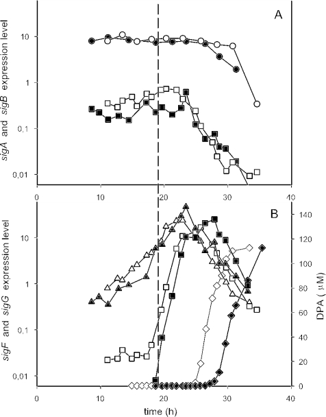
In both media tested, 99% of the cells produced a phase-bright spore and the concentrations of DPA reached about 115 μM, after which the oxygen consumption of the cultures decreased dramatically (data not shown). DPA production and phase brightening of the forespores are indicative of the final phases of sporulation (16). Notably, DPA production and the release of the spores from the mother cells started much earlier after cultivation in YLLG than in YLHG. It is generally assumed that sporulation is a response to starvation (45). In our experiments, however, significant amounts of carbon (d-lactate and glutamate) and nitrogen (NH4+ and glutamate) were present when the cells started to sporulate. Even after sporulation was completed, a significant amount of these C and N sources remained. Nutrient limitation therefore seems an unlikely trigger for sporulation under the conditions we employed. From B. subtilis, it is known that in addition to starvation, quorum sensing can be involved in the initiation of sporulation (46). In silico analysis of the B. cereus ATCC 14579 genome with the ERGO genome and discovery system (36) revealed the presence of the gram-positive quorum-sensing and competence II pathway which is also present in B. subtilis. Thus, although additional experiments in which cell density is carefully manipulated are needed to provide definite evidence, it is tempting to speculate that in our experiments, cell density rather than nutrient limitation may have been the factor that triggered the cells to sporulate. Because extra glutamate did not result in extra biomass, the cell densities in YLHG and YLLG were similar at all times. Indeed, sporulation as judged by the rise in sigF and sigG transcription started at the same moment under both conditions (Fig. 3) (see below).
FIG. 3.
Sigma factor expression levels (calculated as described in the text) of B. cereus bacteria grown and sporulated in YLHG (closed symbols) and YLLG (open symbols). (A) Expression levels of sigA (circles) and sigB (squares). (B) Expression levels of sporulation sigma factors sigF (triangles) and sigG (squares). Diamonds, DPA concentrations. The dashed line indicates the moment of upregulation of sigG transcription.
Gene expression.
The differences we found in the nitrogen metabolism and timing of DPA formation prompted us to check for differences in the genetic programs of sporulation in YLLG and YLHG. Therefore, we measured the expression of genes for four key sigma factors involved in stress response, growth, and sporulation: sigB, sigA, and sigF and sigG, respectively (Fig. 3). The product of sigA, sigma factor σA, is the primary sigma factor. It regulates macromolecular synthesis, plays a key role in the housekeeping functions of the cell (22), and is expressed by B. cereus at a high level throughout growth and sporulation (14); therefore, we used sigA expression as a reference and a positive control. During cultivation in YLLG and YLHG, sigB transcription levels were high compared to the levels of transcription during growth with glucose (14) and showed no clear peak upon entry into the stationary phase (Fig. 3A). It was reported that sigB transcription is increased upon entry in stationary phase (14, 22) and when the cell encounters stress (50), directly affecting cell and carbon metabolisms (40). Our present findings indicate that B. cereus experiences more stress when grown in a medium with lactate instead of glucose and that the sigB peak observed before (14) was caused by a shift from glucose metabolism to other carbon sources rather than by changes in growth rate upon entry into the stationary phase.
During the cultivations in YLLG and YLHG, the expression patterns of sigF and sigG (Fig. 3B) were comparable to those on glucose (14) in that we observed a high peak of sigF transcription, followed by a sigG peak. However, with lactate as a carbon source, sigF expression showed a sharp peak, while during sporulation with glucose, we observed a broad peak (14). The product of sigF, sporulation sigma factor σF, is produced in the predivisional cell (37) well before the first irreversible step of sporulation (15). After septation, when the cells have become committed to sporulate, σF is released from its anti-sigma factor to become active in the forespore only (37). The gene encoding σG, sigG, is under the control of σF and expressed in the forespore compartment just after the appearance of the sporulation septum (15). Thus, an increase of sigG transcription indicates σF activity. σG plays a role in the final stages of sporulation, regulating the synthesis of SASP and Ger proteins (2, 22, 24). Interestingly, the times at which the maximum sigF and sigG expressions occurred were almost the same under both conditions, with only a slight delay of 30 min for the YLHG cultivation. This suggested that there was no considerable difference in the programming of sporulation, judging by the temporal expression of the key sigma factors we measured. Nevertheless, B. cereus started to produce DPA and phase-bright spores in YLLG more than 3 h earlier than in YLHG, indicating that the length of the sporulation process was increased by glutamate. In contrast, Kennedy et al. (29) reported that lower glutamate concentrations led to a longer sporulation time. These authors used different media and a different strain, however, which may explain this difference. DPA synthesis is regulated by σK (16), a sporulation-specific sigma factor expressed by the mother cell compartment in response to σG activity in the forespore during the final stages of sporulation (37). It is possible that although sigG expressions are parallel in YLLG and YLHG, the actual σG activity in YLHG is delayed, or that σG activity is also parallel but σK activity in the mother cell is delayed. Either way, our findings suggest that there is a certain degree of plasticity in the tightly regulated sporulation program.
Spore properties.
Important spore properties are germinability and heat resistance. Therefore, we determined the resistance of the spores from YLHG and YLLG to 95°C, and their germination response to two well-known germinants for B. cereus: l-alanine and inosine. Spores from YLHG were more resistant to 95°C than those from YLLG (Table 2). Because heat resistance has been associated with the core water content (4, 38), we measured the core density, which is linearly correlated with the water content (38). The whole-spore density and the DPA content of spores from YLLG were rather similar to those of spores from YLHG, but the core density was somewhat higher (Table 2). This was in contrast to our expectations, because core density is generally believed to be positively correlated with heat resistance. Indeed, as was stated by Atrih and Foster (1), complex cooperative phenomena are involved in the assembly of spores, and a multicomponent system is the basis for spore heat resistance.
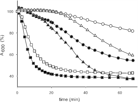
We measured the germinability of the spores without a prior heat activation step, in order to prevent any possible interference by compounds released from the spores themselves (39). Furthermore, spores from YLLG tended to germinate spontaneously after a heat shock for 15 min at 75°C, which reflects their heat sensitivity. Clear differences in germination speed and extent between spores from YLHG and YLLG were observed, as can be seen in Fig. 4. The response to l-alanine in spores from YLLG was severely limited, while these spores responded faster to inosine but much slower still than spores from YLHG. The most powerful germinant known for B. cereus is a combination of l-alanine and inosine (13). Although the difference was less pronounced, even with a combination of l-alanine and inosine, spores from YLHG germinated quicker and more completely than spores from YLLG. Thus, with every germinant tested, spores from YLHG germinated faster and to a greater extent than spores from YLLG. It is known that spores from different media may have different responses to l-alanine because of differences in the alanine racemase content (A. Moir, personal communication), an enzyme that is present in the exosporium of B. cereus (49) and converts the germinant l-alanine into the germination inhibitor d-alanine. Spores produced from YLLG may have higher alanine racemase contents than those from YLHG. In addition to containing alanine racemase, the exosporium of B. cereus contains nucleoside hydrolase, an enzyme that could inhibit inosine-induced germination (49). The presence of higher levels of nucleoside hydrolase in spores from YLLG could therefore explain the differences in inosine-induced germination that we observed. We tried germination in the presence of d-cycloserine, a known alanine racemase inhibitor (6), but this resulted in an overall lower response in all cases. Experiments with other alanine racemase inhibitors (e.g., O-carbamyl-d-serine; 28) and nucleoside hydrolase inhibitors could bring more clarity to those issues.
FIG. 4.
Germination measured by the decrease in A600 of B. cereus spores from YLHG (closed symbols) and YLLG (open symbols). A600 at time zero was 0.8 to 1.0 in all cases; curves are averages from four independent experiments. The following germinants were used: l-alanine, 10 mM (circles); inosine, 5 mM (triangles); and inosine in combination with l-alanine, 0.5 mM (each) (squares).
Clearly, the glutamate concentration affected the sporulation process and spore properties. Indeed, glutamate has been reported to have a large impact on sporulation as well as spore properties of bacilli. In Bacillus thuringiensis, a close relative of B. cereus (21), glutamate appears to regulate TCA cycle activity as well as influence spore heat resistance and DPA content (35). B. cereus has been reported to metabolize glutamate primarily as an energy source during the time of sporogenesis (10). Several studies have attempted to determine the fate of the carbon skeleton of glutamate, and it was concluded that glutamate used during sporulation is respired almost completely to CO2, largely via the TCA cycle (10, 35). Our results confirm that glutamate is used largely during sporulation when present in a high concentration, but an interesting outcome is that glutamate metabolism during sporulation does not result in an increase of NH4+ in the medium. This implies that the glutamate served as an nitrogen donor during the sporulation process, not only as a source of energy and reducing power, as was stated by Charba and Nakata (10). Indeed, in YLLG, NH4+ was taken up by the cells during sporulation, but d-lactate, which can serve as a source of reducing potential and energy, partly remained in the supernatant.
In conclusion, we have found that B. cereus is able to form spores on a chemically defined medium without glucose but with lactate as a main carbon source. Sporulation was not induced by nutrient limitation. Glutamate delayed the final stages of sporulation but not the initiation. Clearly, the glutamate concentration influences such key spore properties as heat resistance and germination. Our defined approach to sporulation and spore properties is highly suitable for gene expression analysis employing DNA microarrays, which we are developing at this moment. Future experiments coupling defined sporulation conditions with full transcriptome analysis and spore properties will allow us to carefully dissect the molecular basis of such important spore properties as heat resistance and germination.
REFERENCES
- 1.Atrih, A., and S. J. Foster. 2001. Analysis of the role of bacterial endospore cortex structure in resistance properties and demonstration of its conservation amongst species. J. Appl. Microbiol. 91:364-372. [DOI] [PubMed] [Google Scholar]
- 2.Barlass, P. J., C. W. Houston, M. O. Clements, and A. Moir. 2002. Germination of Bacillus cereus spores in response to l-alanine and to inosine: the roles of gerL and gerQ operons. Microbiology 148:2089-2095. [DOI] [PubMed] [Google Scholar]
- 3.Barnett, T. A., and J. H. Hageman. 1983. Characterization of a brown pigment from Bacillus subtilis cultures. Can. J. Microbiol. 29:309-315. [Google Scholar]
- 4.Beaman, T. C., T. Koshikawa, H. S. Pankratz, and P. Gerhardt. 1984. Dehydration partitioned within core protoplast accounts for heat resistance of bacterial spores. FEMS Microbiol. Lett. 24:47-51. [Google Scholar]
- 5.Bidingmeyer, B. A., S. A. Cohen, and T. L. Tarvin. 1984. Rapid analysis of amino acids using pre-column derivatization. J. Chromatogr. 336:93-104. [DOI] [PubMed] [Google Scholar]
- 6.Bron, P. A., S. M. Hoffer, I. I. van Swam, W. M. De Vos, and M. Kleerebezem. 2004. Selection and characterization of conditionally active promoters in Lactobacillus plantarum, using alanine racemase as a promoter probe. Appl. Environ. Microbiol. 70:310-317. [DOI] [PMC free article] [PubMed] [Google Scholar]
- 7.Buono, F., R. Testa, and D. G. Lundgren. 1966. Physiology of growth and sporulation in Bacillus cereus. I. Effect of glutamic and other amino acids. J. Bacteriol. 91:2291-2299. [DOI] [PMC free article] [PubMed] [Google Scholar]
- 8.Carlin, F., H. Girardin, M. W. Peck, S. C. Stringer, G. C. Barker, A. Martinez, A. Fernandez, P. Fernandez, W. M. Waites, S. Movahedi, F. van Leusden, M. Nauta, R. Moezelaar, M. D. Torre, and S. Litman. 2000. Research on factors allowing a risk assessment of spore-forming pathogenic bacteria in cooked chilled foods containing vegetables: a FAIR collaborative project. Int. J. Food Microbiol. 60:117-135. [DOI] [PubMed] [Google Scholar]
- 9.Cazemier, A. E., S. F. Wagenaars, and P. F. ter Steeg. 2001. Effect of sporulation and recovery medium on the heat resistance and amount of injury of spores from spoilage bacilli. J. Appl. Microbiol. 90:761-770. [DOI] [PubMed] [Google Scholar]
- 10.Charba, J. F., and H. M. Nakata. 1977. Role of glutamate in the sporogenesis of Bacillus cereus. J. Bacteriol. 130:242-248. [DOI] [PMC free article] [PubMed] [Google Scholar]
- 11.Choma, C., H. Clavel, H. Dominguez, N. Razafindramboa, H. Soumille, C. Nguyen-The, and P. Schmitt. 2000. Effect of temperature on growth characteristics of Bacillus cereus TZ415. Int. J. Food Microbiol. 55:73-77. [DOI] [PubMed] [Google Scholar]
- 12.Christiansson, A., J. Bertilsson, and B. Svensson. 1999. Bacillus cereus spores in raw milk: factors affecting the contamination of milk during the grazing period. J. Dairy Sci. 82:305-314. [DOI] [PubMed] [Google Scholar]
- 13.Clements, M. O., and A. Moir. 1998. Role of the gerI operon of Bacillus cereus 569 in the response of spores to germinants. J. Bacteriol. 180:6729-6735. [DOI] [PMC free article] [PubMed] [Google Scholar]
- 14.de Vries, Y. P., L. M. Hornstra, W. M. de Vos, and T. Abee. 2004. Growth and sporulation of Bacillus cereus ATCC 14579 under defined conditions: temporal expression of genes for key sigma factors. Appl. Environ. Microbiol. 70:2514-2519. [DOI] [PMC free article] [PubMed] [Google Scholar]
- 15.Driks, A. 1999. Bacillus subtilis spore coat. Microbiol. Mol. Biol. Rev. 63:1-20. [DOI] [PMC free article] [PubMed] [Google Scholar]
- 16.Errington, J. 1993. Bacillus subtilis sporulation: regulation of gene expression and control of morphogenesis. Microbiol. Rev. 57:1-33. [DOI] [PMC free article] [PubMed] [Google Scholar]
- 17.Evans, R. I., N. J. Russell, G. W. Gould, and P. J. McClure. 1997. The germinability of spores of a psychrotolerant, non-proteolytic strain of Clostridium botulinum is influenced by their formation and storage temperature. J. Appl. Microbiol. 83:273-280. [DOI] [PubMed] [Google Scholar]
- 18.Gonzalez, I., M. Lopez, S. Matrinez, A. Bernardo, and J. Gonzalez. 1999. Thermal inactivation of Bacillus cereus spores formed at different temperatures. Int. J. Food Microbiol. 51:81-84. [DOI] [PubMed] [Google Scholar]
- 19.Granum, P. E. 2001. Bacillus cereus, p. 373-381. In M. P. Doyle, L. R. Beuchat, and T. J. Montville (ed.), Food microbiology: fundamentals and frontiers, 2nd ed. ASM Press, Washington, D.C.
- 20.Guinebretiere, M. H., H. Girardin, C. Dargaignaratz, F. Carlin, and C. Nguyen-The. 2003. Contamination flows of Bacillus cereus and spore-forming aerobic bacteria in a cooked, pasteurized and chilled zucchini puree processing line. Int. J. Food Microbiol. 82:223-232. [DOI] [PubMed] [Google Scholar]
- 21.Helgason, E., O. A. Okstad, D. A. Caugant, H. A. Johansen, A. Fouet, M. Mock, I. Hegna, and Kolsto. 2000. Bacillus anthracis, Bacillus cereus, and Bacillus thuringiensis—one species on the basis of genetic evidence. Appl. Environ. Microbiol. 66:2627-2630. [DOI] [PMC free article] [PubMed] [Google Scholar]
- 22.Helmann, J. D., and C. P. Moran, Jr. 2002. RNA polymerase and sigma factors, p. 289-313. In A. L. Sonenshein, J. A. Hoch, and R. Losick (ed.), Bacillus subtilis and its closest relatives: from genes to cells. ASM Press, Washington, D.C.
- 23.Hindle, A. A., and E. A. H. Hall. 1999. Dipicolinic acid (DPA) assay revisited and appraised for spore detection. Analyst 124:1599-1604. [DOI] [PubMed] [Google Scholar]
- 24.Hornstra, L. M., Y. P. de Vries, T. Abee, and W. M. de Vos. 2004. gerR, a novel ger operon involved in l-alanine- and inosine-initiated germination of Bacillus cereus ATCC 14579. Appl. Environ. Microbiol. 71:774-781. [DOI] [PMC free article] [PubMed] [Google Scholar]
- 25.Ireton, K., S. Jin, A. D. Grossman, and A. L. Sonenshein. 1995. Krebs cycle function is required for activation of the Spo0A transcription factor in Bacillus subtilis. Proc. Natl. Acad. Sci. USA 92:2845-2849. [DOI] [PMC free article] [PubMed] [Google Scholar]
- 26.Ivanova, N., A. Sorokin, I. Anderson, N. Galleron, B. Candelon, V. Kapatral, A. Bhattacharyya, G. Reznik, N. Mikhailova, A. Lapidus, L. Chu, M. Mazur, E. Goltsman, N. Larsen, M. D'Souza, T. Walunas, Y. Grechkin, G. Pusch, R. Haselkorn, M. Fonstein, S. D. Ehrlich, R. Overbeek, and N. Kyrpides. 2003. Genome sequence of Bacillus cereus and comparative analysis with Bacillus anthracis. Nature 423:87-91. [DOI] [PubMed] [Google Scholar]
- 27.Janssen, F. W., A. J. Lund, and L. E. Anderson. 1958. Colorimetric assay for dipicolinic acid in bacterial spores. Science 127:26-27. [DOI] [PubMed] [Google Scholar]
- 28.Jones, A., and G. W. Gould. 1968. Stimulation of germination of bacterial spores by analogues of d-alanine. J. Gen. Microbiol. 53:383-394. [DOI] [PubMed] [Google Scholar]
- 29.Kennedy, R. S., F. J. Malveaux, and J. J. Cooney. 1971. Effects of glutamic acid on sporulation of Bacillus cereus and on spore properties. Can. J. Microbiol. 17:511-519. [DOI] [PubMed] [Google Scholar]
- 30.Lindsay, J. A., T. C. Beaman, and P. Gerhardt. 1985. Protoplast water content of bacterial spores determined by buoyant density sedimentation. J. Bacteriol. 163:735-737. [DOI] [PMC free article] [PubMed] [Google Scholar]
- 31.Lund, T., M. L. De Buyser, and P. E. Granum. 2000. A new cytotoxin from Bacillus cereus that may cause necrotic enteritis. Mol. Microbiol. 38:254-261. [DOI] [PubMed] [Google Scholar]
- 32.Mazas, M., I. Gonzalez, M. Lopez, and R. Martin. 1995. Effects of sporulation media and strain on thermal resistance of Bacillus cereus spores. Int. J. Food Sci. Technol. 30:71-78. [Google Scholar]
- 33.Melly, E., P. C. Genest, M. E. Gilmore, S. Little, D. L. Popham, A. Driks, and P. Setlow. 2002. Analysis of the properties of spores of Bacillus subtilis prepared at different temperatures. J. Appl. Microbiol. 92:1105-1115. [DOI] [PubMed] [Google Scholar]
- 34.Nauta, M. J., S. Litman, G. C. Barker, and F. Carlin. 2003. A retail and consumer phase model for exposure assessment of Bacillus cereus. Int. J. Food Microbiol. 83:205-218. [DOI] [PubMed] [Google Scholar]
- 35.Nickerson, K. W., J. De Pinto, and L. A. Bulla, Jr. 1974. Sporulation of Bacillus thuringiensis without concurrent derepression of the tricarboxylic acid cycle. J. Bacteriol. 117:321-323. [DOI] [PMC free article] [PubMed] [Google Scholar]
- 36.Overbeek, R., N. Larsen, T. Walunas, M. D'Souza, G. Pusch, E. Selkov, Jr., K. Liolios, V. Joukov, D. Kaznadzey, I. Anderson, A. Bhattacharyya, H. Burd, W. Gardner, P. Hanke, V. Kapatral, N. Mikhailova, O. Vasieva, A. Osterman, V. Vonstein, M. Fonstein, N. Ivanova, and N. Kyrpides. 2003. The ERGO genome analysis and discovery system. Nucleic Acids Res. 31:164-171. [DOI] [PMC free article] [PubMed] [Google Scholar]
- 37.Piggot, P. J., and R. Losick. 2002. Sporulation genes and intercompartmental regulation, p. 483-519. In A. L. Sonenshein, J. A. Hoch, and R. Losick (ed.), Bacillus subtilis and its closest relatives: from genes to cells. ASM Press, Washington, D.C.
- 38.Popham, D. L., S. Sengupta, and P. Setlow. 1995. Heat, hydrogen peroxide, and UV resistance of Bacillus subtilis spores with increased core water content and with or without major DNA-binding proteins. Appl. Environ. Microbiol. 61:3633-3638. [DOI] [PMC free article] [PubMed] [Google Scholar]
- 39.Preston, R. A., and H. A. Douthit. 1984. Stimulation of germination of unactivated Bacillus cereus spores by ammonia. J. Gen. Microbiol. 130:1041-1050. [DOI] [PubMed] [Google Scholar]
- 40.Price, C. W. 2002. General stress response, p. 369-385. In A. L. Sonenshein, J. A. Hoch, and R. Losick (ed.), Bacillus subtilis and its closest relatives: from genes to cells. ASM Press, Washington, D.C.
- 41.Raso, J., G. Barbosa-Canovas, and B. G. Swanson. 1998. Sporulation temperature affects initiation of germination and inactivation by high hydrostatic pressure of Bacillus cereus. J. Appl. Microbiol. 85:17-24. [DOI] [PubMed] [Google Scholar]
- 42.Raso, J., M. M. Gongora-Nieto, G. V. Barbosa-Canovas, and B. G. Swanson. 1998. Influence of several environmental factors on the initiation of germination and inactivation of Bacillus cereus by high hydrostatic pressure. Int. J. Food Microbiol. 44:125-132. [DOI] [PubMed] [Google Scholar]
- 43.Rickwood, D., T. Ford, and J. Graham. 1982. Nycodenz: a new nonionic iodinated gradient medium. Anal. Biochem. 123:23-31. [DOI] [PubMed] [Google Scholar]
- 44.Sarrías, J. A., M. Valero, and M. C. Salmerón. 2002. Enumeration, isolation and characterization of Bacillus cereus strains from Spanish raw rice. Food Microbiol. 19:589-595. [Google Scholar]
- 45.Setlow, P. 2003. Spore germination. Curr. Opin. Microbiol. 6:1-7. [DOI] [PubMed] [Google Scholar]
- 46.Sonenshein, A. L. 2000. Control of sporulation initiation in Bacillus subtilis. Curr. Opin. Microbiol. 3:561-566. [DOI] [PubMed] [Google Scholar]
- 47.te Giffel, M. C., A. Wagendorp, A. Herrewegh, and F. Driehuis. 2002. Bac-terial spores in silage and raw milk. Antonie Leeuwenhoek 81:625-630. [DOI] [PubMed] [Google Scholar]
- 48.Tisa, L. S., T. Koshikawa, and P. Gerhardt. 1982. Wet and dry bacterial spore densities determined by buoyant sedimentation. Appl. Environ. Microbiol. 43:1307-1310. [DOI] [PMC free article] [PubMed] [Google Scholar]
- 49.Todd, S. J., A. J. Moir, M. J. Johnson, and A. Moir. 2003. Genes of Bacillus cereus and Bacillus anthracis encoding proteins of the exosporium. J. Bacteriol. 185:3373-3378. [DOI] [PMC free article] [PubMed] [Google Scholar]
- 50.van Schaik, W., M. H. Tempelaars, J. A. Wouters, W. M. de Vos, and T. Abee. 2004. The alternative sigma factor σB of Bacillus cereus: response to stress and role in heat adaptation. J. Bacteriol. 186:316-325. [DOI] [PMC free article] [PubMed] [Google Scholar]
- 51.Vary, J. C. 1973. Germination of Bacillus megaterium spores after various extraction procedures. J. Bacteriol. 116:797-802. [DOI] [PMC free article] [PubMed] [Google Scholar]


