Abstract
Replicative oriC plasmids were recently developed for several mollicutes, including three Mycoplasma species belonging to the mycoides cluster that are responsible for bovine and caprine diseases: Mycoplasma mycoides subsp. mycoides small-colony type, Mycoplasma mycoides subsp. mycoides large-colony type, and Mycoplasma capricolum subsp. capricolum. In this study, oriC plasmids were evaluated in M. capricolum subsp. capricolum as genetic tools for (i) expression of heterologous proteins and (ii) gene inactivation by homologous recombination. The reporter gene lacZ, encoding β-galactosidase, and the gene encoding spiralin, an abundant surface lipoprotein of the related mollicute Spiroplasma citri, were successfully expressed. Functional Escherichia coli β-galactosidase was detected in transformed Mycoplasma capricolum subsp. capricolum cells despite noticeable codon usage differences. The expression of spiralin in M. capricolum subsp. capricolum was assessed by colony and Western blotting. Accessibility of this protein at the cell surface and its partition into the Triton X-114 detergent phase suggest a correct maturation of the spiralin precursor. The expression of a heterologous lipoprotein in a mycoplasma raises potentially interesting applications, e.g., the use of these bacteria as live vaccines. Targeted inactivation of gene lppA encoding lipoprotein A was achieved in M. capricolum subsp. capricolum with plasmids harboring a replication origin derived from S. citri. Our results suggest that the selection of the infrequent events of homologous recombination could be enhanced by the use of oriC plasmids derived from related mollicute species. Mycoplasma gene inactivation opens the way to functional genomics in a group of bacteria for which a large wealth of genome data are already available and steadily growing.
Mycoplasmas are small bacteria from the class Mollicutes that lack a cell wall and are characterized by a genome with a low percent G+C (for a review, see reference 27). In contrast to the large wealth of data extracted from the analysis of their genome sequences (2), there is still a general lack of efficient genetic tools for the functional genomics of these bacteria. Transposon-based strategies have been used to generate random insertion mutants in a few mycoplasma species, but the attempts to develop cloning vectors from endogenous plasmids and viruses have encountered limited success (for a review, see reference 28). Recently, oriC-based replicative plasmids were developed for three mycoplasmas that cause economically important diseases in ruminants and belong to the mycoides cluster: Mycoplasma mycoides subsp. mycoides large-colony type, Mycoplasma mycoides subsp. mycoides small-colony type, and Mycoplasma capricolum subsp. capricolum (20). As previously shown for Mycoplasma pulmonis (6) and for another mollicute, Spiroplasma citri (38), the oriC plasmids that harbor the chromosomal dnaA gene and the adjacent DnaA box sequences were efficiently replicated in their respective hosts. Moreover, by heterologous transformation of these mollicutes with the different oriC plasmids, it was shown that the large- and small-colony forms of M. mycoides subsp. mycoides which are closely related, could tolerate plasmids with each other's oriC sequences. More strikingly, M. capricolum subsp. capricolum could be transformed by oriC plasmids from the three species belonging to the mycoides cluster but also by the S. citri oriC plasmid (20).
The aim of this study was to evaluate the usefulness of these vectors as genetic tools. Because of its relatively fast growth and its ability to replicate a wide spectrum of oriC plasmids, M. capricolum subsp. capricolum was chosen in this work. Two types of applications were investigated. First, the M. capricolum subsp. capricolum oriC plasmid was used as a genetic vector for expressing heterologous proteins, which is indeed required for functional genomics as it allows the complementation of mutants or the study of gene regulation via a reporter gene. Second, targeted gene inactivation was attempted with M. capricolum subsp. capricolum. Production of mutants by gene disruption is a crucial step in the understanding of protein function and involvement in complex processes such as pathogenesis. In mollicutes, the inactivation of target genes through homologous recombination has been described for Acholeplasma laidlawii (12), Mycoplasma gallisepticum (5), and Mycoplasma genitalium (7, 8). In these cases, the plasmid vector used could not replicate in the host, and drug-resistant transformants could only be obtained via an integration of the plasmid into the chromosome. In S. citri (9, 16, 21) and M. pulmonis (6), for which no gene inactivation could ever be obtained with nonreplicating plasmids, oriC plasmids have been successfully used to drive homologous recombination events. To develop tools for genetic investigations in M. capricolum subsp. capricolum, oriC plasmids were evaluated as genetic vectors for gene targeting experiments.
MATERIALS AND METHODS
Bacterial strains and culture conditions.
Mycoplasma capricolum subsp. capricolum California KidT strain (referred to here as M. capricolum subsp. capricolum) was used in this study. This bacterium was grown at 37°C in modified Hayflick medium (14) without thallium acetate and supplemented with BBL IsoVitalex Enrichment (Becton Dickinson, Sparks, MD). Spiroplasma citri R8A2 strain (ATCC 27556) was grown in SP-4 medium at 32°C (35). For cloning procedures and propagation of plasmids, Escherichia coli strain DH10B [F′-mcrA Δ(mrr-hsdRMS-mcrBC) Φ80dlacZΔM15 ΔlacX74 deoR recA1 endA1 araD139 Δ(ara leu)7697 galU galKl− rpsL nupG] (Stratagene) was used. E. coli cells were grown in LB broth at 37°C. β-Galactosidase activity was detected by plating mycoplasmas on solid medium spread with 200 μl of 5-bromo-4-chloro-3-indolyl-β-d-galactopyranoside (X-Gal) at a concentration of 4 mg/ml.
Plasmid construction.
The pMCO3 plasmid contains the chromosomal oriC region of M. capricolum subsp. capricolum and the selection marker tet(M) under the control of the spiralin promoter (20). The lacZ gene from E. coli was first amplified by PCR from the pβgal-Basic (BD Biosciences Clontech) using the primers lacZF (5′ AGGCAGATCTATGGACACCAGCAAGGAGCTG 3′) and lacZR (5′ TCGAAGATCTTGGGGTGTTGTAACAATATCG 3′). The amplification product (3,206 bp) was cleaved by BglII at the sites included in the primer sequence (underlined nucleotides) and cloned at the BglII site located downstream of the spiralin promoter of pSRT2 (21) to generate the pWZ1 plasmid (W. Maccheroni and J. Renaudin, unpublished data). An expression cassette containing the lacZ gene under control of the spiralin promoter was then amplified using the primers PS4237E1 (5′ GGAGAATTCGCGCAATTTATTTGG 3′; the EcoRI site is underlined) and LacZ1 (5′ TCGAGAATTCTGGGGTGTTGTAACAATATC 3′). The amplification product was cloned at the EcoRI site of the pMCO3 to generate pPSlacZ. The S. citri spiralin gene, under control of its own promoter, was amplified from S. citri genomic DNA using PS1 (5′ GCGATATCCGATCGGCAATTTATTTGGAAAATC 3′) and PS2 (5′ GCGATATCCGATCGAGTTGATATTCTAAGATTG 3′) as primers and cloned in the PCR cloning vector TOPO 2.1 (Invitrogen). The insert was isolated after cleavage by EcoRI and cloned at the EcoRI site of pMCO3 to obtain pSPI. An internal fragment of the lppA gene was amplified from M. capricolum subsp. capricolum genomic DNA using the oligonucleotides MCLA1 (5′ GATCGAATTCGGGCCCCCATAAAACCTGAAGATTC 3′; the EcoRI site is underlined) and MCLA2 (5′ GATCGAATTCCCGCGGGTAATTCTAGTATGGAAAGG 3′). After cleavage by EcoRI, the amplification product was cloned into the EcoRI sites of pMCO3 and pSD4 to generate the pMCO6 and the pSD6 constructs, respectively.
Transformation of M. capricolum subsp. capricolum.
Polyethylene glycol-mediated transformation of M. capricolum subsp. capricolum was performed as described previously (11). Ten micrograms of plasmid DNA was used for each transformation. After being plated on selective solid medium containing 5 μg/ml of tetracycline, the cultures were kept at 37°C and examined for colony development from the third day of incubation. Transformants were then picked up and subcultured in Hayflick broth medium supplemented with 20 μg of tetracycline/ml. Cloning of M. capricolum subsp. capricolum transformed with pSPI or pPSlacZ was achieved by three cycles of picking colonies obtained after plating cultures submitted to filtration using 0.45-μm-pore-size filters to eliminate lumps of cells (30).
DNA isolation and Southern blot hybridization.
Mycoplasma genomic DNA was prepared from 10-ml cultures using the Wizard genomic DNA purification kit (Promega). For Southern blot hybridization, 1.5 μg of genomic DNA or 15 ng of plasmid DNA was digested by the appropriate restriction enzyme and submitted to electrophoresis in a 0.8% agarose gel. After alkali transfer of the DNA fragments to a positively charged nylon membrane (Nytran Super Charge; Schleicher and Schuell), hybridization was performed in the presence of 20 ng/ml of digoxigenin-labeled DNA probes. Detection of hybridized probes was achieved using Fab fragments of anti-digoxigenin antibodies coupled to alkaline phosphatase and the fluorescent substrate 2-hydroxy-3-naphthoic acid-2′-phenylanilide phosphate (Roche Molecular Biochemicals). Chemifluorescence was detected by using a high-resolution camera (Fluor-S; Bio-Rad) and Quantity One, a dedicated software for image acquirement (Bio-Rad).
Protein extraction.
Exponentially growing mycoplasma cells were collected by centrifugation (9,500 × g for 15 min; 4°C). The pellet was dispersed in 1× phosphate-buffered saline (PBS 1×) (13.7 mM NaCl, 0.27 mM KCl, 0.15 mM KH2PO4, 0.8 mM Na2HPO4, 12 H2O [pH 7.4]) and washed three times in the same buffer. Cells were then dispersed in lysis solution (PBS 1×, 0.5% sodium dodecyl sulfate [SDS], and 1 mM Pefabloc [Roche Applied Science]) in the volume required to concentrate the sample 100 times. Lysis was facilitated and viscosity was reduced by a 15-s sonication using a microprobe (Vibra cell; Branson). The sample was then heated at 60°C for 10 min and centrifuged to sediment cellular debris (18,000 × g for 5 min; room temperature). The protein concentration of the supernatant was measured by the bicinchoninic acid method (31) with bovine serum albumin as a standard. To isolate a membrane protein-enriched fraction, mycoplasma cells were submitted to an extraction with Triton X-114 (adapted from a previously published procedure) (4). Briefly, after centrifugation of mycoplasma cultures, the cells were washed three times in PBS 1× and resuspended in 100 μl of Triton X-114 diluted with 900 μl of Tris/NaCl buffer (10 mM Tris-HCl [pH 7.4], 154 mM NaCl). After a 15-s sonication, the sample was mixed by rotary agitation at 4°C for 40 min. The separation of the detergent and aqueous phases was obtained by incubation of the mixture for 10 min at 37°C and centrifugation (14,000 × g for 5 min; room temperature). The aqueous and detergent phases were washed three times with 10% Triton X-114 and with Tris/NaCl buffer, respectively. The aqueous phase was stored at −20°C. Nine volumes of methanol was added to the detergent phase, and the sample was then incubated overnight at −80°C to precipitate the proteins. After centrifugation (14,000 × g for 10 min; 4°C), the pellet of proteins was washed once in 70% ethanol and then resuspended in 100 μl of PBS 1×, 0.5% SDS, and 1 mM Pefabloc (Roche Applied Science).
Protein electrophoresis and immunoblotting.
Proteins were separated by electrophoresis as previously described (19) in 10% or 12% polyacrylamide gels. For staining with Coomassie brilliant blue (R250; Bio-Rad), 60 μg of total cell protein was loaded into each well. For spiralin immunodetection, 0.01 μg and 1.5 μg of total protein from S. citri and M. capricolum subsp. capricolum/pSPI were loaded, respectively. For both aqueous and Triton X-114 phases from M. capricolum subsp. capricolum, 0.15 μg of protein were loaded. Proteins were electroblotted onto a nitrocellulose membrane at 10 V for 1.5 h in a semidry transfer unit (Amersham Biosciences) using a Tris-glycine transfer buffer (25 mM Tris-HCl, 192 mM glycine, 10% methanol, 0.1% SDS, pH 8.3) (34). After saturation in TBS buffer (25 mM Tris-HCl, 125 mM NaCl, pH 8.0) supplemented with 5% defatted dry milk and 0.1% Tween 20, the membrane was incubated for 1 h at room temperature with the primary antibodies. Spiralin was detected using a polyclonal, monospecific serum (diluted 1:1,000) obtained after immunization of two rabbits with purified spiralin (3). The lipoprotein LppA was revealed with a polyclonal mouse antiserum (diluted 1:500) specifically directed against M. capricolum subsp. capricolum LppA (23). After being washed three times in saturating buffer, the membrane was incubated with the alkaline phosphatase-conjugated secondary antibody (diluted 1:2,000). Depending on the primary antibodies used, secondary antibodies were either goat anti-rabbit or goat anti-mouse immunoglobulin G (Bio-Rad). Alkaline phosphatase was revealed using Nitro Blue Tetrazolium/5-bromo-4-chloro-3-indolylphosphate (Sigma) as a substrate.
RESULTS
Expression of heterologous proteins in M. capricolum subsp. capricolum. (i) Expression of β-galactosidase.
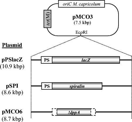
The lacZ gene encoding E. coli β-galactosidase was chosen in a first attempt to express a reporter gene in M. capricolum subsp. capricolum. This gene was cloned under the control of the S. citri spiralin promoter into the pMCO3 plasmid to generate pPSlacZ (Fig. 1). Plasmid pMCO3, which harbors the tet(M) selection marker and the chromosomal origin of replication (oriC) from M. capricolum subsp. capricolum, has been shown to replicate efficiently in its original host (20). M. capricolum subsp. capricolum was transformed with plasmid pPSlacZ. In every polyethylene glycol-mediated transformation assay, plasmid pMCO3 was used as a positive control. After 3 to 5 days of incubation on tetracycline-supplemented medium, colonies were observed for both pPSlacZ and pMCO3 transformation assays. Transformation efficiencies for pPSlacZ (2 × 10⁻⁷ transformant CFU−1 μg−1) and for pMCO3 (9 × 10⁻⁷ transformant CFU−1 μg−1) were similar. A pure culture of a M. capricolum subsp. capricolum/pPSlacZ transformant was obtained by filter cloning (see Materials and Methods). The M. capricolum subsp. capricolum/pPSlacZ transformant was plated on solid medium spread with the chromogenic substrate of β-galactosidase (X-Gal). A deep blue coloration was observed for all colonies, indicating that β-galactosidase was expressed and functional in M. capricolum subsp. capricolum (Fig. S1).
FIG. 1.
Structure of the pMCO3-derived plasmids used in this study. Various inserts were cloned at the EcoRI site of plasmid pMCO3. PS, spiralin gene promoter; lacZ, coding region of E. coli β-galactosidase-encoding gene; ΔlppA, internal region of the lppA gene from M. capricolum subsp. capricolum (1,238 nucleotides [nt]). Truncated regions of the gene (165 nt upstream and 190 nt downstream) are represented by dashed open boxes. Plasmid pSPI contains the spiralin gene under control of its own promoter. Genes are not drawn to scale; plasmid sizes are indicated in brackets.
(ii) Spiralin expression.
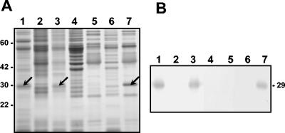
With the aim of expressing a heterologous protein at the cell surface of M. capricolum subsp. capricolum, we chose S. citri spiralin for three reasons. First, this lipoprotein is exposed at the cell surface of spiroplasmas (37). Second, S. citri and M. capricolum subsp. capricolum are members of the same phylogenetic group (17). Third, the spiralin promoter can be used to drive gene expression in M. capricolum subsp. capricolum (20). The spiralin gene driven by its own promoter was cloned at the EcoRI site of pMCO3 (Fig. 1). The resulting plasmid pSPI was amplified in E. coli, and the integrity of the spiralin coding region was checked by sequencing. After transformation of M. capricolum subsp. capricolum with pSPI, cells were spread on tetracycline-supplemented medium and incubated at 37°C. In these experimental conditions, no transformant could be obtained, despite several attempts. Transformation was then reiterated at 32°C, a temperature which also supports the growth of M. capricolum subsp. capricolum. Despite a low transformation efficiency (1 × 10⁻⁹ transformant CFU−1 μg−1 for pSPI compared to 2 × 10⁻⁷ transformant CFU−1 μg−1 for pMCO3), five transformants were obtained on solid medium. Transformants were isolated and grown in liquid medium at 37°C. After being cloned, one of the pSPI transformants was analyzed for spiralin expression and cellular localization. A Triton X-114 extraction was performed for M. capricolum subsp. capricolum and the transformant M. capricolum subsp. capricolum/pSPI. A polypeptide of 29 kDa (a mass close to that of spiralin) (13, 37), specific to the M. capricolum subsp. capricolum/pSPI transformant (Fig. 2A, lane 7), was found among the major proteins of the Triton X-114 phase. The amphiphilicity of this protein was confirmed as it was not found in the aqueous phase (Fig. 2A, lane 6), and its identity was confirmed by immunolabeling using a monospecific polyclonal anti-spiralin serum (Fig. 2B). Moreover, colony-blotting experiments with this transformant with the same anti-spiralin serum indicated spiralin accessibility to antibodies at the M. capricolum subsp. capricolum cell surface (Fig. S2).
FIG. 2.
Expression of the spiralin gene in M. capricolum subsp. capricolum. Total proteins (lane 1, S. citri; lane 2, M. capricolum subsp. capricolum, lane 3, M. capricolum subsp. capricolum/pSPI) and proteins separated by Triton X-114 extraction from M. capricolum subsp. capricolum (lane 4, aqueous phase; lane 5, Triton X-114 phase) and M. capricolum subsp. capricolum/pSPI (lane 6, aqueous phase; lane 7, Triton X-114 phase) were separated by SDS-polyacrylamide gel electrophoresis. (A) Coomassie brilliant blue staining. (B) Immunodetection of the spiralin using a monospecific polyclonal anti-spiralin serum. The position of the spiralin is indicated by black arrows. Molecular masses are indicated in kilodaltons.
Altogether, these results show that spiralin was abundantly expressed in the M. capricolum subsp. capricolum/pSPI transformant and, as expected, exposed at the cell surface.
Targeted gene inactivation in M. capricolum subsp. capricolum. (i) Homologous oriC plasmid as a disruption vector.
To evaluate oriC plasmids as tools for targeted gene inactivation in M. capricolum subsp. capricolum, an internal fragment of the lppA gene (1,241 bp) was cloned at the EcoRI site of plasmid pMCO3, which contains the replication origin from M. capricolum subsp. capricolum. LppA (57 kDa) is a major surface lipoprotein that is found with minor variations in other members of the mycoides cluster (15, 23, 24). The recombinant plasmid pMCO6 (Fig. 1) and the control plasmid pMCO3 were used to transform M. capricolum subsp. capricolum. After plating and 3 days of incubation, tetracycline-resistant transformants were obtained with an equivalent efficiency for both plasmids (2 × 10⁻⁶ transformants CFU−1 μg−1). Ten pMCO6-transformants were subcultured for 15 passages in tetracycline-containing broth medium. Southern blot analysis of the clones using an lppA probe revealed that no integration event occurred in any of the clones even after 15 passages (data not shown): plasmid pMCO6 remained as a free molecule, suggesting that the integration events at the lppA locus were rather rare.
(ii) Heterologous oriC plasmid as a disruption vector.
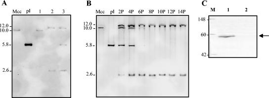
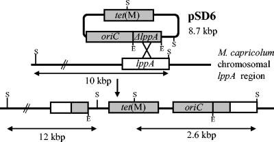
To help in selection of recombinant cells among transformants, the internal fragment of the lppA gene was cloned into plasmid pSD4 that harbors the replication origin from S. citri to generate pSD6. The heterologous pSD4 oriC plasmid was previously shown to transform M. capricolum subsp. capricolum but with a low efficiency, suggesting a reduced fitness (20). Three transformations of M. capricolum subsp. capricolum with 20 μg of pSD6 were performed, and only three tetracycline resistant clones were obtained. After 15 passages in selective medium, the genomic DNAs of these clones were extracted, ScaI digested, and analyzed by Southern blot hybridization with an lppA probe (Fig. 3A). Only the 10-kbp chromosomal copy of lppA was detected for clone 1 (Fig. 4), suggesting that this clone either underwent a deletion of lppA on the plasmid or that it was a spontaneous tetracycline-resistant colony. In clone 2, the lppA probe revealed two bands (2.6 kbp and 12 kbp), indicating that an integration event had occurred into the target gene, lppA. A third clone showed a hybridization pattern as clone 2 but contained a third 5.8-kbp fragment hybridizing, indicating the presence of free plasmid. To determine more precisely the integration process of pSD6 in clone 2 (McapΔlppAcl2), the total genomic DNA of this clone was extracted at passages 2, 4, 6, 8, 10, 12, and 14 and analyzed as described above (Fig. 3B). The presence of 2.6-kbp and 12.0-kbp bands from the second passage indicated that plasmid integration into the target gene had started to occur early, at least in some cells. From passage 6, the bands corresponding to the wild-type chromosomal lppA gene (10 kbp) and to the free plasmid (5.8 kbp) were not detected anymore, suggesting that the cells harboring an integrated plasmid in their genome had been positively selected. To verify the inactivation of the lppA gene in McapΔlppAcl2, total proteins were extracted and probed with a monospecific polyclonal anti-LppA serum (Fig. 3C). A single band corresponding to the predicted 57-kDa LppA was detected for the untransformed control but not for the mutant McapΔlppAcl2, suggesting the lack of LppA. Interestingly, although truncated lppA mRNAs were evidenced by reverse transcription-PCR in agreement with the integration scheme (data not shown), no truncated form of the protein could be immunodetected, suggesting that it was degraded. Colony-blotting experiments with the same serum confirmed the lack of LppA on the cell surface of the mutant (data not shown). This result shows that targeted gene inactivation was obtained with M. capricolum subsp. capricolum by using a heterologous oriC plasmid.
FIG. 3.
lppA gene inactivation in M. capricolum subsp. capricolum using the heterologous oriC plasmid pSD6. (A) Southern blot hybridization between ScaI-digested DNAs extracted from M. capricolum subsp. capricolum (lane Mcc) and from three tetracycline-resistant clones (lanes 1, 2, and 3) obtained after transformation of M. capricolum subsp. capricolum with pSD6. The ΔlppA fragment was used as a probe. Lane pl, plasmid pSD6. (B) Southern blot hybridization of ScaI-digested DNAs extracted from clone 2 at passage 2, 4, 6, 8, 10, 12, and 14. Lane pl, pSD6 plasmid DNA; Mcc, genomic DNA from M. capricolum subsp. capricolum. Sizes are indicated in kilobase pairs. (C) Immunodetection of the lipoprotein LppA in the mutant McapΔlppAcl2. Total proteins (lane 1, M. capricolum subsp. capricolum; lane 2, McapΔlppAcl2) were separated by SDS-polyacrylamide gel electrophoresis. The lipoprotein LppA was revealed with a monospecific polyclonal anti-LppA serum. M, molecular mass marker (in kilodaltons). The position of LppA is indicated by an arrow.
FIG. 4.
Schematic representation of the pSD6 plasmid integration in the lppA gene by homologous recombination. tet(M), tetracycline resistance gene; oriC, replication origin from S. citri; ΔlppA, 1,241-bp internal lppA fragment; E, EcoRI; S, ScaI. Elements are not drawn to scale. The pSD6 ScaI fragment containing the ΔlppA sequence is 5.8 kbp in size.
DISCUSSION
Heterologous protein expression.
In this study, two heterologous proteins were successfully expressed in M. capricolum subsp. capricolum. A first experiment was performed using the reporter gene lacZ from E. coli under control of the promoter of the spiralin gene. Some examples of expression of the lacZ gene in mollicutes have been described for M. pulmonis, Mycoplasma arthritidis (10) and for S. citri (W. Macheroni and J. Renaudin, unpublished results). Although a relatively high concentration of X-Gal was required to obtain a blue coloration of the transformants, our data show that the expression of this reporter gene is also possible in M. capricolum subsp. capricolum, despite many guanosin-and cytosin-rich codons that are rarely found in mycoplasma genomes (27). Previous reports, based on in vitro translation experiments and analysis of available genome sequences, have suggested that the CGG codon (Arg) was unassigned or nonsense in M. capricolum subsp. capricolum (1, 25). On the contrary, other reports (18, 20) showed, as does the present work, that the tet(M) and the lacZ genes which contain three and seven CGG codons, respectively, could be expressed in this mycoplasma. These results indicate that CGG codons do not stop the translation in vivo and suggest that the tRNAArg (ICG), which has been proposed to decode the three other arginine codons (CGU, CGC, and CGA) (1), may also recognize the CGG codon. A similar proposal can been formulated for the Mycoplasma mycoides subsp. mycoides small-colony type, which possesses only 10 CGG codons within all the predicted genes and one tRNAArg (ACG) to decode the four CGN codons (36). Thus, expression of heterologous proteins is possible in mycoplasmas despite significant differences in codon usage.
The spiralin gene, encoding a spiroplasma lipoprotein, was chosen to demonstrate the feasibility of expressing and exposing a heterologous protein at the cell surface of a mycoplasma. To our knowledge, although the Spiroplasma phoeniceum spiralin was previously expressed in S. citri (29), there was no example of heterologous lipoprotein expression in a Mycoplasma species. Problems encountered when M. capricolum subsp. capricolum transformation was performed at 37°C suggest that the expression of spiralin is somewhat deleterious for cell viability, at least initially. However, the transformant obtained at 32°C grows at 37°C and forms normally shaped colonies on solid medium. In S. citri, spiralin is a particularly abundant protein (20 to 30% of the mass of the membrane proteins) (37), suggesting that its expression in M. capricolum subsp. capricolum might lead to a transient perturbation of the cell membrane and that this effect could be reduced by lowering the temperature during the transformation. Similar temperature effects have been described in E. coli; lowering the temperature has been used to reduce the toxic effects observed during the expression of fusion proteins artificially addressed to the membrane (32). Although several vaccines are based on attenuated strains of mycoplasma (26, 33), these bacteria have not yet been used to deliver heterologous protective antigens. Such strategy is already applied with other Bacteria species (22), and the expression of a foreign lipoprotein in a species of the mycoides cluster is promising for the use of mycoplasmas as live vaccine. More specifically, the ability of several mycoplasma species to colonize the respiratory tract of animals makes them attractive to stimulate mucosal responses (22).
Gene targeting with oriC plasmids.
In M. capricolum subsp. capricolum, several attempts to inactivate the lppA gene using nonreplicative vectors have been performed without success (not shown). Moreover, no integration in the targeted lppA gene was observed when a plasmid harboring a complete and homologous oriC was used. In contrast, the use of the heterologous oriC plasmid pSD4 from S. citri as a vector led to the desired mutant. It should be noticed that transformation efficiency with the pSD4-derived plasmid was very low, in accordance with previous results (20). Thus, it seems that reducing the fitness of the plasmid lowers its replication capacity and, consequently, favors the selection of the rare recombinant cells. Alternative strategies based on plasmids harboring reduced homologous oriC have also given interesting results with S. citri (21) and with M. pulmonis (6). In these cases, the reduction of sequence homology of the oriC region limits the background integration events at the chromosomal replication origin.
In conclusion, this work shows that oriC plasmids can be used in M. capricolum subsp. capricolum as vectors for the expression of heterologous cytoplasmic and membrane proteins and for targeted gene inactivation. Considering the growing number of available genome sequences of mollicutes and the importance of these bacteria as pathogenic agents, the development of efficient tools for the functional genomics of these bacteria is a real challenge. From that point of view, the demonstration that oriC plasmids can be used as genetic vectors in a mycoplasma from the mycoides cluster constitutes significant progress, which could also be applied to other mycoplasma species.
Supplementary Material
Acknowledgments
This research was supported by grants from the INRA, the Région Aquitaine, the University Victor Segalen Bordeaux 2, and the CIRAD.
We thank Géraldine Gourgues for technical assistance, Joël Renaudin for helpful discussions, and Walter Maccheroni and Sybille Duret-Nurbel for constructing the plasmids pWZ1 and pSD4, respectively.
Footnotes
Supplemental material for this article may be found at http://aem.asm.org/.
REFERENCES
- 1.Andachi, Y. F., F. Yamao, A. Muto, and S. Osawa. 1989. Codon recognition patterns as deduced from sequences of the complete set of transfer RNA species in Mycoplasma capricolum. J. Mol. Biol. 209:37-54. [DOI] [PubMed] [Google Scholar]
- 2.Barré, A., A. de Daruvar, and A. Blanchard. 2004. MolliGen, a database dedicated to the comparative genomics of mollicutes. Nucleic Acids Res. 32:D307-D310. [DOI] [PMC free article] [PubMed] [Google Scholar]
- 3.Béven, L., and H. Wróblewski. 1997. Effect of natural amphipathic peptides on viability, membrane potential, cell shape and motility of mollicutes. Res. Microbiol. 148:163-175. [DOI] [PubMed] [Google Scholar]
- 4.Bordier, C. 1981. Phase separation of integral membrane proteins in Triton X-114 solution. J. Biol. Chem. 256:1604-1607. [PubMed] [Google Scholar]
- 5.Cao, J., P. A. Kapke, and F. C. Minion. 1994. Transformation of Mycoplasma gallisepticum with Tn916, Tn4001, and integrative plasmid vectors. J. Bacteriol. 176:4459-4462. [DOI] [PMC free article] [PubMed] [Google Scholar]
- 6.Cordova, C. M., C. Lartigue, P. Sirand-Pugnet, J. Renaudin, R. A. Cunha, and A. Blanchard. 2002. Identification of the origin of replication of the Mycoplasma pulmonis chromosome and its use in oriC replicative plasmids. J. Bacteriol. 184:5426-5435. [DOI] [PMC free article] [PubMed] [Google Scholar]
- 7.Dhandayuthapani, S., M. W. Blaylock, C. M. Bebear, W. G. Rasmussen, and J. B. Baseman. 2001. Peptide methionine sulfoxide reductase (MsrA) is a virulence determinant in Mycoplasma genitalium. J. Bacteriol. 183:5645-5650. [DOI] [PMC free article] [PubMed] [Google Scholar]
- 8.Dhandayuthapani, S., W. G. Rasmussen, and J. B. Baseman. 1999. Disruption of gene mg218 of Mycoplasma genitalium through homologous recombination leads to an adherence-deficient phenotype. Proc. Natl. Acad. Sci. USA 96:5227-5232. [DOI] [PMC free article] [PubMed] [Google Scholar]
- 9.Duret, S., J. L. Danet, M. Garnier, and J. Renaudin. 1999. Gene disruption through homologous recombination in Spiroplasma citri: an scm1-disrupted motility mutant is pathogenic. J. Bacteriol. 181:7449-7456. [DOI] [PMC free article] [PubMed] [Google Scholar]
- 10.Dybvig, K., C. T. French, and L. L. Voelker. 2000. Construction and use of derivatives of transposon Tn4001 that function in Mycoplasma pulmonis and Mycoplasma arthritidis. J. Bacteriol. 182:4343-4347. [DOI] [PMC free article] [PubMed] [Google Scholar]
- 11.Dybvig, K., and L. L. Voelker. 1996. Molecular biology of mycoplasmas. Annu. Rev. Microbiol. 50:25-57. [DOI] [PubMed] [Google Scholar]
- 12.Dybvig, K., and A. Woodard. 1992. Construction of recA mutants of Acholeplasma laidlawii by insertional inactivation with a homologous DNA fragment. Plasmid 28:262-266. [DOI] [PubMed] [Google Scholar]
- 13.Foissac, X., C. Saillard, J. Gandar, L. Zreik, and J. M. Bové. 1996. Spiralin polymorphism in strains of Spiroplasma citri is not due to differences in posttranslational palmitoylation. J. Bacteriol. 178:2934-2940. [DOI] [PMC free article] [PubMed] [Google Scholar]
- 14.Freund, E. A. 1983. Culture media for classic mycoplasmas, p. 127. In S. Razin and J. G. Tully (ed.), Methods in mycoplasmology, vol. 1. Academic Press, San Diego, Calif. [Google Scholar]
- 15.Frey, J., X. Cheng, M. P. Monnerat, E. M. Abdo, M. Krawinkler, G. Bolske, and J. Nicolet. 1998. Genetic and serological analysis of the immunogenic 67-kDa lipoprotein of Mycoplasma sp. bovine group 7. Res. Microbiol. 149:55-64. [DOI] [PubMed] [Google Scholar]
- 16.Gaurivaud, P., F. Laigret, E. Verdin, M. Garnier, and J. M. Bové. 2000. Fructose operon mutants of Spiroplasma citri. Microbiology 146:2229-2236. [DOI] [PubMed] [Google Scholar]
- 17.Johansson, K.-E., and B. Pettersson. 2002. Taxonomy of Mollicutes, p. 1-30. In S. Razin and R. Herrmann (ed.), Molecular biology and pathogenicity of mycoplasmas. Kluwer Academic/Plenum Publisher, New York, N.Y.
- 18.King, K. W., and K. Dybvig. 1994. Transformation of Mycoplasma capricolum and examination of DNA restriction modification in M. capricolum and Mycoplasma mycoides subsp. mycoides. Plasmid 31:308-311. [DOI] [PubMed] [Google Scholar]
- 19.Laemmli, U. K. 1970. Cleavage of structural proteins during the assembly of the head of bacteriophage T4. Nature 227:680-685. [DOI] [PubMed] [Google Scholar]
- 20.Lartigue, C., A. Blanchard, J. Renaudin, F. Thiaucourt, and P. Sirand-Pugnet. 2003. Host specificity of mollicutes oriC plasmids: functional analysis of replication origin. Nucleic Acids Res. 31:6610-6618. [DOI] [PMC free article] [PubMed] [Google Scholar]
- 21.Lartigue, C., S. Duret, M. Garnier, and J. Renaudin. 2002. New plasmid vectors for specific gene targeting in Spiroplasma citri. Plasmid 48:149-159. [DOI] [PubMed] [Google Scholar]
- 22.Medina, E., and C. A. Guzman. 2001. Use of live bacterial vaccine vectors for antigen delivery: potential and limitations. Vaccine 19:1573-1580. [DOI] [PubMed] [Google Scholar]
- 23.Monnerat, M. P., F. Thiaucourt, J. Nicolet, and J. Frey. 1999. Comparative analysis of the lppA locus in Mycoplasma capricolum subsp. capricolum and Mycoplasma capricolum subsp. capripneumoniae. Vet. Microbiol. 69:157-172. [DOI] [PubMed] [Google Scholar]
- 24.Monnerat, M. P., F. Thiaucourt, J. B. Poveda, J. Nicolet, and J. Frey. 1999. Genetic and serological analysis of lipoprotein LppA in Mycoplasma mycoides subsp. mycoides LC and Mycoplasma mycoides subsp. capri. Clin. Diagn. Lab. Immunol. 6:224-230. [DOI] [PMC free article] [PubMed] [Google Scholar]
- 25.Oba, T., Y. Andachi, A. Muto, and S. Osawa. 1991. CGG: an unassigned or nonsense codon in Mycoplasma capricolum. Proc. Natl. Acad. Sci. USA 88:921-925. [DOI] [PMC free article] [PubMed] [Google Scholar]
- 26.Papazisi, L., L. K. Silbart, S. Frasca, D. Rood, X. Liao, M. Gladd, M. A. Javed, and S. J. Geary. 2002. A modified live Mycoplasma gallisepticum vaccine to protect chickens from respiratory disease. Vaccine 20:3709-3719. [DOI] [PubMed] [Google Scholar]
- 27.Razin, S., D. Yogev, and Y. Naot. 1998. Molecular biology and pathogenicity of mycoplasmas. Microbiol. Mol. Biol. Rev. 62:1094-1156. [DOI] [PMC free article] [PubMed] [Google Scholar]
- 28.Renaudin, J. 2002. Extrachromosomal elements and gene transfer, p. 347-371. In S. Razin and R. Herrmann (ed.), Molecular biology and pathogenicity of mycoplasmas. Kluwer Academic/Plenum Publisher, New York, N.Y.
- 29.Renaudin, J., A. Marais, E. Verdin, S. Duret, X. Foissac, F. Laigret, and J. M. Bové. 1995. Integrative and free Spiroplasma citri oriC plasmids: expression of the Spiroplasma phoeniceum spiralin in Spiroplasma citri. J. Bacteriol. 177:2870-2877. [DOI] [PMC free article] [PubMed] [Google Scholar]
- 30.Rosengarten, R., and K. S. Wise. 1990. Phenotypic switching in mycoplasmas: phase variation of diverse surface lipoproteins. Science 247:315-318. [DOI] [PubMed] [Google Scholar]
- 31.Smith, P. F., R. I. Krohn, G. T. Hermanson, A. K. Mallia, F. H. Gartner, and E. Coll. 1985. Measurement of protein using bicinchoninic acid. Anal. Biochem. 150:76-85. [DOI] [PubMed] [Google Scholar]
- 32.Stathopoulos, C., G. Georgiou, and C. F. Earhart. 1996. Characterization of Escherichia coli expressing an Lpp′OmpA(46-159)-PhoA fusion protein localized in the outer membrane. Appl. Microbiol. Biotechnol. 45:112-119. [DOI] [PubMed] [Google Scholar]
- 33.Thiaucourt, F., A. Yaya, H. Wesonga, O. J. Huebschle, J. J. Tulasne, and A. Provost. 2000. Contagious bovine pleuropneumonia. A reassessment of the efficacy of vaccines used in Africa. Ann. N. Y. Acad. Sci. 916:71-80. [DOI] [PubMed] [Google Scholar]
- 34.Towbin, H., T. Staehelin, and J. Gordon. 1979. Electrophoretic transfer of proteins from polyacrylamide gels to nitrocellulose sheets: procedure and some applications. Proc. Natl. Acad. Sci. USA 76:4350-4354. [DOI] [PMC free article] [PubMed] [Google Scholar]
- 35.Tully, J., R. Whitcomb, H. Clarck, and D. Williamson. 1977. Pathogenic mycoplasmas: cultivation and vertebrate pathogenicity of a new spiroplasma. Science 195:892-894. [DOI] [PubMed] [Google Scholar]
- 36.Westberg, J., A. Persson, A. Holmberg, A. Goesmann, J. Lundeberg, K.-E. Johansson, B. Pettersson, and M. Uhlen. 2004. The genome sequence of Mycoplasma mycoides subsp. mycoides SC type strain PG1T, the causative agent of contagious bovine pleuropneumonia (CBPP). Genome Res. 14:221-227. [DOI] [PMC free article] [PubMed] [Google Scholar]
- 37.Wróblewski, H., K.-E. Johansson, and S. Hjertén. 1977. Purification and characterization of spiralin, the main protein of the Spiroplasma citri membrane. Biochim. Biophys. Acta 465:275-289. [DOI] [PubMed] [Google Scholar]
- 38.Ye, F., J. Renaudin, J. M. Bové, and F. Laigret. 1994. Cloning and sequencing of the replication origin (oriC) of the Spiroplasma citri chromosome and construction of autonomously replicating artificial plasmids. Curr. Microbiol. 29:23-29. [DOI] [PubMed] [Google Scholar]
Associated Data
This section collects any data citations, data availability statements, or supplementary materials included in this article.


