



Abstract
Global mortality data indicates cancer as the second-leading cause of death worldwide. Therefore, there’s a pressing need to innovate effective treatments to address this significant medical and societal challenge. In recent years, X-ray-induced photodynamic therapy (X-PDT) has emerged as a promising advancement, revolutionizing traditional photodynamic therapy (PDT) for deeply entrenched malignancies by harnessing penetrating X-rays as external stimuli. Recent developments in X-ray photodynamic therapy have shown a trend toward minimizing radiation doses to remarkably low levels after the proof-of-concept demonstration. Early detection and real-time monitoring are crucial aspects of effective cancer treatment. Sophisticated X-ray imaging techniques have been enhanced by the introduction of X-ray luminescence nano-agents, alongside contrast nanomaterials based on X-ray attenuation. X-ray luminescence-based in vivo imaging offers excellent detection sensitivity and superior image quality in deep tissues at a reasonable cost, due to unhindered penetration and unimpeded auto-fluorescence of X-rays. This review emphasizes the significance of X-ray responsive theranostics, exploring their mechanism of action, feasibility, biocompatibility, and promising prospects in imaging-guided therapy for deep-seated tumors. Additionally, it discusses promising applications of X-PDT in treating breast cancer, liver cancer, lung cancer, skin cancer, and colorectal cancer.
Graphical Abstract

Keywords: Theranostic, X-PDT, Deep tumors, X-ray responsive imaging, ROS
Introduction
Cancer has been affecting millions of people worldwide every year [1], encompassing a spectrum of diseases characterized by rapid and uncontrolled cellular proliferation due to genetic and epigenetic abnormalities [2]. According to the recent reports, breast cancer [3], liver cancer [4], lung cancer [5], thyroid cancer [6], prostate cancer [7], cervical cancer [8], bladder cancer [9], colon cancer [10], endometrial cancer [11], kidney cancer [12], multiple myeloma [13], lymphoma [14], pancreatic cancer [15], leukemia [16], melanoma [17], non-Hodgkin lymphoma [18] are the common global challenges. Enhancing the survival rate of cancer patients is feasible by timely diagnosis and a systematic therapeutic approach [19].
In the realm of medicine, where hope meets innovation, theranostics emerges as a game-changer, fusing cutting-edge technology with the art of healing. Imagine tiny nanoparticles, smaller than a grain of sand, stealthily navigating the bloodstream, pinpointing cancerous cells with laser-like precision. With the power to deliver potent therapies directly to tumors while simultaneously providing real-time imaging, these nanoscopic warriors transform the battlefield of disease. By utilizing nanoscale materials, researchers aim to develop multifunctional systems that can simultaneously diagnose diseases, deliver drugs, and monitor therapeutic responses. For example, magnetic nanoplatforms [20], gold nanoparticles with targeting agents [21], functionalized silica nanoparticles [22], polymeric nanocarriers [23], and quantum dots [24].
These theranostics can be categorized into X-ray responsive and non-X-ray responsive theranostics. X-ray responsive theranostics combine X-ray imaging and targeted therapy, utilizing methods like radiotherapy and radiolabeled imaging agents to precisely locate and treat tumors. These approaches offer high spatial resolution but come with challenges like radiation exposure and equipment requirements [25]. In contrast, non-X-ray responsive theranostics employ alternative imaging techniques such as MRI, ultrasound, or optical imaging, paired with therapies like chemotherapy or targeted drug delivery. While they reduce radiation risks but they may face limitations in spatial resolution or tissue penetration [26, 27]. Both modalities contribute significantly to personalized medicine, enhancing diagnostic and therapeutic outcomes.
It presents a compelling and innovative approach to addressing the challenges associated with developing personalized medicine of the future [28]. In this context, medical imaging has gained importance for early detection, diagnosis, and proper therapy [29]. In contemporary medicine, X-ray imaging is the most commonly used diagnostic imaging technique, and various new, cutting-edge variations of this technology have lately surfaced. Established X-ray contrast agents currently in clinical use include iodinated molecules and barium sulfate solutions, although these agents offer limited information, are not suitable for novel X-ray imaging techniques, and pose safety concerns. Consequently, the rapid development of nanoparticles as X-ray contrast agents over the past 15 years has been noteworthy [30]. Nanoparticle-based X-ray contrast agents have emerged as promising alternatives for improved X-ray imaging diagnostics due to their small, customizable sizes, ease of surface modification, and effective X-ray absorption capabilities [31]. Moreover, the advancement of X-ray imaging has been aided by X-ray luminescence nano-agents, which enable highly sensitive, cost-effective, and high-quality in vivo imaging of deep tissues [32, 33].
A nanostructure-based method called X-ray Photodynamic Therapy (X-PDT) allows for the treatment of tumors in deep tissues that are inaccessible to NIR or visible light. Under radiotherapy (RT) irradiation, X-PDT employs nanostructures that are capable of generating reactive oxygen species (ROS) for type-I PDT or type-II PDT. The resulting oxidative stress modifies the metabolism of cancer cells, reducing their viability and enhancing the efficacy of RT [34]. In this way, X-PDT overcomes the issue of tissue penetration depth that traditional PDT encounters and enables the management of systemic metastases. Even though deep and large tumors often suffer from limited oxygen availability, the chemistry of type I PDT circumvents this potential technological limitation as the production of hydroxyl radicals is not heavily reliant on molecular oxygen levels. Moreover, certain X-ray-excited nanosensitizers employed in X-PDT exhibit long-lasting after-glows, further bolstering the treatment’s effectiveness [35].
In this study, we focus on the domain of X-PDT responsive cancer theranostics, exploring both X-ray-based tumor imaging and the application of X-PDT across different malignancies. While prior research has touched on elements such as X-PDT, X-ray imaging techniques, dosage considerations, and standardization efforts, none have provided a comprehensive examination of cancer diagnosis and treatment through the lens of X-PDT. Essentially, our study offers a thorough review of X-PDT responsive cancer theranostics, detailing their mechanisms of action and their applicability across diverse cancer types. Additionally, we discuss the potential and future prospects of X-PDT in this context.
Mechanism of X-ray interaction
Generally, the dependence of X-PDT is on X-ray responsive photosensitizer for X-ray induced excitations or UV–Vis emitting semiconductors, quantum dots, and radio-luminophores [36]. In accordance, these photosensitizers possess the capability to directly absorb X-rays or through the molecules and nanomaterials attached to them. Photosensitizers that harness X-ray energy and are activated directly by X-ray irradiation are known as mono-formulations, while those combined with non-covalently interacting nanomaterials are termed X-PDT-responsive nanocomposites. The illuminating sources for photosensitizers in composites comprise X-ray luminous nanoparticles (NPs) or quantum dots (QDs), along with X-ray-activated semiconductors that engender electron–hole pairs, wherein the electrons subsequently engage with oxygen (O2) and water (H2O) to yield reactive oxygen species (ROS) [35]. A schematic illustration is presented in Fig. 1a showcasing the potential physical reactions of X-ray responsive nanomaterial under the X-ray exposure. Briefly, the Rayleigh scattering of X-ray does not provide any energy transfer due to the elastic nature. In contrast, Compton scattering (inelastic scattering) of X-ray would transfer a portion of the incident proton’s energy to an ejected electron. Additionally, the photoelectric effect of X-ray would eject electrons and generate excess energy which is subsequently released in the form of Auger electrons (short-range secondary electrons) or fluorescent photons [37].
Fig. 1.

Schematic representation of a various physical reactions that take place when X-rays come into contact with an X-ray responsive nanomaterial, b radio-sensitization caused by X-ray irradiation and a generic scintillation process
Likewise, a scintillation process in which the high-energy photons are converted to low-energy photons is utilized for X-ray luminescence or X-ray excited optical luminescence (XEOL). Furthermore, owing to their ability to provide both functional and anatomical information concurrently through the use of nano-scintillators, cutting-edge imaging methodologies such as X-ray luminescence imaging and X-ray luminescence computed tomography (CT) have garnered significant interest.
A common scintillation leading to a radio-sensitization mechanism is presented in Fig. 1b and completed in four steps. Firstly, the photoelectric effect and Compton scattering cause a large number of free electrons and holes to be produced in the inner shell of atoms during X-ray irradiation. In addition, an event cascade occurs by Auger electron, X-ray fluorescence emission as well as thermal relaxation while filling up the vacancies in the inner shell. Secondly, the electrons and holes get migrated to the lowest unoccupied molecular orbital (LUMO) and the highest occupied molecular orbital (HOMO), correspondingly. Accordingly, LUMO and HOMO are referred to as the conduction band and valance band, respectively. Thirdly, the intrinsic quantum yield of the scintillator determines the radiative recombination of electrons and holes that occurs concurrently with the non-radiative transition to produce X-ray illumination. Fourthly, these X-rays are used for the functional activation of targeted nanomaterials by Förster resonance energy transfer (FRET) [38, 39]. The resulting electrons and holes are captured by electron acceptor (O2) and electron donor (H2O) molecules, respectively, to form ROS species (O2−⋅, HO⋅) that can boost the PDT effect [40, 41].
X-PDT responsive theranostic materials
Commonly, X-ray-responsive nanomaterials are comprised of elements with higher atomic numbers (Z) that possess the potential to act as photosensitizers to enhance the effects of radiation doses [42]. Moreover, these high-Z elements have been well-documented for their applications in X-ray imaging, such as CT imaging, up-conversion fluorescence imaging, and positron emission tomography (PET), thus supporting their promising imaging-guided therapeutic effect [43–46]. These X-ray responsive theranostic nanomaterials can be either organic, inorganic, or metal-based hybrid, with or without the inclusion of photosensitizers (PSs) [47]. An overview of some X-PDT responsive theranostics is provided in Table 1.
Table 1.
A summary of various types of theranostics
| Category | Nanomaterial | Functionality | References |
|---|---|---|---|
| Nanomaterials with PSs | CeF3:Tb3+@SiO2-PpIX | Radioluminescence, X-PDT | [48] |
| Copper-cysteamine complex (Cu-Cy) | Photosensitization, immunotherapy, oxidative therapy, radiotherapy | [49, 50] | |
| Co, Cu and ZnS based NPs with tetrabromorhodamine-123 (TBrRh123) | X-ray luminescence, X-PDT | [51, 52] | |
| Y2.99Pr0.01Al5O12 (YAG:Pr) with ZnO and protoporphyrin IX (PpIX) | Dual-photosensitization, type I and II X-PDT | [53] | |
| β-NaGdF4:Tb3+ and Rose Bengal (RB) | Ultra-high FRET efficacy, X-PDT | [54] | |
| LiF3:Tb and meso-tetra (4-carboxyphenyl) porphyrin (MTCP) or Rose Bengal (RB) | CT, X-PDT | [55, 56] | |
| Tb2O3@SiO2 and porphyrin | X-PDT | [57] | |
| GdEuC12 and Hypericin | X-PDT, MRI | [58] | |
| NaLuF4:Tb3+ and Rose Bengal (RB) | X-ray excited optical luminescence (XEOL), X-PDT | [59] | |
| Lu3Al5O12:Pr3+@SiO2 and protoporphyrin IX (PpIX) | Radio-photoluminescence, X-PDT | [60] | |
| ZnO or ZnMgO or ZnCdO:Ga@SiO2 and protoporphyrin IX (PpIX) | Tunable luminescence, X-PDT | [61, 62] | |
| SrAl2O4:Eu2+ and MC540 | Bioluminescence imaging (BLI) and X-PDT | [63] | |
| La0.6Ce0.2Tb0.2F3-PEG and Rose Bengal | Photoluminescence, X-PDT | [64] | |
| LaF3:Ce3+ and PpIX encapsulated in poly (lactide-co-glycolide) PLGA | Fluorescence imaging, MRI, X-PDT | [65] | |
| Nanomaterials without PSs | Praseodymium-doped yttrium aluminum garnet (YAG) nanoparticles | X-PDT | [66] |
| HfO2:Eu3+ | Luminescence imaging, X-PDT | [67] | |
| NaYF4:Gd/Tb | X-ray induced optical bioimaging, X-PDT | [68] | |
| ZnO/SiO2 nanoparticles | X-ray-induced radiotherapy | [69] |
Regarding these studies, it can be summarized that rare earth element-based nanoparticles and composites with photosensitizers are promising for imaging-guided X-PDT. Moreover, the small NPs exhibit the capability to traverse the cellular membrane, rendering them ideal for tumor targeting.
X-ray responsive photodynamic therapy of cancers
Over the past few years, there have been significant advancements in the viability of X-PDT as a unique cancer treatment approach. X-PDT is an innovative treatment approach that combines X-ray radiation with photodynamic therapy (PDT) to enhance the effectiveness of cancer treatment, particularly for deep tumors. Unlike conventional radiotherapy, which primarily focuses on the direct ionizing effects of radiation, X-PDT activates photosensitizing agents selectively accumulated in tumor tissues using X-rays, leading to the production of reactive oxygen species (ROS) that induce cell death and damage tumor vasculature. This method leverages the penetrating power of X-rays to treat larger or deeply seated tumors, where normal PDT may be limited due to insufficient light penetration. The importance of X-PDT lies in its enhanced tumor targeting and reduced side effects, as it minimizes damage to surrounding healthy tissues while improving treatment outcomes for challenging cancers. Table 2 is presenting a brief summary of merits of X-PDT upon other photothermal therapy (PTT) and photodynamic therapy (PDT).
Table 2.
A summary of merits and demerits of PTT, PDT and X-PDT
| Therapeutic mode | Merits | Demerits |
|---|---|---|
| Photothermal therapy (PTT) |
High efficiency in converting light to heat Selective targeting of tumors |
Limited penetration depth of light Requires specific light sources Potential for overheating healthy tissues |
| Photodynamic therapy (PDT) |
Activation by low-intensity light, minimizing side effects Can induce both localized and systemic effects Versatile in application (e.g., cancers, infections) |
Dependence on oxygen availability Limited tissue penetration of light Potential for phototoxicity and skin sensitivity post-treatment |
| X-ray responsive photodynamic therapy (X-PDT) |
Better tissue penetration with low-dose X-rays Combines imaging and therapy effectively Enhanced targeting of tumors with minimal damage to surrounding tissues Potential for reduced side effects compared to higher-dose X-ray treatments |
Risks of radiation exposure to healthy tissues, though mitigated with low doses Requires advanced imaging and treatment facilities Increased complexity in treatment planning |
X-ray Photodynamic Therapy (X-PDT) has emerged as a promising therapeutic approach in the battle against various cancers, harnessing the unique properties of X-rays to activate photosensitizers for targeted tumor destruction. The subsequent instances illustrate various types of cancers that have been addressed with X-ray-responsive theranostics, as depicted in Fig. 2.
Fig. 2.
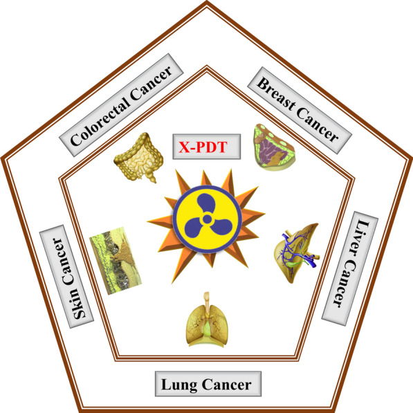
A graphical representation of applications of X-PDT against various types of cancer
Breast cancer
Breast cancer is the most prevalent kind of cancer globally and the primary cause of mortality associated with it. Despite this, there is little undergraduate and graduate exposure to breast cancer, which hampers the ability of clinicians to accurately identify, assess, and refer appropriate patients [70]. Gene expression profiling, a new molecular taxonomy, revealed that breast cancer comprises several unique biologic entities that are only partially recapitulated by ER- and HER2 clinical assays. Unsupervised gene expression analysis was used to create several molecular pictures of breast cancer, which were initially classified as luminal, HER2-enriched, basal-like (BL), and normal-like breast cancer. Each of these molecular subtypes can be found in clinical subsets; luminal A and B subtypes express keratins 8/18 and ER-related gene clusters, the BL has overexpression of keratin 5, 17, and EGFR-related genes, and the HER2-Enriched subtype is characterized by expression of Erb-B2-related genes [71, 72]. On the other hand, triple-negative breast cancer (TNBC) commonly manifests as invasive ductal carcinoma, although certain TNBC histologies require specific attention because of differing biology and diagnostic parameters [73]. In a study conducted by Wang et al. [74], a spherical-shaped organic phosphorescent nano-scintillator was developed for X-PDT of 4T1 cancer cells both in vitro and in vivo utilizing low-dose X-rays.
The model scintillator, 9,9′-(6-iodophenoxy-1,3,5-triazine-2,4-diyl)bis(9H-carbazole) (ITC), underwent modifications involving oxygen (O), nitrogen (N), and iodine (I) atoms to enhance n–π* transitions. Furthermore, the organic scintillator was transformed into a water-soluble nano-scintillator with the aid of a triblock co-polymer (F-127). The results demonstrated minimal side effects along with the high potential for X-PDT of deep-seated tumors, as presented in Fig. 3. In a recent study, Zhang et al. [75] reported X-PDT and ferroptosis of breast cancer using a specially engineered nanocarrier (D-NPVR). Herein, they engineered a nanocarrier with a hyperbranched co-polymer bridge containing 1,2-bis(2-hydroxyethylthio)-ethylene bonds and incorporated a photosensitizer (Verteporfin = VP) along with RSL3 (a ferroptosis inducer). Moreover, X-PDT facilitated the 1O2-induced glutathione (GHS) depletion, while RSL3 deactivated glutathione peroxide 4 (GPX4), leading to 4T1 cancer cell death through ferroptosis. This study demonstrated the remarkable efficacy of combined X-PDT and ferroptosis therapy in combating tumors. Jain et al. [76] developed an X-PDT responsive nanocomposite utilizing magnetic-luminescent nanoparticles composed of Gd2.98Ce0.02Al5O12 (GAG) with a mesoporous silica (mSiO2) coating, incorporating Rose Bengal (RB) as a photosensitizer. The GAG@mSiO2@RB nanocomposite exhibited significant activity against human breast cancer cells (MDA-MB-231), showing their potential as MR-imaging and X-PDT-responsive nanomaterials. Similarly, Maiti et al. [77] fabricated Rose Bengal (RB) decorated NaYF4:Tb NPs for low-dose X-PDT against luciferase-expressing murine epithelial breast cancer (4T1-luc). The synthesis involved a solvothermal approach to produce 7.6 nm NaYF4:Tb NPs, which were then coated with poly(maleicanhydride-alt-1-octedecene)-poly(ethylene glycol) (2000 MW) (C18PMH-PEG2k). Subsequently, RB was conjugated as a photosensitizer to create NaYF4:Tb@RB. The outcomes revealed efficient ROS generation under low dose (0.5 Gy) X-ray exposure, resulting in cancer cell death through severe DNA breaks. Likewise, Maiti et al. [78] developed coordination nanocrystals of Terbium Rose Bengal (T-RBNs) for low-dose X-PDT against 4T1-luc breast cancer cells. The T-RBNs with a nanosize of 7 nm were prepared, which are suitable for rapid renal clearance to minimize systematic toxicity. Moreover, surface modification was carried out by PEGylation to improve the in vivo behavior. The results presented an excellent X-PDT effect under low-dose (0.5 Gy) X-rays. In another study, Chen et al. [79] reported copper-cysteamine (Cu-Cy) based X-PDT for HepG2, SK-HEP-1, Li-7, and 4T1 cancer cell lines and its effects on cell proliferation, as presented in Fig. 4. Furthermore, they mimicked the clinical settings for X-PDT of deep-seated cancer using Cu-Cy NPs as therapeutic agents. MR imaging was utilized to evaluate the response of tumors treated with NPs compared to untreated tumors under X-ray irradiation. The results indicated the promising efficacy of Cu-Cy NPs for X-PDT with inhibition of cancer cell proliferation as well as migration.
Fig. 3.

a Schematic representation of in vivo therapy mechanism of deep seated tumor by X-PDT, b confocal laser scanning (CLSM) images of; (i) 4T1 cells co-staining analysis by Calcein-AM/PI, (live cells = green, dead cells = red); (ii) SOSG stained 4T1 cells; (iii) lipo-peroxides in 4T1 cells (ROS generation with BIODIPY C-11 staining is presented by green fluorescence); (iv) mitochondrial membrane potential of 4T1 cells (positive membrane potential = red fluorescence, decrease in membrane potential = green fluorescence), c representation of in vivo assessment of nano-scintillators; (i) 4T1 tumor growth curves; (ii) tumors weight after 14 days treatment; (iii) H&E-stained images of tumor slices. Reused with the permission of [74]. Copyright, The Author(s) 2022 under the license (http://creativecommons.org/licenses/by/4.0/)
Fig. 4.

a Schematic illustration of X-PDT influence on cancer cells, b MR-imaging for tumoricidal effect; (i) volume changes; (ii) body weight changes, c H & E staining of representative organs (kidney, heart, liver, lung and spleen), d immunohistochemistry (H & E, E-cadherin and PCNA staining of tumors) and morphological analysis; percentages positive expression of (i) E-cadherin; (ii) PCNA, (* p < 0.05 vs. control). Reused with the permission of [79]. Copyright, The Authors 2021, Publishing services by Elsevier B.V. under the license (http://creativecommons.org/licenses/by/4.0/)
An interesting study reported by Zhang et al. [80] showed a groundbreaking approach to combat breast cancer cells (MDA-MB-231) using X-PDT and hypoxia-activated chemotherapy facilitated by an intelligent nanoplatform (DATAT-NPVT). The cancer cellular uptake was enhanced by the accumulation of DATAT-NPVT and the masking of TAT ligands with the reactivation of targeting ability due to the acidic pH within tumors. ROS generation was induced by verteporfin (VP) under low-dose X-ray irradiation, resulting in a remarkable X-PDT effect against breast cancer. Additionally, hypoxic cancer cells were effectively targeted through the creation of a hypoxic environment triggered by the release of cascaded tirapazamine (TPZ) and the degradation of ditelluride-bridged bonds. A comprehensive schematic illustrating the mechanism of hypoxic cancer cell destruction is presented in Fig. 5.
Fig. 5.

A schematic diagram of cancer-killing under hypoxic environment by tirapazamine (TPZ)
Sengar et al. [81] developed Y2.99Pr0.01Al5O12-based (YP) mesoporous silica-coated nanocomposites (YPMS), which were further modified with protoporphyrin IX (PpIX) and folic acid (FA) for targeted X-PDT treatment of breast and prostate cancers. The distinctive structure of these nanocomposites allowed for the attachment of interchangeable targeting ligands, offering flexibility in targeting various types of cancer cells with distinct molecular markers on their surfaces. An admirable study was reported by Yu et al. [41] for dual-modal imaging (MRI/CT) guided X-PDT against 4T1 breast cancer cells by using Merocyanine 540-coupled Gd2(WO4)3:Tb nanoscintillator. The results demonstrated a highly effective synergistic approach for monitoring deep tumor therapy, employing MRI and CT imaging techniques. Similarly, Jiang et al. [82] synthesized CsI(Na)@MgO NPs and incorporated 5-aminolevulinic acid (5-ALA) for X-PDT treatment of 4T1 cancer cells, offering a safe and effective X-PDT strategy, with potential applications in clinical trials.
Based on the findings of these studies, it can be inferred that X-ray-mediated photodynamic therapy holds promise for the treatment of breast cancer. However, the selection of the appropriate therapeutic agent, with or without photosensitizers, as well as targeting agents and environmental factors, is crucial. Furthermore, the amalgamation of nanotechnology and pharmaceuticals has the potential to enhance the precision of treatment targeting and activation by low-dose X-rays in deep tissue, thereby advancing X-ray-mediated photodynamic therapy.
Liver cancer
One of the growing health challenges is liver cancer, which is impacting millions of people worldwide. It is forecasted that around 1 million individuals will be afflicted by liver cancer in 2030 [83]. The most abundant type of live cancer is hepatocellular carcinoma (HCC), which accounts for 90% of cases [84, 85]. An example of HCC can be seen in Fig. 6a under the observations of MRI. The second type of liver cancer is intrahepatic cancer (IHC), a variant of cholangiocarcinoma. IHC is characterized by cancer of the bile ducts within the liver, accounting for 10–20% of liver cancer cases [86]. Additionally, there exists a rare form of liver cancer known as angiosarcoma, originating in the blood cell lining of the liver and found in 1% of liver cancer cases [87]. The most common etiology of HCC is the hepatitis B virus (HBV), responsible for 50% of HCC cases [88]. The second risk factor for HCC is hepatitis C virus (HCV) [89]. In the contemporary era, despite advancements in research and understanding of the disease’s pathogenesis and heterogeneity, there remains a pressing need for therapeutic interventions to advance clinical practices and trial methodologies. Several studies have been conducted utilizing X-ray-responsive materials for the diagnosis and treatment of liver cancer. For instance, Chen et al. [79] investigated the therapeutic efficacy of copper-cysteamine (Cu-Cy) NPs against liver (HepG2, SK-HEP-1, Li-7) and breast (4T1) cancer cell lines under low-dose (2 Gy) X-ray irradiations. The findings demonstrated a remarkable effect of nominating Cu-Cy NPs as a perspective candidate with respect to X-PDT in clinical application, as presented in Fig. 6b–d. A similar kind of study was reported by Zhen et al. [90], and they utilized Cu-Cy NPs with potassium iodide (KI) and assessed the photodynamic effect, effectively inhibiting the growth, migration, and clone formation of liver cancer (HepG2, Hep3B, and Huh7) cells. Although UV irradiations were employed for PDT evaluation, reports suggest that Cu-Cy-based NPs could be utilized for deep tumor therapy under X-ray irradiation. Shi and co-workers developed X-ray responsive nanocomposites from the precursors Zn3Ga2GeO8: Cr3+ 1%, Yb3+ 5%, Er3+ 0.5% using a mesoporous silica (mSiO2) template to generate mZGGOs. Subsequently, it was modified by NHS-PEG3400-COOH and silicon phthalocyanine (Si-Pc) to form Si-Pc@PEG-mZGGOs. The X-ray-excited persistent luminescence (XEPL) and bioluminescence-guided X-PDT were carried out against orthotopic hepatic tumors, demonstrating long-lasting luminescence even through tissues with a thickness of 1 cm, along with effective X-PDT against HepG2 tumor cells.
Fig. 6.

a MRI of HCC in two different patients, first (i–iii) and second (iv–vi). Reused with the permission of [91]. Copyright, The Authors 2019, under the license (http://creativecommons.org/licenses/by/4.0/). b Transwell assays for the assessment of HepG2 and SK-Hep-1 migration, after the X-PDT with Cu-Cy NPs low dose (50 mg/L) as well as high dose (100 mg/L), c,d migratory cells counting (* p < 0.05 vs. control). Reused with the permission of [79]. Copyright, The Authors 2021, Publishing services by Elsevier B.V. under the license (http://creativecommons.org/licenses/by/4.0/)
From these studies, it can be concluded that X-ray excited theranostic techniques offer a way to overcome tissue attenuation and light penetration limitations in order to achieve deep tumor treatment. Furthermore, coinage metals (Cu, Ag, and Au) possess excellent antiviral ability [92], thus coinage metals doped nanomaterials hold promise for addressing HCV and HBV using X-PDT and X-ray-based imaging techniques.
Lung cancer
Lung cancer is one of the most common malignancies to be diagnosed and also accounts for the majority of cancer-related deaths globally [93]. There are two main types of lung cancer known as non-small cell lung cancer (NSCLC) and small cell lung cancer (SCLC). NSCLC comprises 80% of lung cancers and is further categorized into two predominant types (adenocarcinoma and squamous cell carcinoma) as well as two rarer types (adenosquamous carcinoma and sarcomatoid carcinoma). Similarly, SCLC includes oat cell carcinoma (small cell carcinoma) and combined small cell carcinoma [94]. In recent years, lung cancer has been managed through various methods such as surgery [95], radiotherapy [96], targeted chemotherapy [97], and immunotherapy (blocking PD-L1 and PD-L2 receptors pathways) [98]. Photodynamic therapy has been used for the treatment of lung cancer, however, shallow tissue penetration of light presents a significant challenge. To address this issue, low-dose X-rays are emerging as a promising approach for treating deeply located tumors through X-PDT. Chen et al. [99] developed X-ray responsive LiGa5O8:Cr (LGO:Cr) based nano-theranostics combined with cetuximab to combat H1299 orthotopic NSCLC. Specifically, LGO:Cr with 2,3-phthalocyanine (a photosensitizer) was encapsulated by silica, followed by conjugation with cetuximab. After the X-ray irradiation, the nanoscintillator converted X-ray photons to visible photons by the phenomenon of X-ray excited luminescence (XEOL), leading to the activation of the photosensitizer and the generation of ROS in the nano-system. Because of the deep penetrations of X-rays, this nano-assembly was utilized for imaging-guided therapy of lung cancer with remarkable suppression of cancer cells and minimal side effects on normal tissues. In another study, Rossi et al. [100] performed low dose (0.4–2 Gy) X-PDT on lung adenocarcinoma cells (the human NSCLC cell line A549) by using core–shell SiC/SiOx nanowires and an organic photosensitizer tetracarboxyphenyl porphyrin derivative. The results exhibited a 75% reduction in cancer cell viability compared to the control group at 2 Gy low-dose X-ray irradiations. Wang et al. [63] reported a study in which they synthesized MC540-SAO:Eu@mSiO2 nanoparticles and conducted X-PDT against NSCLC H1299 cells. The findings demonstrated X-PDT to be an effective imaging-guided therapy for tumor suppression within deep tissues, as shown in Fig. 7. The X-PDT monitoring was carried out by bioluminescence imaging (BLI), revealing higher efficiency compared to simple radiotherapy (RT).
Fig. 7.

Therapeutic outcomes in vivo. a BLI results of mice X-PDT with radiation dose of 5 Gy, b tumor growth analysis under BLI monitoring, c ex vivo BLI after dissection with left to right organs arrangement of top row: intestine, spleen, liver and skin; bottom row: muscle, brain, lung, heart and kidneys, d lungs BLI signal, based on ROI, e lung images of control and X-PDT groups, f H&E staining analysis (scale bar is 100 μm). Reused with the permission of [63]. Copyright, Ivyspring International Publisher under the terms and conditions (http://ivyspring.com/terms)
In a recent investigation, Yang et al. [101] assessed the radio dynamic therapeutic approach for C57BL/6 mouse model of KP1 SCLC cells by using 5-aminolevulinic acid (5-ALA) photosensitizer. In their study, a significant synergistic effect of X-ray based photodynamic therapy was observed and tumor growth was delayed up to 58% as compared to the control group. The results highlight X-PDT as an effective modality for tumor treatment. Likewise, Yang et al. [97] synthesized Ce-CaCO3 porous particles with an average pore size of 500 nm and assessed X-PDT against A549 lung cancer cells, revealing a remarkable ROS generation after X-ray irradiations, leading to the eradication of cancer cells.
The aforementioned studies illustrated the efficacy and potency of X-PDT in combating lung cancer. Hence, it is conceivable that the fabrication of X-ray responsive nanomaterials under controlled conditions and with specific morphology holds promise in combating various forms of lung neoplasms.
Skin cancer
The three main forms of skin cancer are squamous cell carcinoma (SCC), melanoma, and basal cell carcinoma (BCC). SCC is a rapidly proliferating neoplasm that typically manifests as crusted, erythematous nodules or scaly plaques upon solar exposure. Under severe circumstances, ulceration and hemorrhage are possible. Left untreated, SCC may progress into a formidable mass, rendering it perilous albeit less so than melanoma. Conversely, BCC is characterized by a pearly translucence transitioning into a fleshy hue with telangiectasia (dilated blood vessels). BCC is deemed the least lethal skin cancer and can be eradicated without leaving scars through appropriate intervention [102], on the other hand, stands as the most aggressive form of skin cancer, afflicting millions of people worldwide. It frequently presents asymmetrical borders with variegated pigmentation (often ranging from brown to black, occasionally pink, red, or flesh-colored) and typically exceeds 6 mm in diameter. The management of melanoma poses a substantial challenge in clinical practice [103, 104]. Shi et al. [105] applied X-PDT for XL50 cells (SCC) and B16F10 cells (melanoma) using copper-cysteamine (Cu-Cy) NPs, indicating an effective and safe treatment for SCC as compared to melanoma. The studies by Ma et al. [106] and Panday et al. [107] further support the X-PDT efficacy of Cu-Cy NPs and their potential utility in cancer therapy through the robust generation of ROS upon exposure to X-rays. Figure 8 illustrates the application of X-PDT in conjunction with immunotherapy using Cu-Cy NPs. In another study, Takahashi et al. [108] reported the treatment of B16-BL6 melanoma cells by using 5-aminolevulinic acid (5-ALA) as a radiosensitizer and X-ray irradiation. This approach hinged on the production of ROS facilitated by the accumulation of protoporphyrin IX (PpIX) derived from 5-ALA. Clinically, 5-ALA has been utilized in photodynamic therapy and fluorescence imaging-guided surgical procedures. Given melanoma’s well-established resistance to radiation, which poses a risk of toxicity, this remains a daunting challenge for researchers. In contrast, 5-ALA possesses low toxicity and high selectivity for tumor cells. In light of these characteristics, clinical trials are imperative to combat melanomas using low-dose X-PDT with 5-ALA or analogous agents.
Fig. 8.

a A graphical presentation of synergistic therapy by Cu-Cy NPs, b images to present the morphology of B16 (melanoma) cells before and after X-ray irradiations and with and without Cu-Cy NPs treatment, respectively, c a presentation of cellular viability at different concentrations of Cu-Cy NPs with low dose (2.5 Gy) X-rays irradiation, d intracellular ROS generation by DCFH-DA assay, presenting increase in fluorescence intensity after exposure to X-rays, e,f measurement of rates of cell apoptosis and/or necrosis after treatment and irradiation, g tumor volumes measurement, h tumor growth curves, i–k detection of infiltrative immune cells changes by flow cytometry. Reused with the permission of [49]. Copyright, The Authors 2020, under the license (http://creativecommons.org/licenses/by/4.0/)
Additionally, Hasegawa et al. [109] explored 5-ALA-based X-PDT against B16 (melanoma) cells. The results revealed the therapeutic efficacy of 5-ALA through the accumulation of PpIX, followed by ROS production, which nominated 5-ALA as a promising option for radio-resistant tumors.
These studies highlighted the significance of X-PDT in the management of skin cancers. Although there are some shortcomings in material selection, irradiation power, and time, X-PDT is still making its way into clinical trials by virtue of its high efficiency and low toxicity.
Colorectal cancer
Colorectal cancer (CRC) stands as the fourth most prevalent fatal malignancy, with a rising incidence each passing year. The primary instigators of CRC are genetic aberrations, predominantly affecting tumor suppressor genes, oncogenes, and genes associated with DNA mending. CRC is categorized into sporadic (70%), familial (25%), and hereditary (5%) types based on these mutations [110]. Studies have identified mutations in genes such as TP53, SMAD4, AXIN, NQO1, CYP2E, and MTHFR, translocations in genes like p16, p53, p14, E-cadherin, APC, β-catenin, TGF-β, SMADs, MSH2, MSH6, AXIN, PMS2, PTEN, STK11, KRAS, and DCC, and chromosomal alterations in genes including APC (5q21), β-Catenin (3p21), p16 and p15 (9p), p53 (17p), retinoblastoma (13q), BRCA1 (17q), SMAD4 and DCC (18q), and less frequently, E-cadherin (16q) [111]. Treatment modalities have progressed from the use of 5-fluorouracil (5-FU) as a standalone therapy to combination regimens incorporating 5-FU with irinotecan, oxaliplatin, or both [112]. There are still shortcomings related to drug toxicity, off-target effects, and poor growth inhibition for cancer cells, etc. In an effort to address these limitations, Sang et al. [113] developed a lipid-polymer nanoplatform (FA-LPNPs-VP-5-FU) for X-PDT against human colorectal cancer (CRC). This innovative approach involved the creation of lipid-polymer hybrid nanoparticles (LPNPs) functionalized with folic acid (FA), and loaded with verteporfin (VP) and 5-fluorouracil (5-FU). Folic acid served as a targeting ligand, VP acted as a photosensitizer, and 5-FU worked as a therapeutic agent, thus establishing a synergistic targeted X-PDT and chemotherapeutic regimen. The results demonstrated significant growth and proliferation inhibition of HCT116 cells, with minimal drug toxicity and adverse effects, as presented in Fig. 9.
Fig. 9.

a (i) A graphical presentation of targeted X-PDT; (ii) TEM image of FA-LPNPs-VP-5-FU, b confocal images of cellular viability of HCT116 cells (green fluorescence = dead cells); (i) quantitative analysis by ImageJ; (ii) calculated cellular viability by MTS assay after 24 h, c confocal images of apoptosis/necrosis assay; quantitative measurements of percentage changes at apoptosis/necrosis. Reproduced with the permission of [113]. Copyright, The Authors 2022, Published by Elsevier Masson SAS, under the license (https://creativecommons.org/licenses/by-nc-nd/4.0/)
Another study was reported by Lan et al. [114], in which they synthesized metal–organic layers (MOLs) constructed from [Hf6O4(OH)4(HCO2)6] secondary building units (SBUs) and produced tricarboxylate ligands derived from Ir[bpy(ppy)2]+- or [Ru(bpy)3]2+. Hf-BPY-Ru and Hf-BPY-Ir MOLs demonstrated outstanding X-PDT efficacy against colon cancer. Within these MOLs, Hf atoms absorbed X-ray energies and transferred them to Ir[bpy(ppy)2]+- or [Ru(bpy)3]2+ moieties to initiate ROS generation, leading to the eradication of colon cancer cells in deep tissues. These results illustrated the potential of a new class of 2D materials, MOLs, in the X-PDT for cancer treatment. Likewise, Deng et al. [115] engineered mitochondrial-targeting nanostructures using biodegradable poly(lactic-co-glycolic acid) as a nanocarrier, verteporfin as a photosensitizer, 2–5 nm gold (Au) NPs as the radiation enhancers, and triphenyl phosphonium as a mitochondrial targeting agent to perform X-PDT against rectal cancer. The average size of these nanostructures was 160 nm. After X-ray irradiation, these nanostructures generated ROS within the mitochondria, resulting in the loss of membrane potential and apoptosis in cancer cells. This low dose (4 Gy) X-ray-induced PDT effectively managed tumor growth, leading to an improved survival rate in the colorectal cancer mice model. Furthermore, in vivo results demonstrated X-PDT as an antiproliferative, cytoreductive, and profibrotic therapeutic strategy.
In summary, these reports suggest that the treatment modality is contingent upon the stage of the ailment, the molecular composition of the neoplasm, and the functional capacity of the patient. Despite these challenges, X-PDT appears to be a promising strategy for the treatment of profound neoplasms.
Conclusion and perspectives
Traditional photodynamic therapy (PDT) has shown potential in the treatment of single metastases as well as primary tumors. However, widely dispersed, numerous metastases are the primary cause of cancer mortality and have emerged as the main focus of oncology. Photothermal therapy (PTT) and photodynamic therapy (PDT) are laser-irradiation-dependant methodologies that exhibit inadequate responses to deeply situated tumors due to the limited penetrations of laser irradiations. In addition, drug loading and release, as well as the creation of delivery vector, are the main obstacles to multidrug combination delivery. These issues are among the reasons why co-delivery systems based on nanocarriers have not been used in clinics. Further causes include the insufficient safety evaluation of preparation protocols and the complexities associated with implementing the preparation in large-scale clinical applications. To effectively apply the PDT concept to deep tumors and dispersed metastases, X-ray-induced photodynamic therapy (X-PDT) represents a superb alternative. The development of agents that are X-ray-excited and emit visible light to activate photosensitizers, results in the generation of reactive oxygen species and subsequent cancer cell death. Besides that, real-time monitoring and early diagnosis play a key role in enhancing the effectiveness of cancer therapy. In this context, X-ray responsive theranostics have been exhibiting dual functionalities in imaging as well as therapeutics through the driving force of X-rays. According to the reports discussed in this review, it can be seen that low dose X-ray-induced photodynamic therapy in combination with X-ray responsive nanomaterials, photosensitizers, and targeting agents holds promise for the treatment of breast, liver, lung, skin, and colorectal cancers.
In the future, X-PDT may prove to be a promising therapeutic option for a variety of tumors in clinical settings, owing to its efficacy, safety, and feasibility. Prior to its clinical transformation, further elucidation is necessary regarding the safety of nanoparticles in humans and the impact of immune activation on X-PDT. Once X-PDT is executed in clinics, it can be utilized for the treatment of diverse malignancies, including those affecting the nasal passages, throat, bladder, osteosarcoma, prostate, kidney, pancreas, and so forth. Furthermore, due to the synergistic efficacy, the combination of immune checkpoint therapy and X-PDT for melanoma may develop into a focal point of research in the coming years. Besides, X-PDT can enhance tumor targeting by developing precise targeting methods and advancements in imaging modalities, like a combination of X-ray imaging with molecular imaging. Advances in molecular profiling and imaging technologies may enable personalized approaches to X-PDT. By characterizing individual tumors at the molecular level, clinicians could tailor treatment regimens to target specific genetic mutations or biomarkers associated with tumor aggressiveness. Moreover, non-invasive delivery of X-rays to deep-seated tumors can be facilitated by novel delivery methods, such as magnetic targeting or focused ultrasound, that will minimize damage to surrounding healthy tissue and improve patient comfort and safety. Likewise, X-PDT can be integrated with other therapeutic techniques, such as immunotherapy and chemotherapy, to generate a synergistic effect on deep tumors. This synergistic therapeutic approach can lead to enhanced treatment with reduced risk.
Acknowledgements
This work has been acknowledged to National Natural Science Foundation of China and Zhejiang University.
Author contributions
A.M. contributed to study concept, collect the literature, and wrote the first draft. W.S. supervised and finallize the draft. M.Z.I. and J.T. edited the English version. W.S. approved the submitted version after modification. All authors contributed to the article and approved the submitted version. All authors read and approved the final manuscript.
Funding
This work was supported by the National Natural Science Foundation of China (T2293753), the 2023 Hangzhou West Lake Pearl Project Leading Innovative Youth Team Project (TD2023017), and the Postdoctoral funding Grant (No. 02080100-K3F113001).
Availability of data and materials
No datasets were generated or analysed during the current study.
Declarations
Ethics approval and consent to participate
Not applicable.
Consent for publication
Not applicable.
Competing interests
The authors declare no competing interests.
Footnotes
Publisher's Note
Springer Nature remains neutral with regard to jurisdictional claims in published maps and institutional affiliations.
References
- 1.Siegel RL, Miller KD, Fuchs HE, Jemal A. Cancer statistics, 2022. CA Cancer J Clin. 2022;72:7–33. [DOI] [PubMed] [Google Scholar]
- 2.Mattiuzzi C, Lippi G. Current Cancer Epidemiology glossary. J Epidemiol Glob Health. 2019;9:217–22. [DOI] [PMC free article] [PubMed] [Google Scholar]
- 3.Debien V, De Caluwé A, Wang X, Piccart-Gebhart M, Tuohy VK, Romano E, et al. Immunotherapy in breast cancer: an overview of current strategies and perspectives. NPJ Breast Cancer. 2023;9:1–10. [DOI] [PMC free article] [PubMed] [Google Scholar]
- 4.Li X, Ramadori P, Pfister D, Seehawer M, Zender L, Heikenwalder M. The immunological and metabolic landscape in primary and metastatic liver cancer. Nat Rev Cancer. 2021;21:541–57. [DOI] [PubMed] [Google Scholar]
- 5.Oudkerk M, Liu SY, Heuvelmans MA, Walter JE, Field JK. Lung cancer LDCT screening and mortality reduction—evidence, pitfalls and future perspectives. Nat Rev Clin Oncol. 2021;18:135–51. [DOI] [PubMed] [Google Scholar]
- 6.Schlumberger M, Leboulleux S. Current practice in patients with differentiated thyroid cancer. Nat Rev Endocrinol. 2021;17:176–88. [DOI] [PubMed] [Google Scholar]
- 7.Gandaglia G, Leni R, Bray F, Fleshner N, Freedland SJ, Kibel A, et al. Epidemiology and prevention of prostate cancer. Eur Urol Oncol. 2021;4:877–92. [DOI] [PubMed] [Google Scholar]
- 8.Singh D, Vignat J, Lorenzoni V, Eslahi M, Ginsburg O, Lauby-Secretan B, et al. Global estimates of incidence and mortality of cervical cancer in 2020: a baseline analysis of the WHO Global Cervical Cancer Elimination Initiative. Lancet Glob Health. 2023;11:e197–206. [DOI] [PMC free article] [PubMed] [Google Scholar]
- 9.Van HLMC, Vrieling A, Van Der HAG, Kogevinas M, Richters A, Kiemeney LA. Global trends in the epidemiology of bladder cancer : challenges for public health and clinical practice. Nat Rev Clin Oncol. 2023;20:287–304. [DOI] [PubMed] [Google Scholar]
- 10.Esmeeta A, Adhikary S, Dharshnaa V, Swarnamughi P, Ummul Maqsummiya Z, Banerjee A, et al. Plant-derived bioactive compounds in colon cancer treatment: an updated review. Biomed Pharmacother. 2022;153: 113384. [DOI] [PubMed] [Google Scholar]
- 11.Mirza MR, Chase DM, Slomovitz BM, dePont CR, Novák Z, Black D, et al. Dostarlimab for primary advanced or recurrent endometrial cancer. N Engl J Med. 2023;388:2145–58. [DOI] [PubMed] [Google Scholar]
- 12.Choueiri TK, Albiges L, Atkins MB, Bakouny Z, Bratslavsky G, Braun DA, et al. From basic science to clinical translation in kidney cancer: a report from the second kidney cancer research summit. Clin Cancer Res. 2022;28:831–9. [DOI] [PMC free article] [PubMed] [Google Scholar]
- 13.Rodriguez-Otero P, Ailawadhi S, Arnulf B, Patel K, Cavo M, Nooka AK, et al. Ide-cel or standard regimens in relapsed and refractory multiple myeloma. N Engl J Med. 2023;388:1002–14. [DOI] [PubMed] [Google Scholar]
- 14.Armitage JO, Long DL. Mantle-cell lymphoma. N Engl J Med. 2022;386:2495–506. [DOI] [PubMed] [Google Scholar]
- 15.Hingorani SR. Epithelial and stromal co-evolution and complicity in pancreatic cancer. Nat Rev Cancer. 2023;23:57–77. [DOI] [PMC free article] [PubMed] [Google Scholar]
- 16.Brown JR, Eichhorst B, Hillmen P, Jurczak W, Kaźmierczak M, Lamanna N, et al. Zanubrutinib or Ibrutinib in relapsed or refractory chronic lymphocytic leukemia. N Engl J Med. 2023;388:319–32. [DOI] [PubMed] [Google Scholar]
- 17.Patel SP, Othus M, Chen Y, Wright GP, Yost KJ, Hyngstrom JR, et al. Neoadjuvant–adjuvant or adjuvant-only pembrolizumab in advanced melanoma. N Engl J Med. 2023;388:813–23. [DOI] [PMC free article] [PubMed] [Google Scholar]
- 18.Nakhoda S, Vistarop A, Wang YL. Resistance to Bruton tyrosine kinase inhibition in chronic lymphocytic leukaemia and non-Hodgkin lymphoma. Br J Haematol. 2023;200:137–49. [DOI] [PMC free article] [PubMed] [Google Scholar]
- 19.Pashayan N, Pharoah PDP. The challenge of early detection in cancer. Science. 1979;2020(368):589–90. [DOI] [PubMed] [Google Scholar]
- 20.Li X, Bao Y, Li Z, Teng P, Ma L, Zhang H, et al. Employing antagonistic C-X-C motif chemokine receptor 4 antagonistic peptide functionalized NaGdF4 nanodots for magnetic resonance imaging-guided biotherapy of breast cancer. Sci Rep. 2024;14:15764. [DOI] [PMC free article] [PubMed] [Google Scholar]
- 21.Kumar Sarangi M, Padhi S, Rath G, Sekhar Nanda S, Kee YD. Advances in immunological and theranostic approaches of gold nanoparticles—a review. Inorg Chem Commun. 2023;153:110858. [Google Scholar]
- 22.Dutta Gupta Y, Mackeyev Y, Krishnan S, Bhandary S. Mesoporous silica nanotechnology: promising advances in augmenting cancer theranostics. Cancer Nanotechnol. 2024;15:1–44. [Google Scholar]
- 23.Hosseini SM, Mohammadnejad J, Salamat S, Beiram Zadeh Z, Tanhaei M, Ramakrishna S. Theranostic polymeric nanoparticles as a new approach in cancer therapy and diagnosis: a review. Mater Today Chem. 2023;29: 101400. [Google Scholar]
- 24.Yukawa H, Sato K, Baba Y. Theranostics applications of quantum dots in regenerative medicine, cancer medicine, and infectious diseases. Adv Drug Deliv Rev. 2023;200:114863. [DOI] [PubMed] [Google Scholar]
- 25.Ma Q, Cao Y, Ge X, Zhang Z, Gao S, Song J. X-ray excited luminescence materials for cancer diagnosis and theranostics. Laser Photon Rev. 2024;18:2300565. [Google Scholar]
- 26.Al-Thani AN, Jan AG, Abbas M, Geetha M, Sadasivuni KK. Nanoparticles in cancer theragnostic and drug delivery: a comprehensive review. Life Sci. 2024;352: 122899. [DOI] [PubMed] [Google Scholar]
- 27.Rahimkhoei V, Alzaidy AH, Abed MJ, Rashki S, Salavati-Niasari M. Advances in inorganic nanoparticles-based drug delivery in targeted breast cancer theranostics. Adv Colloid Interface Sci. 2024;329: 103204. [DOI] [PubMed] [Google Scholar]
- 28.Jin GQ, Chau CV, Arambula JF, Gao S, Sessler JL, Zhang JL. Lanthanide porphyrinoids as molecular theranostics. Chem Soc Rev. 2022;51:6177–209. [DOI] [PMC free article] [PubMed] [Google Scholar]
- 29.Domingues I, Pereira G, Martins P, Duarte H, Santos J, Abreu PH. Using deep learning techniques in medical imaging: a systematic review of applications on CT and PET. Artif Intell Rev. 2020;53:4093–160. [Google Scholar]
- 30.Hsu JC, Nieves LM, Betzer O, Sadan T, Noël PB, Popovtzer R, et al. Nanoparticle contrast agents for X-ray imaging applications. Wiley Interdiscip Rev Nanomed Nanobiotechnol. 2020;12:1–26. [DOI] [PMC free article] [PubMed] [Google Scholar]
- 31.Wu Q, Zheng Q, He Y, Chen Q, Yang H. Emerging nanoagents for medical x-ray imaging. Anal Chem. 2023;95:33–48. [DOI] [PubMed] [Google Scholar]
- 32.Sun W, Zhou Z, Pratx G, Chen X, Chen H. Nanoscintillator-mediated X-ray induced photodynamic therapy for deep-seated tumors: from concept to biomedical applications. Theranostics. 2020;10:1296–318. [DOI] [PMC free article] [PubMed] [Google Scholar]
- 33.Yu X, Liu X, Yang K, Chen X, Li W. Pnictogen semimetal (Sb, Bi)-based nanomaterials for cancer imaging and therapy: a materials perspective. ACS Nano. 2021;15:2038–67. [DOI] [PubMed] [Google Scholar]
- 34.Pires L, Calcada C, Grafinger O, Juneja R, Mar S, Leong H, et al. Photosensitizers as radiosensitizers for metastatic and deep-seated tumors: in vitro and ex-ovo preclinical studies. Photodiagnosis Photodyn Ther. 2023;41: 103422. [Google Scholar]
- 35.Belanova A, Chmykhalo V, Beseda D, Belousova M, Butova V, Soldatov A, et al. A mini-review of X-ray photodynamic therapy (XPDT) nonoagent constituents’ safety and relevant design considerations. Photochem Photobiol Sci. 2020;19:1134–44. [DOI] [PubMed] [Google Scholar]
- 36.Fan W, Huang P, Chen X. Overcoming the Achilles’ heel of photodynamic therapy. Chem Soc Rev. 2016;45:6488–519. [DOI] [PubMed] [Google Scholar]
- 37.Chen X, Song J, Chen X, Yang H. X-ray-activated nanosystems for theranostic applications. Chem Soc Rev. 2019;48:3073–101. [DOI] [PubMed] [Google Scholar]
- 38.Kirakci K, Kubát P, Fejfarova K, Martinčík J, Nikl M, Lang K. X-ray inducible luminescence and singlet oxygen sensitization by an octahedral molybdenum cluster compound: a new class of nanoscintillators. Inorg Chem. 2016;55:803–9. [DOI] [PubMed] [Google Scholar]
- 39.Lei L, Wang Y, Kuzmin A, Hua Y, Zhao J, Xu S, et al. Next generation lanthanide doped nanoscintillators and photon converters. eLight. 2022;2:1–24. [Google Scholar]
- 40.Bulin A-L, Vasil’ev A, Belsky A, Amans D, Ledoux G, Dujardin C. Modelling energy deposition in nanoscintillators to predict the efficiency of the X-ray-induced photodynamic effect. Nanoscale. 2015;7:5744–51. [DOI] [PubMed] [Google Scholar]
- 41.Yu X, Liu X, Wu W, Yang K, Mao R, Ahmad F, et al. CT/MRI-guided synergistic radiotherapy and X-ray inducible photodynamic therapy using Tb-doped Gd-W-nanoscintillators. Angew Chem. 2019;131:2039–44. [DOI] [PubMed] [Google Scholar]
- 42.Song G, Cheng L, Chao Y, Yang K, Liu Z. Emerging nanotechnology and advanced materials for cancer radiation therapy. Adv Mater. 2017;29:1–26. [DOI] [PubMed] [Google Scholar]
- 43.Shen S, Jiang D, Cheng L, Chao Y, Nie K, Dong Z, et al. Renal-clearable ultrasmall coordination polymer nanodots for chelator-free 64Cu-labeling and imaging-guided enhanced radiotherapy of cancer. ACS Nano. 2017;11:9103–11. [DOI] [PMC free article] [PubMed] [Google Scholar]
- 44.Zhang XD, Chen J, Min Y, Park GB, Shen X, Song SS, et al. Metabolizable Bi2Se3 nanoplates: biodistribution, toxicity, and uses for cancer radiation therapy and imaging. Adv Funct Mater. 2014;24:1718–29. [Google Scholar]
- 45.Gong T, Li Y, Lv B, Wang H, Liu Y, Yang W, et al. Full-process radiosensitization based on nanoscale metal-organic frameworks. ACS Nano. 2020;14:3032–40. [DOI] [PubMed] [Google Scholar]
- 46.Yang B, Chen Y, Shi J. Reactive oxygen species (ROS)-based nanomedicine. Chem Rev. 2019;119:4881–985. [DOI] [PubMed] [Google Scholar]
- 47.Kirsanova DY, Gadzhimagomedova ZM, Maksimov AY, Soldatov AV. Nanomaterials for deep tumor treatment. Mini Rev Med Chem. 2021;21:677–88. [DOI] [PubMed] [Google Scholar]
- 48.Popovich K, Procházková L, Pelikánová IT, Vlk M, Palkovský M, Jarý V, et al. Preliminary study on singlet oxygen production using CeF3:Tb3+@SiO2-PpIX. Radiat Meas. 2016;90:325–8. [Google Scholar]
- 49.Zhang Q, Guo X, Cheng Y, Chudal L, Pandey NK, Zhang J, et al. Use of copper-cysteamine nanoparticles to simultaneously enable radiotherapy, oxidative therapy and immunotherapy for melanoma treatment. Signal Transduct Target Ther. 2020;5:4–6. [DOI] [PMC free article] [PubMed] [Google Scholar]
- 50.Ma L, Zou X, Chen W. A new X-ray activated nanoparticle photosensitizer for cancer treatment. J Biomed Nanotechnol. 2014;10:1501–8. [DOI] [PubMed] [Google Scholar]
- 51.Ma L, Zou X, Bui B, Chen W, Song KH, Solberg T. X-ray excited ZnS:Cu, Co afterglow nanoparticles for photodynamic activation. Appl Phys Lett. 2014;105: 013702. [Google Scholar]
- 52.Gadzhimagomedova Z, Zolotukhin P, Kit O, Kirsanova D, Soldatov A. Nanocomposites for X-ray photodynamic therapy. Int J Mol Sci. 2020;21:1–15. [DOI] [PMC free article] [PubMed] [Google Scholar]
- 53.Sengar P, Garcia-Tapia K, Chauhan K, Jain A, Juarez-Moreno K, Borbón-Nuñez HA, et al. Dualphotosensitizer coupled nanoscintillator capable of producing type I and type II ROS for next generation photodynamic therapy. J Colloid Interface Sci. 2019;536:586–97. [DOI] [PubMed] [Google Scholar]
- 54.Zhang W, Zhang X, Shen Y, Shi F, Song C, Liu T, et al. Ultra-high FRET efficiency NaGdF4: Tb3+-Rose Bengal biocompatible nanocomposite for X-ray excited photodynamic therapy application. Biomaterials. 2018;184:31–40. [DOI] [PubMed] [Google Scholar]
- 55.Chen MH, Jenh YJ, Wu SK, Chen YS, Hanagata N, Lin FH. Non-invasive photodynamic therapy in brain cancer by use of Tb3+-doped LaF3 nanoparticles in combination with photosensitizer through X-ray irradiation: a proof-of-concept study. Nanoscale Res Lett. 2017;12:1–6. [DOI] [PMC free article] [PubMed] [Google Scholar]
- 56.Elmenoufy AH, Tang Y, Hu J, Xu H, Yang X. A novel deep photodynamic therapy modality combined with CT imaging established via X-ray stimulated silica-modified lanthanide scintillating nanoparticles. Chem Commun. 2015;51:12247–50. [DOI] [PubMed] [Google Scholar]
- 57.Bulin A-L, Truillet C, Chouikrat R, Lux F, Frochot C, Amans D, et al. X-ray-induced singlet oxygen activation with nanoscintillator-coupled porphyrins. J Phys Chem C. 2013;117:21583–9. [Google Scholar]
- 58.Kaščáková S, Giuliani A, Lacerda S, Pallier A, Mercère P, Tóth É, et al. X-ray-induced radiophotodynamic therapy (RPDT) using lanthanide micelles: beyond depth limitations. Nano Res. 2015;8:2373–9. [Google Scholar]
- 59.Zhang X, Lan B, Wang S, Gao P, Liu T, Rong J, et al. Low-dose X-ray excited photodynamic therapy based on NaLuF4:Tb3+-Rose Bengal nanocomposite. Bioconjug Chem. 2019;30:2191–200. [DOI] [PubMed] [Google Scholar]
- 60.Popovich K, Tomanová K, Čuba V, Procházková L, Pelikánová IT, Jakubec I, et al. LuAG:Pr3+-porphyrin based nanohybrid system for singlet oxygen production: toward the next generation of PDTX drugs. J Photochem Photobiol B. 2018;179:149–55. [DOI] [PubMed] [Google Scholar]
- 61.Procházková L, Pelikánová IT, Mihóková E, Dědic R, Čuba V. Novel scintillating nanocomposite for X-ray induced photodynamic therapy. Radiat Meas. 2019;121:13–7. [Google Scholar]
- 62.Procházková L, Čuba V, Beitlerová A, Jarý V, Omelkov S, Nikl M. Ultrafast Zn(Cd, Mg)O: Ga nanoscintillators with luminescence tunable by band gap modulation. Opt Express. 2018;26:29482–94. [DOI] [PubMed] [Google Scholar]
- 63.Wang GD, Nguyen HT, Chen H, Cox PB, Wang L, Nagata K, et al. X-ray induced photodynamic therapy: a combination of radiotherapy and photodynamic therapy. Theranostics. 2016;6:2295–305. [DOI] [PMC free article] [PubMed] [Google Scholar]
- 64.Bekah D, Cooper D, Kudinov K, Hill C, Seuntjens J, Bradforth S, et al. Synthesis and characterization of biologically stable, doped LaF3 nanoparticles co-conjugated to PEG and photosensitizers. J Photochem Photobiol A Chem. 2016;329:26–34. [Google Scholar]
- 65.Zou X, Yao M, Ma L, Hossu M, Han X, Juzenas P, et al. X-ray-induced nanoparticle-based photodynamic therapy of cancer. Nanomedicine. 2014;9:2339–51. [DOI] [PubMed] [Google Scholar]
- 66.Sapre AA, Novitskaya E, Vakharia V, Cota A, Wrasidlo W, Hanrahan SM, et al. Optimized scintillator YAG: Pr nanoparticles for X-ray inducible photodynamic therapy. Mater Lett. 2018;228:49–52. [DOI] [PMC free article] [PubMed] [Google Scholar]
- 67.Kaszewski J, Olszewski J, Rosowska J, Witkowski B, Wachnicki Ł, Wenelska K, et al. HfO2: Eu nanoparticles excited by X-rays and UV-visible radiation used in biological imaging. J Rare Earths. 2019;37:1176–82. [Google Scholar]
- 68.Li X, Xue Z, Jiang M, Li Y, Zeng S, Liu H. Soft X-ray activated NaYF4:Gd/Tb scintillating nanorods for in vivo dual-modal X-ray/X-ray-induced optical bioimaging. Nanoscale. 2018;10:342–50. [DOI] [PubMed] [Google Scholar]
- 69.Generalov R, Kuan WB, Chen W, Kristensen S, Juzenas P. Radiosensitizing effect of zinc oxide and silica nanocomposites on cancer cells. Colloids Surf B Biointerfaces. 2015;129:79–86. [DOI] [PubMed] [Google Scholar]
- 70.Zagami P, Carey LA. Triple negative breast cancer: pitfalls and progress. NPJ Breast Cancer. 2022;8:1–10. [DOI] [PMC free article] [PubMed] [Google Scholar]
- 71.Perou CM, Sorlie T, Eisen MB, Van De RM, Jeffrey SS, Rees CA, et al. Molecular portraits of human breast tumours. Nature. 2000;533:747–52. [DOI] [PubMed] [Google Scholar]
- 72.Hoadley KA, Yau C, Hinoue T, Wolf DM, Lazar AJ, Drill E, et al. Cell-of-origin patterns dominate the molecular classification of 10,000 tumors from 33 types of cancer. Cell. 2019;173:291–304. [DOI] [PMC free article] [PubMed] [Google Scholar]
- 73.Schmid P, Cortes J, Dent R, Pusztai L, McArthur H, Kümmel S, et al. Event-free survival with pembrolizumab in early triple-negative breast cancer. N Engl J Med. 2022;386:556–67. [DOI] [PubMed] [Google Scholar]
- 74.Wang X, Sun W, Shi H, Ma H, Niu G, Li Y, et al. Organic phosphorescent nanoscintillator for low-dose X-ray-induced photodynamic therapy. Nat Commun. 2022;13:1–9. [DOI] [PMC free article] [PubMed] [Google Scholar]
- 75.Zhang B, Liu H, Wang Y, Zhang Y, Cheng J. Application of singlet oxygen-activatable nanocarriers to boost X-ray-induced photodynamic therapy and cascaded ferroptosis for breast cancer treatment. J Mater Chem B. 2023;11:9685–96. [DOI] [PubMed] [Google Scholar]
- 76.Jain A, Koyani R, Muñoz C, Sengar P, Contreras OE, Juárez P, et al. Magnetic-luminescent cerium-doped gadolinium aluminum garnet nanoparticles for simultaneous imaging and photodynamic therapy of cancer cells. J Colloid Interface Sci. 2018;526:220–9. [DOI] [PubMed] [Google Scholar]
- 77.Maiti D, Yu H, Kim BS, Naito M, Yamashita S, Kim HJ, et al. Rose Bengal decorated NaYF4: Tb nanoparticles for low dose X-ray-induced photodynamic therapy in cancer cells. ACS Appl Bio Mater. 2022;5:5477–86. [DOI] [PubMed] [Google Scholar]
- 78.Maiti D, Yu H, Mochida Y, Won S, Yamashita S, Naito M, et al. Terbium-Rose Bengal coordination nanocrystals-induced ROS production under low-dose X-rays in cultured cancer cells for photodynamic therapy. ACS Appl Bio Mater. 2023;6:2505–13. [DOI] [PubMed] [Google Scholar]
- 79.Chen X, Liu J, Li Y, Pandey NK, Chen T, Wang L, et al. Study of copper-cysteamine based X-ray induced photodynamic therapy and its effects on cancer cell proliferation and migration in a clinical mimic setting. Bioact Mater. 2022;7:504–14. [DOI] [PMC free article] [PubMed] [Google Scholar]
- 80.Zhang B, Xue R, Lyu J, Gao A, Sun C. Tumor acidity/redox hierarchical-activable nanoparticles for precise combination of X-ray-induced photodynamic therapy and hypoxia-activated chemotherapy. J Mater Chem B. 2022;10:3849–60. [DOI] [PubMed] [Google Scholar]
- 81.Sengar P, Juárez P, Meza AV, Arellano DL, Jain A, Chauhan K, et al. Development of a functionalized UV-emitting nanocomposite for the treatment of cancer using indirect photodynamic therapy. J Nanobiotechnol. 2018;16:1–19. [DOI] [PMC free article] [PubMed] [Google Scholar]
- 82.Jiang F, Lee C, Zhang W, Jiang W, Cao Z, Chong HB, et al. Radiodynamic therapy with CsI(na)@MgO nanoparticles and 5-aminolevulinic acid. J Nanobiotechno. 2022;20:1–15. [DOI] [PMC free article] [PubMed] [Google Scholar]
- 83.Villanueva A. Hepatocellular carcinoma. N Engl J Med. 2019;380:1450–62. [DOI] [PubMed] [Google Scholar]
- 84.Yang JD, Roberts LR. Hepatocellular carcinoma: a global view. Nat Rev Gastroenterol Hepatol. 2010;7:448–58. [DOI] [PMC free article] [PubMed] [Google Scholar]
- 85.Sia D, Villanueva A, Friedman SL, Llovet JM. Liver cancer cell of origin, molecular class, and effects on patient prognosis. Gastroenterology. 2017;152:745–61. [DOI] [PMC free article] [PubMed] [Google Scholar]
- 86.Bridgewater J, Galle PR, Khan SA, Llovet JM, Park JW, Patel T, et al. Guidelines for the diagnosis and management of intrahepatic cholangiocarcinoma. J Hepatol. 2014;60:1268–89. [DOI] [PubMed] [Google Scholar]
- 87.Cao J, Wang J, He C, Fang M. Angiosarcoma: a review of diagnosis and current treatment. Am J Cancer Res. 2019;9:2303–13. [PMC free article] [PubMed] [Google Scholar]
- 88.Fitzmaurice C, Akinyemiju T, Abera S, Ahmed M, Alam N, Alemayohu MA, et al. The burden of primary liver cancer and underlying etiologies from 1990 to 2015 at the global, regional, and national level results from the global burden of disease study 2015. JAMA Oncol. 2017;3:1683–91. [DOI] [PMC free article] [PubMed] [Google Scholar]
- 89.Celsa C, Stornello C, Giuffrida P, Giacchetto CM, Grova M, Rancatore G, et al. Direct-acting antiviral agents and risk of Hepatocellular carcinoma: critical appraisal of the evidence. Ann Hepatol. 2022;27: 100568. [DOI] [PubMed] [Google Scholar]
- 90.Zhen X, Pandey NK, Amador E, Hu W, Liu B, Nong W, et al. Potassium iodide enhances the anti-hepatocellular carcinoma effect of copper-cysteamine nanoparticle mediated photodynamic therapy on cancer treatment. Mater Today Phys. 2022;27: 100838. [Google Scholar]
- 91.Vasuri F, Renzulli M, Fittipaldi S, Brocchi S, Clemente A, Cappabianca S, et al. Pathobiological and radiological approach for hepatocellular carcinoma subclassification. Sci Rep. 2019;9:1–10. [DOI] [PMC free article] [PubMed] [Google Scholar]
- 92.Mushtaq A, Iqbal MZ, Kong X. Antiviral effects of coinage metal-based nanomaterials to combat COVID-19 and its variants. J Mater Chem B. 2022;10:5323–43. [DOI] [PubMed] [Google Scholar]
- 93.Thai AA, Solomon BJ, Sequist LV, Gainor JF, Heist RS. Lung cancer. The Lancet. 2021;398:535–54. [DOI] [PubMed] [Google Scholar]
- 94.Araujo LH, Horn L, Merritt RE, Shilo K, Xu-Welliver M, Carbone DP. 69—Cancer of the lung: non-small cell lung cancer and small cell lung cancer. In: Niederhuber JE, Armitage JO, Kastan MB, Doroshow JH, Tepper JE, editors. Abeloff’s clinical oncology. Amsterdam: Elsevier; 2020. [Google Scholar]
- 95.Jiang Y, Su Z, Liang H, Liu J, Liang W, He J. Video-assisted thoracoscopy for lung cancer: Who is the future of thoracic surgery? J Thorac Dis. 2020;12:4427–33. [DOI] [PMC free article] [PubMed] [Google Scholar]
- 96.Baker S, Dahele M, Lagerwaard FJ, Senan S. A critical review of recent developments in radiotherapy for non-small cell lung cancer. Radiat Oncol. 2016;11:1–14. [DOI] [PMC free article] [PubMed] [Google Scholar]
- 97.Yang CC, Wang WY, Lin FH, Hou CH. Rare-earth-doped calcium carbonate exposed to X-ray irradiation to induce reactive oxygen species for tumor treatment. Int J Mol Sci. 2019;20:1148. [DOI] [PMC free article] [PubMed] [Google Scholar]
- 98.Herbst RS, Baas P, Kim DW, Felip E, Pérez-Gracia JL, Han JY, et al. Pembrolizumab versus docetaxel for previously treated, PD-L1-positive, advanced non-small-cell lung cancer (KEYNOTE-010): a randomised controlled trial. The Lancet. 2016;387:1540–50. [DOI] [PubMed] [Google Scholar]
- 99.Chen H, Sun X, Wang GD, Nagata K, Hao Z, Wang A, et al. LiGa5O8:Cr-based theranostic nanoparticles for imaging-guided X-ray induced photodynamic therapy of deep-seated tumors. Mater Horiz. 2017;4:1092–101. [DOI] [PMC free article] [PubMed] [Google Scholar]
- 100.Rossi F, Bedogni E, Bigi F, Rimoldi T, Cristofolini L, Pinelli S, et al. Porphyrin conjugated SiC/SiOx nanowires for X-ray-excited photodynamic therapy. Sci Rep. 2015;5:1–6. [DOI] [PMC free article] [PubMed] [Google Scholar]
- 101.Yang DM, Cvetkovic D, Chen L, Ma CMC. Therapeutic effects of in-vivo radiodynamic therapy (RDT) for lung cancer treatment: a combination of 15 MV photons and 5-aminolevulinic acid (5-ALA). Biomed Phys Eng Express. 2022;8: 065031. [DOI] [PMC free article] [PubMed] [Google Scholar]
- 102.Sanchez G, Nova J, Rodriguez-Hernandez AE, Solorzano-Restrepo C, Gonzalez J, Olmos M, et al. Sun protection for preventing basal cell and squamous cell skin cancers. Cochrane Database Syst Rev. 2016;2016:1–32. [DOI] [PMC free article] [PubMed] [Google Scholar]
- 103.Domingues B, Lopes JM, Soares P, Pópulo H. Melanoma treatment in review. Immunotargets Ther. 2018;7:35–49. [DOI] [PMC free article] [PubMed] [Google Scholar]
- 104.Hu W, Fang L, Ni R, Zhang H, Pan G. Changing trends in the disease burden of non-melanoma skin cancer globally from 1990 to 2019 and its predicted level in 25 years. BMC Cancer. 2022;22:1–11. [DOI] [PMC free article] [PubMed] [Google Scholar]
- 105.Shi L, Liu P, Wu J, Ma L, Zheng H, Antosh MP, et al. The effectiveness and safety of X-PDT for cutaneous squamous cell carcinoma and melanoma. Nanomedicine. 2019;14:2027–43. [DOI] [PMC free article] [PubMed] [Google Scholar]
- 106.Ma L, Chen W, Schatte G, Wang W, Joly AG, Huang Y, et al. A new Cu-cysteamine complex: structure and optical properties. J Mater Chem C Mater. 2014;2:4239–46. [Google Scholar]
- 107.Pandey NK, Chudal L, Phan J, Lin L, Johnson O, Xing M, et al. A facile method for the synthesis of copper-cysteamine nanoparticles and study of ROS production for cancer treatment. J Mater Chem B. 2019;7:6630–42. [DOI] [PubMed] [Google Scholar]
- 108.Takahashi J, Murakami M, Mori T, Iwahashi H. Verification of radiodynamic therapy by medical linear accelerator using a mouse melanoma tumor model. Sci Rep. 2018;8:1–9. [DOI] [PMC free article] [PubMed] [Google Scholar]
- 109.Hasegawa T, Takahashi J, Nagasawa S, Doi M, Moriyama A, Iwahashi H. Dna strand break properties of protoporphyrin IX by X-ray irradiation against melanoma. Int J Mol Sci. 2020;21:2302. [DOI] [PMC free article] [PubMed] [Google Scholar]
- 110.Mármol I, Sánchez-de-Diego C, Dieste AP, Cerrada E, Yoldi MJR. Colorectal carcinoma: a general overview and future perspectives in colorectal cancer. Int J Mol Sci. 2017;18:197. [DOI] [PMC free article] [PubMed] [Google Scholar]
- 111.Sameer AS. Colorectal cancer: molecular mutations and polymorphisms. Front Oncol. 2013;3:114. [DOI] [PMC free article] [PubMed] [Google Scholar]
- 112.Stintzing S. Management of colorectal cancer. Prime Rep. 2014;6:108. [DOI] [PMC free article] [PubMed] [Google Scholar]
- 113.Sang R, Deng F, Engel A, Goldys E, Deng W. Lipid-polymer nanocarrier platform enables X-ray induced photodynamic therapy against human colorectal cancer cells. Biomed Pharmacother. 2022;155: 113837. [DOI] [PubMed] [Google Scholar]
- 114.Lan G, Ni K, Xu R, Lu K, Lin Z, Chan C, et al. Nanoscale metal–organic layers for deeply penetrating X-ray-induced photodynamic therapy. Angew Chem. 2017;56:12102–6. [DOI] [PMC free article] [PubMed] [Google Scholar]
- 115.Deng W, McKelvey KJ, Guller A, Fayzullin A, Campbell JM, Clement S, et al. Application of mitochondrially targeted nanoconstructs to neoadjuvant X-ray-induced photodynamic therapy for rectal cancer. ACS Cent Sci. 2020;6:715–26. [DOI] [PMC free article] [PubMed] [Google Scholar]
Associated Data
This section collects any data citations, data availability statements, or supplementary materials included in this article.
Data Availability Statement
No datasets were generated or analysed during the current study.