ABSTRACT
The occurrence of teratoma is 1 in 4,000 live births. Head-and-neck region teratoma (epignathus) is rare. These patients present with facial disfigurement, respiratory problems, and difficulty in feeding. It warrants early excision to avoid any morbidity or mortality due to airway obstruction. We describe the management of a patient with pedunculated epignathus with cervical lymphangioma.
KEYWORDS: Epignathus, lymphangioma, cervical
INTRODUCTION
Epignathus is a rare form of teratoma originating from the base of the skull usually attached to the hard palate or the mandible. Epignathus manifests approximately 1 in 35,000–1 in 200,000 live births, with a slight female predominance with a ratio of 3:1. Teratoma consists of various tissues originating from all three germ cell layers. The occurrence of teratoma is approximately 1 in 4,000 live births. The occurrence of teratoma is approximately 1 in 4,000 live births. In the pediatric population, the sacrococcygeal region is the most frequent site for teratoma, followed by the retroperitoneum, mediastinum, and gonads.[1]
Teratoma in the head-and neck-region accounts only 2%–9%, and malignant transformation is observed in <20% of these cases.[2]
This tumor can vary in size, small ones may pose feeding problems and large-size tumors can lead to significant obstruction of the upper airways, resulting in the respiratory excursion.
CASE REPORT
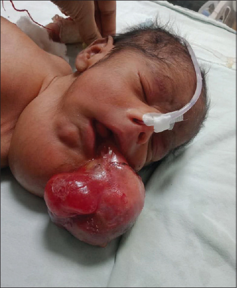
A 2-day male child presented to the emergency of the pediatric surgery department, with a complaint of a mass protruding out of the mouth and swelling on the left side of the neck [Figure 1]. The patient was of the fourth order, twin child, preterm, other baby was normal as stated by the parents, delivered by cesarean section, cried immediately after birth, and there was drooling of saliva without complaint of respiratory distress. The patient was on top feed with some difficulty. The mother of the patient was not on regular antenatal visits and she did not undergo an anomaly scan.
Figure 1.

A pedunculated mass protruding out of the oral cavity and left cervical swelling
On examination, the mass was attached with a hard palate, pedunculated, and variable in consistency; neck swelling was cystic in consistency. Ultrasonography of mass was suggestive of heterogeneous echogenicity, and the cervical region mass was suggestive of cystic lymphangioma. X-rays chest and neck were done as a preanesthetic workup, X-ray chest showed a bilateral clear lung field and no deviation of the airway. Mass was pedunculated, and a screening ultrasound of the cranium did not show intracranial extension so a computed tomography scan was not advised. The patient underwent total excision of the mass, operatively oral endotracheal intubation was done with throat packing in the supine position, mass was attached from the hard palate, without any cleft and it had feeding vessels arising from the ascending palatine artery which was secured before excision. Aspiration and intralesional injection of bleomycin at 0.5 IU/kg dose was done for lymphangioma.[3] Malformations associated with epignathus, such as cleft palate, bifid tongue, and bifid nose are not present.
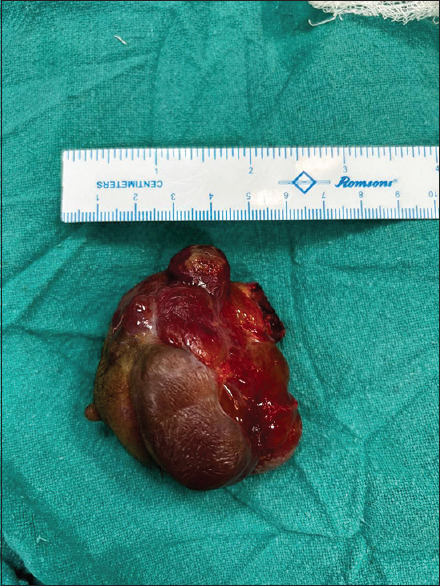
The specimen measured 6.5 cm × 5 cm × 3 cm [Figure 2]. The histopathology report was suggestive of mature teratoma.
Figure 2.

Photograph of the excised mass
The patient was nursed in a supine and lateral position with frequent, low-pressure gentle oral suctioning. The postoperative period was uneventful, wound was healthy, however, the baby expired 10th postoperative day. The patient developed sepsis because of his Surgical neonatal intensive care unit (SNICU) stay, his blood culture report was suggestive of candida pelliculosa, (an unusual isolate).
DISCUSSION
Teratomas are prevalent germ cell tumors in childhood, incorporating tissues from all three germ cell layers. They are frequently observed in the sacrococcygeal regions, anterior mediastinum, retroperitoneum, testes, and ovaries.[4]
The etiology of epignathus remains uncertain. The prevailing theory attributes its origin to the disorganized growth of pluripotent cells in the region of Rathke’s pouch.[5]
A cleft palate stands as the most common malformation associated with epignathus. This condition arises due to teratoma formation during the early stages of fetal development, occurring between 8 and 12 weeks, before the fusion of the palatal shelves. Epignathus has the potential to induce various malformations and complications, especially when the tumor mass is situated between the palatine processes before the 6th week of gestation as it undergoes substantial growth between the 7th and 9th weeks, which may impede the fusion of the nasal septum with the bilateral palatine processes, ultimately leading to the formation of a cleft palate.[6] Notably, malformations associated with epignathus, such as cleft palate, bifid tongue, bifid nose, and duplication of the pituitary gland, were absent in our case.
In recent years, there has been a growing utilization of ultrasonography in prenatal monitoring for congenital anomalies. In addition, magnetic resonance imaging serves as a complementary diagnostic tool for epignathus, aiding in the detection of the tumor’s relationship to the fetal airway and intracranial structures.[7] There was no airway obstruction or intracranial extension in our patient.
Lymphangioma and teratoma are the spectrum of the aberrant proliferation of germ cells during their embryonic development. Various theories have been put forward related to the presence of cleft palate with epignathus, the most accepted one is that epignathus is a parasitic twin arising from the nasopalatine region impairing the fusion of palatal selves in embryonic stages.[8] The simultaneous presence of cervical lymphangioma and epignathus is rare.
CONCLUSION
Epignathus is a rare congenital oropharyngeal teratoma. Large obstructive oral masses that are not diagnosed during the prenatal period carry a significant risk of airway obstruction in the neonatal period, which is why; it should be diagnosed in utero itself to enable the perinatal preparation for emergency management to avoid respiratory obstruction and feeding-related problems in newborns.
Declaration of patient consent
The authors certify that they have obtained all appropriate patient consent forms. In the form, the legal guardian has given his consent for images and other clinical information to be reported in the journal. The guardian understands that names and initials will not be published and due efforts will be made to conceal patient identity, but anonymity cannot be guaranteed.
Financial support and sponsorship
Nil.
Conflicts of interest
There are no conflicts of interest.
REFERENCES
- 1.Tsai YT, Cala Or MA, Lui CC, Wang TJ, Lai JP. Epignathus teratoma with duplication of mandible and tongue: Report of a case. Cleft Palate Craniofac J. 2013;50:363–8. doi: 10.1597/12-015. [DOI] [PubMed] [Google Scholar]
- 2.Rai M, Hegde P, Devaraju UM. Congenital facial teratoma. J Maxillofac Oral Surg. 2012;11:243–6. doi: 10.1007/s12663-011-0186-0. [DOI] [PMC free article] [PubMed] [Google Scholar]
- 3.Sanlialp I, Karnak I, Tanyel FC, Senocak ME, Büyükpamukçu N. Sclerotherapy for lymphangioma in children. Int J Pediatr Otorhinolaryngol. 2003;67:795–800. doi: 10.1016/s0165-5876(03)00123-x. [DOI] [PubMed] [Google Scholar]
- 4.Benson RE, Fabbroni G, Russell JL. A large teratoma of the hard palate: A case report. Br J Oral Maxillofac Surg. 2009;47:46–9. doi: 10.1016/j.bjoms.2007.12.015. [DOI] [PubMed] [Google Scholar]
- 5.Noguchi T, Jinbu Y, Itoh H, Matsumoto K, Sakai O, Kusama M. Epignathus combined with cleft palate, lobulated tongue, and lingual hamartoma: Report of a case. Oral Surg Oral Med Oral Pathol Oral Radiol Endod. 2006;101:481–6. doi: 10.1016/j.tripleo.2005.06.025. [DOI] [PubMed] [Google Scholar]
- 6.Al-Mahdi AH, Al Khurrhi LE, Atto GZ, Dhaher A. Giant epignathus teratoma involving the palate, tongue, and floor of the mouth. J Craniofac Surg. 2013;24:e97–9. doi: 10.1097/SCS.0b013e3182798f25. [DOI] [PubMed] [Google Scholar]
- 7.Aydemir F, Mutaf M, Eryılmaz MA. Giant epignathus (teratoma of palatine tonsil): A case report. Turk Arch Otorhinolaryngol. 2021;59:158–61. doi: 10.4274/tao.2021.2021-4-7. [DOI] [PMC free article] [PubMed] [Google Scholar]
- 8.Menezes Filho MP, Simão NM. Giant epignathus of the palate: A case report. J Bras Patol Med Lab. 2015;51:339–43. [Google Scholar]


