Abstract
Antineutrophil cytoplasmic antibody-associated vasculitis (AAV) is a rare autoimmune disease with an unclear pathogenesis. The present study investigated the associations between autophagy-related protein 16-like 1 (ATG16L1) rs2241880(T300A) and rs4663421 and AAV. A total of 177 patients with AAV and 216 healthy controls were included. Propensity score matching was used to match the two groups of subjects in terms of sex, age and ethnicity. Analyses of the relationships between these genetic polymorphisms and AAV susceptibility, including comparisons of allele and genotype frequency distribution, linkage disequilibrium analysis and analysis of single nucleotide polymorphism (SNP) interactions between two loci were performed. The association between the loci and laboratory test results and renal pathology were also analysed. A total of 154 pairs of patients with AAV and healthy controls was successfully matched. Neither polymorphism was associated with AAV susceptibility. However, SNP interaction in the model constructed with the two loci was statistically significant (P=0.018), and the combination of the AA genotype of rs2241880(T300A) and GG genotype of rs4663421 was associated the highest disease risk. The differences in the Birmingham Vasculitis Activity Score (BVAS), C-reactive protein (CRP) levels and 24-h urine protein level between patients with the rs2241880(T300A) AA + AG genotypes and the GG genotype were statistically significant (P<0.05). Furthermore, significant differences in the severity of glomerulosclerosis and global sclerosis were detected between individuals with the AA + AG genotype and those with the GG genotype at the rs2241880(T300A) locus (P<0.05). Similarly, there were statistically significant differences in degree of segmental sclerosis between individuals with CC + CG genotypes and those with GG genotypes at the rs2243421 locus (P<0.05). In summary, the single gene polymorphisms of these loci were not associated with genetic susceptibility to AAV. However, SNP interactions may serve a role in the risk of AAV. The rs2241880(T300A) polymorphism may be associated with BVAS, CRP levels and 24-h urine protein level in AAV. These SNPs may be associated with glomerulosclerosis and segmental sclerosis.
Keywords: autophagy gene, gene polymorphism, ANCA-associated vasculitis, propensity score matching
Introduction
Antineutrophil cytoplasmic antibody (ANCA)-associated vasculitis (AAV) is an autoimmune disease characterized by necrotizing inflammation of small and medium-sized blood vessels and the presence of ANCAs in circulation. Based on their targeted antigen, ANCAs are classified as proteinase 3-ANCA (PR3-ANCA) or myeloperoxidase-ANCA (MPO-ANCA) (1-3). Among the organs affected by AAV, the kidney is one of the most severely impacted. The pathogenesis of AAV remains unclear. It is currently hypothesized that environmental factors, infection and immune status lead to the development of this disease. A genetic predisposition to AAV has been reported in familial cases and large genome-wide association studies have revealed that susceptibility to AAV is linked to gene variants (4-6).
Autophagy is the process by which intracellular macromolecules, organelles and invasive microorganisms are transported to the lysosome for degradation under regulation of autophagy-related genes (ATGs) (7). Autophagy is key for the realization of cellular metabolic needs, the maintenance of cell and tissue homeostasis and innate and adaptive immunity (8). Autophagy has been implicated in a variety of diseases, including malignant tumours, neurodegenerative disease, metabolic syndrome and autoimmune diseases (9-12). Neutrophil extracellular traps (NETs) are reticular DNA structures combined with immunogenic proteins released by activated neutrophils that not only capture and kill pathogenic microorganisms but also induce tissue damage (13), serving a key role in the initiation and progression of AAV (14). Sha et al (15) reported that human neutrophils treated with ANCA-positive IgG exhibit higher levels of autophagy and release more NETs, which can be enhanced by autophagy inducers, suggesting that autophagic activity may affect ANCA-induced NET formation and release and thus participate in the pathogenesis of AAV.
Autophagy-related 16-like 1 (ATG16L1) is a key component of ATG12-ATG5/ATG16L1, a large protein complex essential for all stages of autophagy (16). ATG16L1 mediates recognition of cellular cargo and activates autophagy-associated enzyme activity required for recruitment to lysosomes (17). ATG16L1 gene polymorphisms [rs2241880(T300A) and rs4663421] may be associated with rheumatoid arthritis, ankylosing spondylitis and inflammatory bowel disease (18-20). The present analysed the associations between ATG16L1 rs2241880(T300A) and rs4663421 polymorphisms and susceptibility in AAV using propensity score matching (PSM) to control for confounding factors. The aim of the present study was to provide a theoretical basis for pathogenesis and therapeutic targets of AAV.
Materials and methods
Subjects
Patients with AAV were recruited from Department of Nephrology, Second Affiliated Hospital of Guangxi Medical University (Guangxi, China) from January 2005 to April 2018. A total of 177 patients were included, 68 male (38.4%) and 109 female (61.6%), with an age range of 18-82 years and a median (IQR) age of 58.0 (43.0-64.0) years. Inclusion criteria were as follows: i) Diagnosis of AAV in strict accordance with the diagnostic criteria for vasculitis formulated at the International Chapel Hill Consensus Conference in 2012(21) and ii) have been born in the Guangxi Zhuang Autonomous Region with no blood relationship with any other subject in the present study. The exclusion criteria were diagnosis of secondary vasculitis, other autoimmune or disease and malignant tumours. The control group consisted of healthy individuals from the Physical Examination Centre of the same hospital during the same period. A total of 216 healthy individuals were included, 84 male (38.9%) and 132 female (61.1%), with an age range of 19-81 years and a median (IQR) age of 51.0 (44.0-59.0) years. Inclusion criteria were as follows: i) Does not meet any of the above classification diagnostic criteria; and ii) have been born in the Guangxi Zhuang Autonomous Region with no blood relationship with any other subject who has participated in this study. The exclusion criteria were diagnosis of autoimmune diseases, hereditary diseases, and other serious diseases. The study was conducted in accordance with the Declaration of Helsinki and approved by the Ethics Committee of the Second Affiliated Hospital of Guangxi Medical University (approval no. 2018 KY-0100). Verbal informed consent was obtained from all participants.
Clinical data
The clinical data collected included sex, age, white blood cell count, 24-h urine protein, albumin and serum creatinine (SCR) levels, estimated glomerular filtration rate (eGFR), C-reactive protein (CRP) levels, erythrocyte sedimentation rate (ESR), MPO-ANCA and PR3-ANCA titre, Birmingham Vasculitis Activity Score (BVAS) and renal pathology (including renal pathological type, normal glomeruli, sclerotic glomeruli, global sclerosis, segmental sclerosis, crescents, cellular crescents, fibrous crescents and cellular fibrous crescents). The renal samples were fixed at 35˚C with 10% buffered formalin for 5 min, embedded in paraffin, sectioned at a thickness of 1.5-2 µm, then stained with hematoxylin and eosin for 10 min and 31 min respectively at room temperature. Finally, observations were made using an light microscope (Olympus Corporation) with x40. Disease activity was assessed using the Birmingham Vasculitis Activity Score (version 3) (22). eGFR was calculated using the Chronic Kidney Disease Epidemiology Collaboration equation (23). ANCA-associated glomerulonephritis was classified according to the histopathological classification proposed by Berden et al (24). The clinical data collected were obtained at the time of diagnosis of patients or during physical examination of healthy individuals.
Single nucleotide polymorphism (SNP) selection
Locus information for the ATG16L1 gene was downloaded from 1000 Genomes database (grch37.ensembl.org/), and SNP loci were filtered out using Haploview v.4.2 software (25). SNP loci meeting the following criteria were selected: i) Located in a functional region; ii) previously reported to be associated with autoimmune or inflammatory disease and iii) having a minor allele frequency ≥0.05. National Center for Bioinformatics (ncbi.nlm.nih.gov/snp/) was used to confirm functional consequence of SNPs and ATG16L1 marker SNPs [rs2241880(T300A) and rs4663421] were selected.
Gene polymorphism detection
Blood samples were collected from all the enrolled patients with AAV and controls between January 2005 to April 2018. EDTA tubes were used for the collection of 5 ml peripheral venous blood samples and genomic DNA was extracted using the Blood DNA Extraction kit [cat. no. DP319-02; Beijing Tiangen Biotech (Beijing) Co., Ltd.] according to the manufacturer's instructions. Nanodrop 2000 spectrophotometer (Thermo Fisher Scientific, Inc.) to measure the concentration of DNA to ensure the adequate amounts of high-quality genomic DNA. The samples with an absorbance value (A260/A280) of 1.7-1.9 and a DNA concentration >25 mg/l were included and the isolated DNA was stored at -80˚C for further study.
Genotyping was performed by Sangon Biotech (Shanghai) Co., Ltd. The sequences of primers were as follows: rs2241880 forward, 5'-TCTCATTTGAGTGAGGGTGCTTTT-3' and reverse, 5'-GTAGCTGGTACCCTCACTTCTTTAC-3' (product size, 274 bp); and rs4663421 forward, 5'-CCCTTCTTCCATGTATCCTGCTT-3' and reverse 5'-CTTCCAGCCAAATCTGCTTTTCC-3'(product size, 274 bp). Library preparation was performed by two-step PCR. First round PCR reaction was set up as follows: DNA (10 ng/µl) 2 µl; amplicon PCR forward primer mix (10 µM) 1 µl; amplicon PCR reverse primer mix (10 µM) 1 µl; 2xPCR Ready Mix 15 µl (total 25 µl). This step was performed with Kapa HiFi Ready Mix [Roche Dianostics (Shanghai) Co., Ltd.). PCR was performed in a thermal instrument (Bio-Rad Laboratories, Inc.; T100TM) using the following program: 1 cycle of denaturing at 98˚C for 5 min, first 8 cycles of denaturing at 98˚C for 30 sec, annealing at 50˚C for 30 sec, elongation at 72˚C for 30 sec, then 25 cycles of denaturing at 98˚C for 30 sec, annealing at 66˚C for 30 sec, elongation at 72˚C for 30 sec and a final extension at 72˚C for 5 min. Finally hold at 4˚C.
The PCR products were checked using electrophoresis in 1 % (w/v) agarose gels in TBE buffer, stained with ethidium bromide and visualized under UV light. Then we used AMPure XP beads to purify the amplicon product. After that, the second round of PCR was performed. The PCR reaction was set up as follows: DNA (10 ng/µl) 2 µl; universal P7 primer with barcode (10 µM) 1 µl; universal P5 primer (10 µM) 1 µl; 2X PCR Ready Mix 15 µl (total 30 µl)(Kapa HiFi Ready Mix). The plate was sealed and PCR was performed as follows: Initial denaturing at 98˚C for 3 min, then 5 cycles of denaturing at 94˚C for 30 sec, annealing at 55˚C for 20 sec, elongation at 72˚C for 30 sec, and a final extension at 72˚C for 5 min. Then we used AMPure XP beads to purify the amplicon product. The libraries were then quantified and pooled. Paired-end sequencing of the library was performed on the HiSeq XTen sequencers (Illumina, Inc.). The samples were sequenced using NovaSeq 6000 S4 Reagent kit v1.5 (300 cycles) (cat. no. 20028312; Illumina Inc.). The loading concentration of the final library was 100 pM. Samtools v 1.18software (htslib.org/) was used to calculate each genotype of target site.
PSM
IBM SPSS Statistics for Windows, Version 26.0 (IBM Corp.) and R 3.5.1 software (26) was used to match sex, age and ethnicity with a matching tolerance of 0.2(27). Jetter plot of propensity score matching was obtained.
Statistical analysis
Hardy-Weinberg equilibrium was checked using SHEsis online software (analysis.biox.cn/myanalysis.php), and linkage disequilibrium tests were performed. The strength of association between genetic models and the risk of AAV was evaluated by odds ratios (ORs) and 95% confidence intervals (CIs) through online SNPstats (https://www.snpstats.net/start.htm). SNP-SNP interactions were assessed using generalized multifactor dimensionality reduction (GMDR). Expression quantitative trait loci (eQTL) database (gtexportal.org/home/eqtlDashboardPage) was used to assess the effect of different genotypes of candidate loci on tissue-specific gene expression. All statistical analyses were performed with IBM SPSS Statistics for Windows, Version 26.0 (IBM Corp.). Normally distributed variables are presented as the mean ± SD of 3 independent experimental repeats and non-normally distributed variables are presented as median and interquartile range (IQR). The categorical variables are expressed as frequency and percentage and comparisons between groups were made using Pearson's χ2 test or Fisher's exact test. P<0.05 was considered to indicate a statistically significant difference.
Results
Demographic characteristics
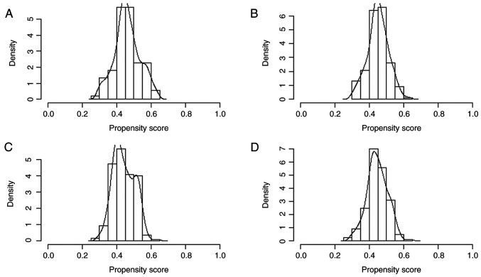
A total of 177 patients and 216 healthy individuals were included. There was a significant age gap between the AAV and control groups before PSM (Table I) but no apparent difference in sex or ethnicity between the groups. Following PSM, 154 patients were successfully matched. There was no significant difference between age of groups and the balance of each covariate was significantly increased (Figs. 1 and 2).
Table I.
Baseline influencing factors before and after PSM.
| Before PSM | After PSM | |||||
|---|---|---|---|---|---|---|
| Characteristic | AAV (n=177) | Control (n=216) | P-value | AAV (n=154) | Control (n=154) | P-value |
| Sex (%) | 0.924 | 0.483 | ||||
| Male | 68 (38.4) | 84 (38.9) | 63 (40.9) | 57 (37.0) | ||
| Female | 109 (61.6) | 132 (61.1) | 91 (59.1) | 97 (63.0) | ||
| Ethnicity (%) | 0.099 | 0.611 | ||||
| Zhuang | 62 (35.0) | 58 (26.9) | 45 (29.2) | 41 (26.6) | ||
| Han | 115 (65.0) | 158 (73.1) | 109 (70.8) | 113 (73.4) | ||
| Median age (IQR), years | 58.0 (43.0-64.0) | 51.0 (44.0-59.0) | 0.005 | 56.0 (42.0-63.0) | 53.0 (45.8-61.0) | 0.445 |
PSM, propensity score matching; AAV, antineutrophil cytoplasmic autoantibody-associated vasculitis.
Figure 1.
Histogram of propensity score matching before and after matching. Antineutrophil cytoplasmic autoantibody-associated vasculitis group (A) before and (B) after matching. Control group (C) before and (D) after matching.
Figure 2.
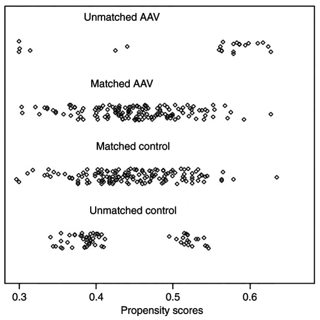
Jetter plot of propensity score matching. AAV, antineutrophil cytoplasmic autoantibody-associated vasculitis.
Genotype distribution, allele frequency, linkage disequilibrium and Hardy-Weinberg equilibrium analysis
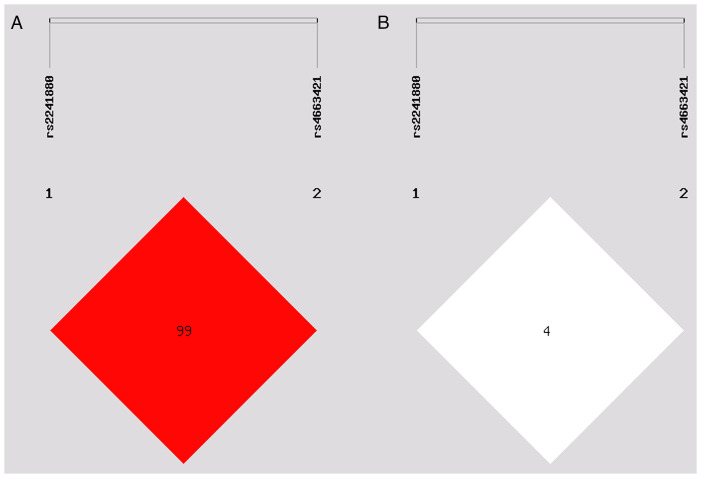
The genotype distribution of rs2241880(T300A) and rs4663421 of ATG16L1 were in accordance with the genetic law of Hardy-Weinberg equilibrium. The linkage disequilibrium was weak between rs2241880(T300A) and rs4663421 loci (Fig. 3; D'=0.998, R2=0.045). Associations between ATG16L1 SNPs and AAV risk were evaluated under different genetic models (Tables II and III). Genotypes of rs2241880 and rs4663421 under different genetic models did not significantly difference between AAV and control groups. And there was no significant difference in the alleles of the two loci between AAV and control groups.
Figure 3.
Linkage disequilibrium analysis of single-nucleotide polymorphisms in the autophagy-related protein 16 like 1 gene. (A) D'=0.998. D', Lewontin's standardised disequilibrium coefficient, 0 ≤ D' ≤ 1, the darker the colour, the bigger D' is. (B) R2=0.045. R2, squared correlation coefficient, 0 ≤ R2 ≤ 1, the darker the color is, the greater the R2 value is.
Table II.
Association between the risk of AAV and single nucleotide polymorphism rs2241880.
| Model | Genetype/Allele | Control (%) | AAV (%) | OR (95% CI) | P-value |
|---|---|---|---|---|---|
| Allele | G | 98 (31.8) | 88 (28.6) | Reference | 0.38 |
| A | 210 (68.2) | 220 (71.4) | 1.17 (0.83-1.65) | ||
| Codominant | AA | 71 (46.1) | 78 (50.6) | Reference | 0.68 |
| AG | 68 (44.2) | 64 (41.6) | 0.86 (0.54-1.37) | ||
| GG | 15 (9.7) | 12 (7.8) | 0.73 (0.32-1.66) | ||
| Dominant | AA | 71 (46.1) | 78 (50.6) | Reference | 0.42 |
| AG-GG | 83 (53.9) | 76 (49.4) | 0.83 (0.53-1.30) | ||
| Recessive | AA-AG | 139 (90.3) | 142 (92.2) | Reference | 0.55 |
| GG | 15 (9.7) | 12 (7.8) | 0.78 (0.35-1.73) | ||
| Overdominant | AA-GG | 86 (55.8) | 90 (58.4) | Reference | 0.65 |
| AG | 68 (44.2%) | 64 (41.6%) | 0.90 (0.57-1.41) |
AAV, antineutrophil cytoplasmic autoantibody-associated vasculitis.
Table III.
Association between risk of AAV and single nucleotide polymorphism rs4663421 in different genotypic models.
| Model | Genetype/Allele | Control (%) | AAV (%) | OR (95% CI) | P-value |
|---|---|---|---|---|---|
| Allele | G | 279 (90.6) | 282 (91.6) | Reference | 0.67 |
| C | 29 (9.4) | 26 (8.4) | 0.89 (0.51-1.55) | ||
| Codominant | GG | 125 (81.2) | 129 (83.8) | Reference | 0.38 |
| GC | 29 (18.8) | 24 (15.6) | 0.80 (0.44-1.45) | ||
| CC | 0 (0.0) | 1 (0.6) | NA (0.00-NA) | ||
| Dominant | GG | 125 (81.2) | 129 (83.8) | Reference | 0.55 |
| GC-CC | 29 (18.8) | 25 (16.2) | 0.84 (0.46-1.51) | ||
| Recessive | GG-GC | 154(100) | 153 (99.3) | Reference | 0.24 |
| CC | 0 (0) | 1 (0.6) | NA (0.00-NA) | ||
| Overdominant | GG-CC | 125 (81.2) | 130 (84.4) | Reference | 0.45 |
| GC | 29 (18.8) | 24 (15.6) | 0.80 (0.44-1.44) |
AAV, antineutrophil cytoplasmic autoantibody-associated vasculitis; NA, not applicable.
SNP interaction analysis
GMDR obtains the best model combination from multiple genes or SNPs and behavioural indicators to analyse gene-gene or SNP-SNP interactions. The interaction between rs2241880(T300A) and rs4663421 was significant. The cross-verifying consistency was 8/10 and the training balance accuracy was 0.5844 after 1,000 permutation tests. The highest risk was found when the AA genotype of rs2241880(T300A) combined with the GG genotype of rs4663421 (Fig. 4).
Figure 4.
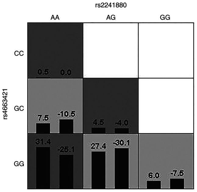
Generalized multifactor dimensionality reduction analysis. A box represents an interaction combination; highrisk boxs are denoted by a dark color, lowrisk boxs are denoted by a light tint. In the same box, the left column is the positive score of the combination, and the right is the negative score; the higher the positive score, the higher the risk of antineutrophil cytoplasmic autoantibody-associated vasculitis (AAV) in the combo.
eQTL analysis
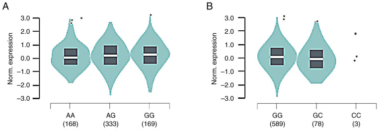
rs2241880/T300A G allele was associated with increased ATG16L1 mRNA in whole-blood samples from 670 healthy donors (median relative expression AA: -0.1331, AG: -0.02428, GG: 0.08790, P=0.0000037; Fig. 5). rs4663421 G allele was associated with increased ATG16L1 mRNA (median relative expression: GC: -0.1746, GG: 0.03176). Therefore, the recessive model of rs2241880(T300A) (AA + AG, GG) and the dominant model of rs4663421 (CC + CG, GG) were used to examine the association between SNPs and clinical characteristics.
Figure 5.
Expression Quantitative Trait Loci (eQTL) analysis of ATG16L1 rs2241880 and rs4663421 in whole blood. (A) ATG16L1 rs2241880-G is associated with increased ATG16L1 mRNA expression. (B) ATG16L1 rs4663421-G is associated with increased ATG16L1 mRNA expression. Norm., normal.
Association between ATG16L1 gene polymorphisms and AAV clinical characteristics
CRP level and BVAS were higher, whereas the 24-h urinary protein level was significantly lower for patients with rs2241880(T300A) AA+AG genotype than for patients with the GG genotype (Table IV). There was no significant association between the rs4663421 gene polymorphism and CRP and 24-h urine protein level, BVAS, ANCA type, white blood cell count, serum creatinine level, ESR, eGFR or serum albumin level in patients with AAV.
Table IV.
Genotypes and clinical characteristics at two single nucleotide polymorphisms of autophagy-related protein 16 like 1 gene.
| rs2241880 | rs4663421 | |||||
|---|---|---|---|---|---|---|
| Variable | AA + AG (n=163) | GG (n=14) | P-value | CC + CG (n=30) | GG (n=147) | P-value |
| ANCA specificity (%) | 0.824 | 0.206 | ||||
| MPO | 102 (77.86) | 10 (76.92) | 16 (69.57) | 96 (79.34) | ||
| PR3 | 15 (11.45) | 1 (7.69) | 2 (8.70) | 14 (11.57) | ||
| None | 14 (10.69) | 2 (15.38) | 5 (21.74) | 11 (9.09) | ||
| Median WBC count, x109/l (IQR) | 8.16 (5.84-10.90) | 8.01 (6.45-10.70) | 0.900 | 7.49 (5.94-11.61) | 8.19 (6.10-10.70) | 0.815 |
| Median 24-h UPR, mg (IQR) | 1,011.90 (352.45-1759.40) | 2116.20 (694.80-4393.00) | 0.024 | 1,369.37 (759.19-2875.45) | 912.10 (371.48-1,786.45) | 0.157 |
| Mean albumin, g/l | 29.99±6.51 | 31.28±8.10 | 0.504 | 28.71±8.11 | 30.37±6.33 | 0.274 |
| Median SCR, µmol/l (IQR) | 259.00 (99.00-486.00) | 317 (141.00-607.00) | 0.378 | 249.00 (68.00-450.00) | 267.00 (110.00-511.00) | 0.597 |
| Median eGFR, ml/min x 1.73 m2 (IQR) | 19.08 (8.46-63.37) | 12.66 (7.79-49.11) | 0.439 | 21.41 (8.96-92.15) | 18.17 (8.03-60.29) | 0.626 |
| Median CRP, mg/l (IQR) | 20.80 (9.10-70.60) | 5.80 (3.14-27.45) | 0.010 | 13.58 (9.69-72.20) | 20.60 (6.67-63.34) | 0.941 |
| Median ESR, mm/h (IQR) | 81.00 (52.00-109.00) | 70.00 (27.50-92.00) | 0.189 | 78.50 (38.50-96.75) | 81.00 (44.50-109.75) | 0.542 |
| Mean BVAS | 16.81±0.39 | 13.50±1.41 | 0.017 | 18.23±4.58 | 16.22±4.56 | 0.060 |
ANCA, antineutrophil cytoplasmic antibody; MPO, myeloperoxidase; PR3, proteinase 3; WBC, white blood cell; UPR, urinary protein; SCR, serum creatinine; eGFR, estimated glomerular filtration rate; CRP, C-reactive protein; ESR, erythrocyte sedimentation rate; BVAS, Birmingham Vasculitis Activity Score.
Analysis of the association of ATG16L1 gene polymorphisms with AAV renal pathology
A total of 68 patients with renal pathology data were included. Based on pathological classification of glomerular burden, 35 patients (51.47%) were classified as having focal-type AAV, 20 (29.41%) as having sclerosing-type AAV, 5 (7.35%) as having crescent-type AAV and 8 (11.76%) as having mixed-type AAV. Compared with the AA + AG genotype, GG genotype of rs2241880(T300A) was significantly associated with more severe glomerulosclerosis and global sclerosis (Table V). For rs4663421, segmental sclerosis was more severe in patients with the CC + CG and GG genotypes. SNPs were not associated with renal pathological type, percentage of normal glomeruli, crescents, cellular crescents, fibrous crescents and cellular fibrous crescents in AAV.
Table V.
Genotypes and renal pathology at two single nucleotide polymorphisms of autophagy-related protein 16-like 1 gene.
| rs2241880 | rs4663421 | |||||
|---|---|---|---|---|---|---|
| Variable | AA + AG (n=61) | GG (n=7) | P-value | CC + CG (n=10) | GG (n=58) | P-value |
| Berden classificatio, n (%) | 0.302 | 0.268 | ||||
| Focal | 32 (52.46) | 3 (42.86) | 5 (50.00) | 30 (51.72) | ||
| Sclerotic | 16 (26.23) | 4 (57.14) | 5 (50.00) | 15 (25.86) | ||
| Crescent | 5 (8.20) | 0 (0.00) | 0 (0.00) | 5 (8.62) | ||
| Mixed | 8 (13.11) | 0 (0.00) | 0 (0.00) | 8 (13.79) | ||
| Median normal glomeruli (IQR) | 52.63 (18.34,81.67) | 20.00 (0.00,81.25) | 0.210 | 43.18 (14.58,66.67) | 51.32 (15.63,81.77) | 0.801 |
| Sclerotic glomeruli, n (%) | 47 (77.05) | 7 (100.00) | 0.330 | 9 (90.00) | 45 (77.59) | 0.370 |
| Degree of disease, % (median, IQR) | 21.43 (4.12,52.28) | 62.50 (18.75,80.00) | 0.042 | 55.00 (19.41,65.42) | 18.47 (4.56,51.14) | 0.082 |
| Global sclerosis, n (%) | 46 (75.41) | 7 (100.00) | 0.334 | 9 (90.00) | 44 (75.86) | 0.319 |
| Degree of disease, % (median, IQR) | 17.43 (3.67,36.20) | 37.50 (18.75,75.00) | 0.040 | 31.19 (12.39,46.59) | 18.18 (4.12,36.93) | 0.185 |
| Segmental sclerosis, n (%) | 22 (36.07) | 3 (42.86) | 0.724 | 8 (80.00) | 17 (29.31) | 0.002 |
| Degree of disease, % (median, IQR) | 0.00 (0.00,7.14) | 0.00 (0.00,25.00) | 0.496 | 10.72 (5.00,22.50) | 0.00 (0.00,5.61) | 0.002 |
| Crescents, n (%) | 37 (60.66) | 2 (28.57) | 0.104 | 5 (50.00) | 34 (58.62) | 0.611 |
| Degree of disease, % (median, IQR) | 15.38 (0.00,27.62) | 0.00 (0.00,25.00) | 0.204 | 3.34 (0.00,21.63) | 13.25 (0.00,29.41) | 0.260 |
| Cellular crescents, n (%) | 12 (19.67) | 0 (0.00) | 0.196 | 2 (20.00) | 10 (17.24) | 0.833 |
| Mean degree of disease | 2.20±5.51 | 0.00±0.00 | 0.202 | 1.38±2.91 | 2.08±5.58 | 0.865 |
| Cellular fibrous crescents, n (%) | 24 (39.34) | 2 (28.57) | 0.579 | 2 (20.00) | 24 (41.38) | 0.199 |
| Degree of disease, % (median, IQR) | 0.00 (0.00,17.92) | 0.00 (0.00,15.00) | 0.541 | 0.00 (0.00,3.57) | 0.00 (0.00,18.64) | 0.166 |
| Fibrous crescents, n (%) | 18 (29.51) | 2 (28.57) | 0.959 | 3 (30.00) | 17 (29.31) | 0.965 |
| Degree of disease, % (median, IQR) | 0.00 (0.00,15.00) | 0.00 (0.00,10.00) | 0.880 | 0.00 (0.00,10.00) | 0.00 (0.00,5.73) | 0.914 |
Discussion
The exact cause of AAV remains uncertain; however, genetic factors serve a significant role in AAV (28). The present study explored the relationship between polymorphisms in ATG16L1 and AAV susceptibility, laboratory examination results and renal pathology in AAV. PSM was used to match AAV and control groups. Before matching, some baseline clinical data differed between the groups, which may have affected the distribution of genes. Following 1:1 matching for sex, age and ethnicity, both groups presented similar demographic characteristics, with no significant differences, indicating PSM significantly increased the balance of covariates.
ANCA is a serum marker for AAV. However, ANCA is also found in patients with inflammatory bowel disease (29) and PR3-ANCA-positive patients with ulcerative colitis have more extensive disease than those who are PR3-ANCA negative (30). The pathogenicity of high-titre ANCA has also been highlighted in case reports of AAV in patients with Crohn's disease (CD) (31,32). Therefore, there may be a common pathogenic mechanism between AAV and inflammatory bowel disease. ATG16L1 is associated with elevated levels of inflammatory factors IL-6, IL-8 and IL-1β (33-34). The release of cytokines IL-8, IL-6 and TNF-α by immune cells leads to excessive activation of neutrophils to induce the formation of NETs, resulting in occurrence of AAV (35,36) and an association with AAV disease activity (37). Here, it was hypothesized that ATG16L1 rs2241880(T300A) and rs4663421 may be involved in the pathogenesis of AAV through effects on NETs and cytokines. rs2241880(T300A) and rs4663421 are associated with inflammatory bowel disease and several studies of polymorphisms in the gene encoding ATG16L1 have revealed that the T300A G allele is associated with a greater risk of IBD development (38-41). Almost all CD risk imposed by the ATG16L1 locus is associated with carrying the T300A variant (42). The ATG16L1 variant T300A affects secretion of TNF-α and IL-1β, promotes inflammatory responses and increases susceptibility to CD (43). Here, the AA genotype (50.6 vs. 46.1) and AG genotype (41.6 vs. 44.2%) of the ATG16L1 rs2241880(T300A) polymorphism were more frequent in patients with AAV than in healthy controls, whereas the GG genotype was less frequent (7.8 vs. 9.7) and the G allele was present in a minority of patients and controls (7.8 vs. 9.7%). A meta-analysis by Zhang et al (44) confirmed that the T300A G allele is positively associated with CD occurrence in Caucasians, but no significant association was found in Asians and the frequency of the G allele differed between Asians and Caucasians, potentially because racial differences play a role in the genetic background. The present study did not find a significant difference in the ATG16L1 T300A genotype or allele frequency between patients and healthy controls (P>0.05), which may be due to the low frequency of G allele and GG genotype. The GG genotype of rs4663421 was predominant in healthy individuals and patients with AAV. By contrast, the CC genotype has rarely been reported, which is consistent with findings in other populations (20,38), but an association between the rs4663421 polymorphism and AAV has not been confirmed. No significant difference was found between cases and controls. Associations between codominant, dominant, recessive and overdominant models with AAV were assessed. However, there was no significant association between these models of ATG16L1 rs2241880(T300A) and rs4663421 and risk of developing AAV. AAV is relatively rare, is not the result of a single gene mutation but is associated with multiple genetic variants and may be associated with gene-gene or SNP-SNP interactions (45). SNP-SNP interaction analysis between ATG16L1 rs2241880(T300A) and rs4663421 found that the combination of the AA genotype of rs2241880 and the GG genotype of rs4663421 increased risk of AAV. Thus, although rs2241880 and rs4663421 did not show any association with AAV individually, the interaction of these two SNPs may promote the development of AAV.
Due to diverse clinical manifestations, courses and outcomes of AAV, the present study investigated the impact of gene polymorphisms on the clinical manifestations of AAV. Higher BVAS indicates more active disease (46). The CRP level is often used as a non-specific marker of inflammation and is associated with disease activity in AAV (47). ATG16L1 T300A variant can affect autophagy (48). rs2241880(T300A), when carried by a GG homozygote, impairs autophagic flux and associated vesicle trafficking (49). Here, GG genotype of rs2241880(T300A) was associated with lower BVAS and CRP level than the AA + AG genotype was. Autophagy regulates formation and release of NETs. The PI3K/AKT/mTOR pathway connects autophagy and NETs, and mTOR inhibitor rapamycin increases the formation of NETs by promoting autophagy (50,51). Therefore, rs2241880(T300A) gene polymorphism may influence the disease activity of AAV by affecting the production and release of NETs and ANCAs. ATG16L1 maintains cell homeostasis by inhibiting inflammatory responses (52). eQTL analysis demonstrated that the rs2241880(T300A) G allele was associated with higher mRNA levels of ATG16L1 than the rs2241880(T300A) A allele. rs2241880(T300A) GG genotype may reduce disease activity and CRP levels due to the upregulation of ATG16L1 expression. Further studies are needed to distinguish the high disease activity and CRP levels in patients with AAV from those with pulmonary infection as there were many confounding factors in the present study.
Renal involvement in AAV can manifest as active lesions, including glomerular crescent formation and necrosis, or as chronic lesions, including glomerulosclerosis. Patients with the GG genotype of rs2241880(T300A) presented more pronounced glomerulosclerosis and global sclerosis than those with the AA + AG genotype. Additionally, 24-h urine protein level was greater in patients with the GG genotype, indicating more severe damage to the glomeruli. When autophagy was impaired, patients exhibited more severe glomerular damage. Patients with AAV carrying CC + CG genotype of rs4663421 had more severe segmental sclerosis. Further investigation of the association between the rs4663421 gene polymorphism and the level of autophagy is needed. Proteinuria specifically reflects renal involvement of AAV and podocyte injury serves a pivotal role in the development of proteinuria. When ATGs are specifically knocked out in podocytes, mice develop proteinuria, progressive glomerulosclerosis, severe renal interstitial lesions and renal failure, thus showing focal segmental glomerulosclerosis (53). These findings indicate the role of autophagy as a key mechanism for maintaining the homeostasis of podocytes and renal epithelial cells, thus preserving their integrity. The present study revealed a connection between autophagy gene variants and glomerulosclerosis and proteinuria in AAV. This association may be connected to the aforementioned mechanism, but further investigation is necessary to develop appropriate animal models.
The present study has numerous limitations. First, some patients had missing data, and the single-centre, small sample size study should be further validated by a larger, more diverse cohort. Second, due to difficulties in sample collection and storage, assays for mRNA expression to complement the genetic association studies were not performed. In vitro experiments or animal models should be used in future to validate the effects of these genetic variants on ATG16L1 expression and other biological processes such as renal pathological changes and disease activity. In addition, the present study only studied the interaction with SNPs in the same gene. In further studies, interactions with SNPs in different genes should be included to provide more insight into the effects of gene interactions in AAV progression.
In summary, rs2241880(T300A) and rs4663421 polymorphisms of ATG16L1 demonstrated regional specificity. The present study did not reveal any connection between these variants and genetic predisposition to AAV. However, the SNP-SNP interaction in the model constructed with the two SNPs was significant. The ATG16L1 rs2241880(T300A) gene polymorphism may be associated with disease activity and glomerulosclerosis in AAV and the rs4663421 gene polymorphism may be associated with segmental sclerosis.
Acknowledgements
Not applicable.
Funding Statement
Funding: The present study was supported by Joint Project on Regional High –Incidence Diseases Research of Guangxi Natural Science Foundation (grant no. 2024GXNSFAA999092), the Guangxi Health Commission self-financial project (No. Z-A20230648), Guangxi Natural Science Foundation Program (grant no. 2018GXNSFAA281122) and the National Natural Science Foundation of China (grant no. 81360117).
Availability of data and materials
The data generated in the present study are not publicly available due to legal constraints but may be requested from the corresponding author.
Authors' contributions
WLT, YRZ and CX conceived the study and interpreted data. WLT and YRZ designed the experiments and wrote the manuscript. WLT and SRL performed experiments. SRL analysed and interpreted data. WLT and YRZ confirm the authenticity of all the raw data. All authors have read and approved the final manuscript.
Ethics approval and consent to participate
The present study was conducted according to the Declaration of Helsinki and approved by the Ethics Committee of the Second Affiliated Hospital of Guangxi Medical University (approval no. 2018 KY-0100). Verbal informed consent for participation was obtained from all participants.
Patient consent for publication
Not applicable.
Competing interests
The authors declare that they have no competing interests.
References
- 1.Cornec D, Cornec-Le Gall E, Fervenza FC, Specks U. ANCA-associated vasculitis-clinical utility of using ANCA specificity to classify patients. Nat Rev Rheumatol. 2016;12:570–579. doi: 10.1038/nrrheum.2016.123. [DOI] [PubMed] [Google Scholar]
- 2.Geetha D, Jefferson JA. ANCA-Associated Vasculitis: Core Curriculum 2020. Am J Kidney Dis. 2020;75:124–137. doi: 10.1053/j.ajkd.2019.04.031. [DOI] [PubMed] [Google Scholar]
- 3.Kitching AR, Anders HJ, Basu N, Brouwer E, Gordon J, Jayne DR, Kullman J, Lyons PA, Merkel PA, Savage COS, et al. ANCA-associated vasculitis. Nat Rev Dis Primers. 2020;6(71) doi: 10.1038/s41572-020-0204-y. [DOI] [PubMed] [Google Scholar]
- 4.Lyons PA, Peters JE, Alberici F, Liley J, Coulson RMR, Astle W, Baldini C, Bonatti F, Cid MC, Elding H, et al. Genome-wide association study of eosinophilic granulomatosis with polyangiitis reveals genomic loci stratified by ANCA status. Nat Commun. 2019;10(5120) doi: 10.1038/s41467-019-12515-9. [DOI] [PMC free article] [PubMed] [Google Scholar]
- 5.Prendecki M, Cairns T, Pusey CD. Familial vasculitides: Granulomatosis with polyangiitis and microscopic polyangiitis in two brothers with differing anti-neutrophil cytoplasm antibody specificity. Clin Kidney J. 2016;9:429–431. doi: 10.1093/ckj/sfw016. [DOI] [PMC free article] [PubMed] [Google Scholar]
- 6.Tanna A, Salama AD, Brookes P, Pusey CD. Familial granulomatosis with polyangiitis: Three cases of this rare disorder in one Indoasian family carrying an identical HLA DPB1 allele. BMJ Case Rep. 2012;2012(bcr0120125502) doi: 10.1136/bcr.01.2012.5502. [DOI] [PMC free article] [PubMed] [Google Scholar]
- 7.Matoba K, Noda NN. A structural catalog of core Atg proteins opens a new era of autophagy research. J Biochem. 2021;169:517–525. doi: 10.1093/jb/mvab017. [DOI] [PubMed] [Google Scholar]
- 8.Yin Z, Pascual C, Klionsky DJ. Autophagy: Machinery and regulation. Microb Cell. 2016;3:588–596. doi: 10.15698/mic2016.12.546. [DOI] [PMC free article] [PubMed] [Google Scholar]
- 9.Arias E, Cuervo AM. Pros and cons of chaperone-mediated autophagy in cancer biology. Trends Endocrinol Metab. 2020;31:53–66. doi: 10.1016/j.tem.2019.09.007. [DOI] [PMC free article] [PubMed] [Google Scholar]
- 10.Celia AI, Colafrancesco S, Barbati C, Alessandri C, Conti F. Autophagy in Rheumatic Diseases: Role in the pathogenesis and therapeutic approaches. Cells. 2022;11(1359) doi: 10.3390/cells11081359. [DOI] [PMC free article] [PubMed] [Google Scholar]
- 11.Pei Y, Chen S, Zhou F, Xie T, Cao H. Construction and evaluation of Alzheimer's disease diagnostic prediction model based on genes involved in mitophagy. Front Aging Neurosci. 2023;15(1146660) doi: 10.3389/fnagi.2023.1146660. [DOI] [PMC free article] [PubMed] [Google Scholar]
- 12.Yang M, Luo S, Chen W, Zhao L, Wang X. Chaperone-Mediated Autophagy: A potential target for metabolic diseases. Curr Med Chem. 2023;30:1887–1899. doi: 10.2174/0929867329666220811141955. [DOI] [PubMed] [Google Scholar]
- 13.Masucci MT, Minopoli M, Del Vecchio S, Carriero MV. The emerging role of neutrophil extracellular traps (NETs) in tumor progression and metastasis. Front Immunol. 2020;11(1749) doi: 10.3389/fimmu.2020.01749. [DOI] [PMC free article] [PubMed] [Google Scholar]
- 14.Arneth B, Arneth R. Neutrophil Extracellular Traps (NETs) and Vasculitis. Int J Med Sci. 2021;18:1532–1540. doi: 10.7150/ijms.53728. [DOI] [PMC free article] [PubMed] [Google Scholar]
- 15.Sha LL, Wang H, Wang C, Peng HY, Chen M, Zhao MH. Autophagy is induced by anti-neutrophil cytoplasmic Abs and promotes neutrophil extracellular trap formation. Innate Immun. 2016;22:658–665. doi: 10.1177/1753425916668981. [DOI] [PubMed] [Google Scholar]
- 16.Romanov J, Walczak M, Ibiricu I, Schuchner S, Ogris E, Kraft C, Martens S. Mechanism and functions of membrane binding by the Atg5-Atg12/Atg16 complex during autophagosome formation. EMBO J. 2012;31:4304–4317. doi: 10.1038/emboj.2012.278. [DOI] [PMC free article] [PubMed] [Google Scholar]
- 17.Gammoh N. The multifaceted functions of ATG16L1 in autophagy and related processes. J Cell Sci. 2020;133(jcs249227) doi: 10.1242/jcs.249227. [DOI] [PubMed] [Google Scholar]
- 18.Salem M, Ammitzboell M, Nys K, Seidelin JB, Nielsen OH. ATG16L1: A multifunctional susceptibility factor in Crohn's disease. Autophagy. 2015;11:585–594. doi: 10.1080/15548627.2015.1017187. [DOI] [PMC free article] [PubMed] [Google Scholar]
- 19.Mo YJ, Zhang W, Wen QW, Wang TH, Qin W, Zhang Z, Huang H, Cen H, Wu XD. doi: 10.1016/j.intimp.2021.108511. Corrigendum to ‘Genetic association analysis of ATG16L1 rs2241880, rs6758317 and ATG16L2 rs11235604 polymorphisms with rheumatoid arthritis in a Chinese population’ [Int. Immunopharmacol 93 (2021) 107378]. Int. Immunopharmacol 104: 108511, 2022. [DOI] [PubMed] [Google Scholar]
- 20.Li X, Chen M, Zhang X, Wang M, Yang X, Xia Q, Han R, Liu R, Xu S, Xu J, et al. Single nucleotide polymorphisms of autophagy-related 16-like 1 gene are associated with ankylosing spondylitis in females: A case-control study. Int J Rheum Dis. 2018;21:322–329. doi: 10.1111/1756-185X.13183. [DOI] [PubMed] [Google Scholar]
- 21.Jennette JC, Falk RJ, Bacon PA, Basu N, Cid MC, Ferrario F, Flores-Suarez LF, Gross WL, Guillevin L, Hagen EC, et al. 2012 revised International Chapel Hill Consensus Conference Nomenclature of Vasculitides. Arthritis Rheum. 2013;65:1–11. doi: 10.1002/art.37715. [DOI] [PubMed] [Google Scholar]
- 22.Mukhtyar C, Lee R, Brown D, Carruthers D, Dasgupta B, Dubey S, Flossmann O, Hall C, Hollywood J, Jayne D, et al. Modification and validation of the Birmingham Vasculitis Activity Score (version 3) Ann Rheum Dis. 2009;68:1827–1832. doi: 10.1136/ard.2008.101279. [DOI] [PubMed] [Google Scholar]
- 23.Stevens LA, Schmid CH, Zhang YL, Coresh J, Manzi J, Landis R, Bakoush O, Contreras G, Genuth S, Klintmalm GB, et al. Development and validation of GFR-estimating equations using diabetes, transplant and weight. Nephrol Dial Transplant. 2010;25:449–457. doi: 10.1093/ndt/gfp510. [DOI] [PMC free article] [PubMed] [Google Scholar]
- 24.Berden AE, Ferrario F, Hagen EC, Jayne DR, Jennette JC, Joh K, Neumann I, Noël LH, Pusey CD, Waldherr R, et al. Histopathologic classification of ANCA-associated glomerulonephritis. J Am Soc Nephrol. 2010;21:1628–1636. doi: 10.1681/ASN.2010050477. [DOI] [PubMed] [Google Scholar]
- 25.Barrett JC, Fry B, Maller J, Daly MJ. Haploview: Analysis and visualization of LD and haplotype maps. Bioinformatics. 2005;21:263–265. doi: 10.1093/bioinformatics/bth457. [DOI] [PubMed] [Google Scholar]
- 26.Huang F, DU C, Sun M, Ning B, Luo Y, An S. Propensity score matching in SPSS. Nan Fang Yi Ke Da Xue Xue Bao. 2015;35:1597–1601. (In Chinese) [PubMed] [Google Scholar]
- 27.Austin PC, Yu AYX, Vyas MV, Kapral MK. Applying propensity score methods in clinical research in neurology. Neurology. 2021;97:856–863. doi: 10.1212/WNL.0000000000012777. [DOI] [PMC free article] [PubMed] [Google Scholar]
- 28.Lyons PA, Rayner TF, Trivedi S, Holle JU, Watts RA, Jayne DR, Baslund B, Brenchley P, Bruchfeld A, Chaudhry AN, et al. Genetically distinct subsets within ANCA-associated vasculitis. N Engl J Med. 2012;367:214–223. doi: 10.1056/NEJMoa1108735. [DOI] [PMC free article] [PubMed] [Google Scholar]
- 29.Tang H, Tan B, Shen BB, Zhang SL, Qian JM. Diagnostic value of different serological markers and correlation analysis with disease phenotype in inflammatory bowel disease. Zhonghua Yi Xue Za Zhi. 2022;102:3743–3748. doi: 10.3760/cma.j.cn112137-20220418-00834. (In Chinese) [DOI] [PubMed] [Google Scholar]
- 30.Laass MW, Ziesmann J, de Laffolie J, Röber N, Conrad K. Anti-Proteinase 3 Antibodies as a Biomarker for Ulcerative Colitis and Primary Sclerosing Cholangitis in Children. J Pediatr Gastroenterol Nutr. 2022;74:463–470. doi: 10.1097/MPG.0000000000003359. [DOI] [PubMed] [Google Scholar]
- 31.Plafkin C, Zhong W, Singh T. ANCA vasculitis presenting with acute interstitial nephritis without glomerular involvement. Clin Nephrol Case Stud. 2019;7:46–50. doi: 10.5414/CNCS109805. [DOI] [PMC free article] [PubMed] [Google Scholar]
- 32.Freeman HJ. Atypical perinuclear antineutrophil cytoplasmic antibodies in patients with Crohn's disease. Can J Gastroenterol. 1997;11:689–693. doi: 10.1155/1997/237085. [DOI] [PubMed] [Google Scholar]
- 33.Mommersteeg MC, Simovic I, Yu B, van Nieuwenburg SAV, Bruno IMJ, Doukas M, Kuipers EJ, Spaander MCW, Peppelenbosch MP, Castaño-Rodríguez N, Fuhler GM. Autophagy mediates ER stress and inflammation in Helicobacter pylori-related gastric cancer. Gut Microbes. 2022;14(2015238) doi: 10.1080/19490976.2021.2015238. [DOI] [PMC free article] [PubMed] [Google Scholar]
- 34.Plantinga TS, Crisan TO, Oosting M, van de Veerdonk FL, de Jong DJ, Philpott DJ, van der Meer JW, Girardin SE, Joosten LA, Netea MG. Crohn's disease-associated ATG16L1 polymorphism modulates pro-inflammatory cytokine responses selectively upon activation of NOD2. Gut. 2011;60:1229–1235. doi: 10.1136/gut.2010.228908. [DOI] [PubMed] [Google Scholar]
- 35.Hao J, Lv TG, Wang C, Xu LP, Zhao JR. Macrophage migration inhibitory factor contributes to anti-neutrophil cytoplasmic antibody-induced neutrophil activation. Hum Immunol. 2016;77:1209–1214. doi: 10.1016/j.humimm.2016.08.006. [DOI] [PubMed] [Google Scholar]
- 36.Takeda S, Watanabe-Kusunoki K, Nakazawa D, Kusunoki Y, Nishio S, Atsumi T. The Pathogenicity of BPI-ANCA in a Patient with Systemic Vasculitis. Front Immunol. 2019;10(1334) doi: 10.3389/fimmu.2019.01334. [DOI] [PMC free article] [PubMed] [Google Scholar]
- 37.Matsumoto K, Suzuki K, Yoshimoto K, Seki N, Tsujimoto H, Chiba K, Takeuchi T. Longitudinal immune cell monitoring identified CD14(++) CD16(+) intermediate monocyte as a marker of relapse in patients with ANCA-associated vasculitis. Arthritis Res Ther. 2020;22(145) doi: 10.1186/s13075-020-02234-8. [DOI] [PMC free article] [PubMed] [Google Scholar]
- 38.Pugazhendhi S, Baskaran K, Santhanam S, Ramakrishna BS. Association of ATG16L1 gene haplotype with inflammatory bowel disease in Indians. PLoS One. 2017;12(e0178291) doi: 10.1371/journal.pone.0178291. [DOI] [PMC free article] [PubMed] [Google Scholar]
- 39.Kee BP, Ng JG, Ng CC, Hilmi I, Goh KL, Chua KH. Genetic polymorphisms of ATG16L1 and IRGM genes in Malaysian patients with Crohn's disease. J Dig Dis. 2020;21:29–37. doi: 10.1111/1751-2980.12829. [DOI] [PubMed] [Google Scholar]
- 40.Teimoori-Toolabi L, Samadpoor S, Mehrtash A, Ghadir M, Vahedi H. Among autophagy genes, ATG16L1 but not IRGM is associated with Crohn's disease in Iranians. Gene. 2018;675:176–184. doi: 10.1016/j.gene.2018.06.074. [DOI] [PubMed] [Google Scholar]
- 41.Zhang J, Chen J, Gu J, Guo H, Chen W. Association of IL23R and ATG16L1 with susceptibility of Crohn's disease in Chinese population. Scand J Gastroenterol. 2014;49:1201–1206. doi: 10.3109/00365521.2014.936031. [DOI] [PubMed] [Google Scholar]
- 42.Hampe J, Franke A, Rosenstiel P, Till A, Teuber M, Huse K, Albrecht M, Mayr G, De La Vega FM, Briggs J, et al. A genome-wide association scan of nonsynonymous SNPs identifies a susceptibility variant for Crohn's disease in ATG16L1. Nat Genet. 2007;39:207–211. doi: 10.1038/ng1954. [DOI] [PubMed] [Google Scholar]
- 43.Salem M, Nielsen OH, Nys K, Yazdanyar S, Seidelin JB. Impact of T300A Variant of ATG16L1 on antibacterial response, risk of culture positive infections, and clinical course of crohn's disease. Clin Transl Gastroenterol. 2015;6(e122) doi: 10.1038/ctg.2015.47. [DOI] [PMC free article] [PubMed] [Google Scholar]
- 44.Zhang HF, Qiu LX, Chen Y, Zhu WL, Mao C, Zhu LG, Zheng MH, Wang Y, Lei L, Shi J. ATG16L1 T300A polymorphism and Crohn's disease susceptibility: Evidence from 13,022 cases and 17,532 controls. Hum Genet. 2009;125:627–631. doi: 10.1007/s00439-009-0660-7. [DOI] [PubMed] [Google Scholar]
- 45.Merkel PA, Xie G, Monach PA, Ji X, Ciavatta DJ, Byun J, Pinder BD, Zhao A, Zhang J, Tadesse Y, et al. Identification of functional and expression polymorphisms associated with risk for antineutrophil cytoplasmic autoantibody-associated vasculitis. Arthritis Rheumatol. 2017;69:1054–1066. doi: 10.1002/art.40034. [DOI] [PMC free article] [PubMed] [Google Scholar]
- 46.Luqmani RA, Bacon PA, Moots RJ, Janssen BA, Pall A, Emery P, Savage C, Adu D. Birmingham Vasculitis Activity Score (BVAS) in systemic necrotizing vasculitis. QJM. 1994;87:671–678. [PubMed] [Google Scholar]
- 47.Hind CR, Winearls CG, Lockwood CM, Rees AJ, Pepys MB. Objective monitoring of activity in Wegener's granulomatosis by measurement of serum C-reactive protein concentration. Clin Nephrol. 1984;21:341–345. [PubMed] [Google Scholar]
- 48.Lassen KG, Kuballa P, Conway KL, Patel KK, Becker CE, Peloquin JM, Villablanca EJ, Norman JM, Liu TC, Heath RJ, et al. Atg16L1 T300A variant decreases selective autophagy resulting in altered cytokine signaling and decreased antibacterial defense. Proc Natl Acad Sci USA. 2014;111:7741–7746. doi: 10.1073/pnas.1407001111. [DOI] [PMC free article] [PubMed] [Google Scholar]
- 49.Le Naour J, Sztupinszki Z, Carbonnier V, Casiraghi O, Marty V, Galluzzi L, Szallasi Z, Kroemer G, Vacchelli E. A loss-of-function polymorphism in ATG16L1 compromises therapeutic outcomes in head and neck carcinoma patients. Oncoimmunology. 2022;11(2059878) doi: 10.1080/2162402X.2022.2059878. [DOI] [PMC free article] [PubMed] [Google Scholar]
- 50.Guo Y, Gao F, Wang X, Pan Z, Wang Q, Xu S, Pan S, Li L, Zhao D, Qian J. Spontaneous formation of neutrophil extracellular traps is associated with autophagy. Sci Rep. 2021;11(24005) doi: 10.1038/s41598-021-03520-4. [DOI] [PMC free article] [PubMed] [Google Scholar]
- 51.Yu Y, Sun B. Autophagy-mediated regulation of neutrophils and clinical applications. Burns Trauma. 2020;8(tkz001) doi: 10.1093/burnst/tkz001. [DOI] [PMC free article] [PubMed] [Google Scholar]
- 52.Hamaoui D, Subtil A. ATG16L1 functions in cell homeostasis beyond autophagy. FEBS J. 2022;289:1779–1800. doi: 10.1111/febs.15833. [DOI] [PubMed] [Google Scholar]
- 53.Kawakami T, Gomez IG, Ren S, Hudkins K, Roach A, Alpers CE, Shankland SJ, D'Agati VD, Duffield JS. Deficient autophagy results in mitochondrial dysfunction and FSGS. J Am Soc Nephrol. 2015;26:1040–1052. doi: 10.1681/ASN.2013111202. [DOI] [PMC free article] [PubMed] [Google Scholar]
Associated Data
This section collects any data citations, data availability statements, or supplementary materials included in this article.
Data Availability Statement
The data generated in the present study are not publicly available due to legal constraints but may be requested from the corresponding author.


