Abstract
Introduction
This is a case of a 32-year-old woman who developed postpartum depression (PPD). She became anxious and depressive about caring for her child, and the Edinburgh Postnatal Depression Scale (EPDS) test showed a score of 9 at 2 weeks after delivery, and at 7 months postpartum, she presented with major melancholic depression followed by mild cognitive decline without any neurological symptoms except cluttering speech.
Case Presentation
Cerebral magnetic resonance imaging showed confluent fluid-attenuated inversion recovery hyperintensities in the periventricular and frontal deep white matter, with multiple spotty calcifications in the frontal white matter by cerebral CT. Genetic testing revealed a mutation in the colony-stimulating factor 1 receptor (CSF1R).
Conclusion
This case report is consistent with evidence that PPD may have organic causes in some cases, including CSF1R mutations. Atypical findings such as mild cognitive decline combined with PPD in psychiatric interview may justify brain imaging to avoid misdiagnosis, since CSF1R-related leukoencephalopathy is probably an under-recognized disease in medical psychiatry. Further investigations are needed to clarify a pathophysiological correlation between CSF1R signaling abnormality and PPD as well as major depression.
Keywords: Magnetic resonance imaging-defined hyperintensities, Microglial activation, Postpartum depression, CSF1R-related leukoencephalopathy, CD68-Positive microglia
Introduction
Accumulating evidence suggests that T2-weighted magnetic resonance imaging-defined hyperintensities, appearing as bright signals, in the deep white matter of the frontal region are more severe in late-onset depression than in age-matched normal controls and young adult-onset depression, evaluated semi-quantitatively by Fazekas criteria. These hyperintensities are assumed to reflect silent lesions of vascular origin in the brain and are speculated to be a vulnerability factor in the development of late-onset depression [1, 2]. Recently, it was proposed that chronic stress and inflammation combine to compromise vascular and brain function. The resulting increases in proinflammatory cytokines and microglial activation drive brain pathology leading to depression, which has been associated with a higher incidence of small vessel disease leading to white matter lesions. Deep cortical white matter is also reported to suffer microstructural abnormalities in normal aging, perhaps indicating why older individuals may be more vulnerable to the development of white matter damage in the setting of depression [3]. Whether fluid-attenuated inversion recovery (FLAIR) hyperintensities in the frontal deep white matter indicate vulnerability for young adult depression remains unknown. Here we present a case of young adult depression in which further diagnostic assessments confirmed colony-stimulating factor-1 receptor (CSF1R)-related leukoencephalopathy. Hereditary diffuse leukoencephalopathy with spheroid (HDLS) being named, according to the pathological characteristics, loss of myelin sheaths and axons, widespread white matter degradation, and numerous neuroaxonal spheroids, is a rare adult-onset autosomal dominant disorder (median age, 43 years) that leads to progressive cognitive decline, anxiety, and depression in the early stages accompanied by motor symptoms, including pyramidal and extrapyramidal signs [4]. Since the discovery of CSF1R gene mutation in families with HDLS, some of HDLS caused by CSF1R mutation are called CSH1R-related leukoencephalopathy. Given that CSF1R mainly expresses in microglia, CSF1R-related leukoencephalopathy is representative of primary microgliopathies. Women tend to develop clinical symptoms at a younger age than men. The median life expectancy after diagnosis is 6.8 years, although this varies among affected patients carrying mutations in the CSF1R gene on chromosome 5 [5]. As of October 2021, at least 106 different CSF1R mutations have been identified in more than 300 cases worldwide [6], and currently, there are no regulatory-approved, disease-modifying therapies for CSF1R-related leukoencephalopathy. The efficacy of allogeneic hematopoietic stem cell transplantation has shown potential effectiveness in small retrospective case reports but has not yet been tested in controlled clinical trials.
This is the first reported case of CSF1R-related leukoencephalopathy mimicking postpartum depression (PPD), although 1 case of HDLS leading to the development of depression, followed by a hypokinetic movement disorder and cognitive decline during pregnancy and postpartum, was previously described [7]. Perinatal depression was initially interpreted, and she was treated by the Department of Psychiatry for 6 months without significant improvement in her symptoms.
Case Report
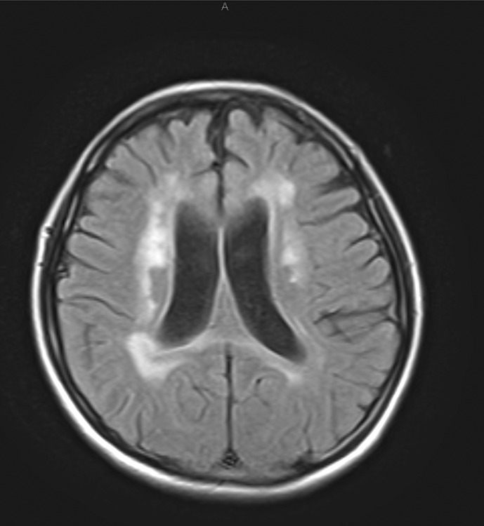
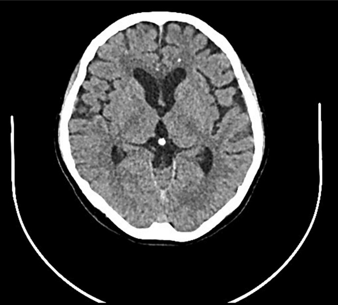
A 32-year-old woman developed PPD. Two weeks after delivery, she became anxious and depressive about caring for her child, and the Edinburgh Postnatal Depression Scale (EPDS) test showed a score of 9. Therefore, the municipal nurse visited weekly for 3 months to support her; at that point, the EPDS test score was 3. However, at 7 months postpartum, she got fed up with herself and did not feel like doing anything, thus she went to the outpatient clinic of psychosomatic medicine and was treated with one of the SSRIs, vortioxetine hydrobromide for several weeks without significant improvement. Therefore, she was referred to the Department of Psychiatry at Hakodate Watanabe Hospital. She reported experiencing persistent feelings of sadness and inadequacy, severe worthlessness, loss of interest, and joy in activities she once enjoyed, decreased appetite, and circadian swings in her depressed mood in the morning, corresponding to a Diagnostic and Statistical Manual of Mental Disorders, Fifth Edition (DSM-5) diagnosis of melancholic depression. She had no history of manic or depressive episodes; however, her uncle was affected by major depression. She showed no seriousness regarding her careless mistakes due to mild cognitive decline, which is an inappropriate symptom for major depression criteria. Magnetic resonance imaging to rule out organic causes of the depression revealed FLAIR hyperintensity in the periventricular and frontal deep white matter (Fig. 1), which led to be suspicious for multiple sclerosis or other neurological disorders. Therefore, we referred the patient immediately to the Department of Neurology, Hakodate Central General Hospital, and the Department of Neurology, Hakodate Municipal Hospital. The shape of confluent FLAIR hyperintensities in the periventricular and frontal deep white matter resembled what was reported in HDLS, and thus, further investigation whether or not cerebral computed tomography (CT) has shown multiple spotty calcifications in the frontal white matter, since calcifications have been described in an increasing number of patients with HDLS. Multiple spotty calcifications in the frontal white matter adjacent to the anterior horns of the lateral ventricles were observed on CT scans (Fig. 2), suggestive of HDLS. Genetic testing revealed an unpublished frameshift mutation in the CSF1R gene, p.Glu24ArgfsTer6, which led to a diagnosis of CSF1R-related leukoencephalopathy. The patient later achieved remission with various antidepressant medications, including venlafaxine hydrochloride and escitalopram oxalate. However, she showed mildly decreased cognitive function without any neurological signs except cluttering speech. She scored 27 points on the Mini-Mental State Examination. She returned to her hometown to recuperate with her husband and 15-month-old baby.
Fig. 1.
Cerebral magnetic resonance imaging (FLAIR) performed at the patientʼs first visit revealing extensive, patchy, and confluent hyperintensities in the periventricular and deep white matter. Compared to age, the enlargement of the body from the anterior horn of the ventricle on both sides and front-cortical atrophy was remarkable.
Fig. 2.
Multiple spotty calcifications in the frontal white matter adjacent to the anterior horns of the lateral ventricles were observed on cerebral CT imaging.
Discussion
An altered mental state is characteristic of psychiatric disorders but may also result from metabolic disorders, infections, and brain lesions. Therefore, a comprehensive differential diagnostic procedure should include neuroimaging in unusual cases. Here we presented the case of a postpartum woman who presented with major melancholic depression, according to DSM-5 criteria, but showed no seriousness regarding her careless mistakes due to mild cognitive decline, which is an inappropriate symptom for major depression criteria. Exactly, confluent FLAIR hyperintensities in the periventricular and deep white matter, multiple spotty calcifications in the frontal white matter, and a CSF1R mutation led to the diagnosis of CSF1R-related leukoencephalopathy. Many diseases or pathological conditions widely involving the cerebral white matter in adult humans have been reported, but among them, only 5 are characterized by extensive spherically swollen axons (spheroids and globules): HDLS, pigmentary orthochromatic leukodystrophy, Nasu-Hakola disease, sudanophilic leukodystrophy, and traumatic diffuse brain injury [8]. Recently, adult-onset leukoencephalopathy with axonal spheroids and pigmented glia (ALSP) was proposed as a comprehensive term encompassing HDLS and pigmentary orthochromatic leukodystrophy because of similar characteristics of clinical and pathological features, and the presence of mutations of CSF1R in the diseases by Rademakers et al. [5]. However, the original HDLS of the Swedish family was recently found to carry a different genetic makeup, with the affected family members displaying the alanyl-transfer (t) RNA synthetase (AARS) gene mutation but not the CSF1R mutation [9]; thus, another class of genetic disorders, called AARS-related leukoencephalopathy, is differentiated from CSF1R-related leukoencephalopathy.
CSF1R is a transmembrane tyrosine kinase receptor that is expressed in mononuclear phagocytic cells and microglia in the brain. Interestingly, a previous study [10] strongly implicated colony-stimulating factor-1 (CSF1) and CSF1R in the trophoblast regulation of placental development. They demonstrated that the concentration of CSF1 in serum and the expression of CSF1R mRNA in placental tissue were significantly increased during pregnancy. Therefore, abnormal CSF1-CSF1R signaling might contribute to the clinical manifestation of CSF1R-related leukoencephalopathy during pregnancy. Moreover, it is known that the proinflammatory cytokine, granulocyte-macrophage CSF1, was shown to be significantly upregulated in the gray matter on post-mortem analysis of brain tissue from patients with CSF1R-related leukoencephalopathy as well as the adhesion-related molecules derived peripheral blood monocytes were also significantly upregulated, suggesting widespread immune dysfunction.
Many researchers have speculated that PPD is caused, at least in part, by rapid changes in the reproductive hormone, estradiol, and progesterone before and immediately after delivery and the dysregulation of hypothalamic-pituitary-adrenal axis, associated with the altered GABA signaling, as well as the occurrence of stressful life events, such as life changes associated with caring for a new baby [11]. This case report can add the CSF1R mutation as a novel mechanism of PPD pathogenesis.
Accumulating evidence suggests a therapeutic application of CSF1R antagonists in tackling microglial activation, the progression of several neurodegenerative conditions [12]. It may be able to speculate that CSF1R antagonist may be therapeutic for FLAIR hyperintensity, since the increased CSF1 and pathologically activated microglia, which can disrupt brain capillary vessels and may produce the leukoencephalopathy in the CNS of CSF1R mutant, might be blocked by CSF1R antagonist. It is also reported that stress-induced neuronal CSF1 provokes microglia-mediated neuronal remodeling, which may result in the vulnerability factor to develop major depression. Although the molecular basis of FLAIR hyperintensities in the frontal deep white matter in major depression is unclear, this case may suggest that the regulation of CSF1R signaling might be a novel therapeutic strategy to treat late-onset depression with FLAIR hyperintensities in the frontal deep white matter. We demonstrated that the transplantation of brain microvascular endothelial cells (MVECs) stimulated remyelination in the white matter infarct of the internal capsule induced by endothelin-1 injection and MVEC transplantation repressed the inflammatory response in the lesion, so there was a possibility that MVECs reduce apoptotic death of oligodendrocyte precursor cells indirectly by inhibition of the inflammatory response [13]. It is also known that cytokines released by infiltrating microglia/macrophages induce apoptotic death of oligodendrocyte precursor cells in demyelinating lesions [14]. However, the molecular basis of the demyelination repair was still unclear.
Oyanagi et al. [8] reported that one difference between the clinical pictures of ALSP and N-HD, both of which are characterized by extensive spherically swollen axons, as mentioned above, was that comorbid depression was observed in five of 10 cases of ALSP versus zero of eight cases of N-HD. They also reported that the neuropathological differences between ALSP and N-HD consisted of CD68-positive microglia in the early stages of ALSP but not N-HD; even in stages II–IV, CD68-positive microglia were far more obvious in ALSP than N-HD. Thus, it may be able to speculate that CD68-positive microglia may play a key role in developing depression in CSF1R-related leukoencephalopathy. Ikeda [15] preliminarily demonstrated that CD68-positive microglia were observed to a greater degree in the frontal deep white matter of patients with late-onset depression than in age-matched healthy controls. Therefore, CD68-positive microglia may exert pathogenic effects in patients with CSF1R-related leukoencephalopathy and late-onset depression.
Conclusion
This case report is consistent with evidence that PPD may have organic causes in some cases, including CSF1R mutations. Atypical findings such as mild cognitive decline combined with PPD in psychiatric interview may justify brain imaging to avoid misdiagnosis, since CSF1R-related leukoencephalopathy is probably an under-recognized disease in medical psychiatry. Further investigations are needed to clarify a pathophysiological correlation between CSF1R signaling abnormality and PPD as well as major depression. The CARE Checklist has been completed by the authors for this case report, attached as online supplementary material (for all online suppl. material, see https://doi.org/10.1159/000541551).
Statement of Ethics
The present study was approved by the Institutional Review Board of Hakodate Watanabe Hospital (Approval No.: WEC 202408-01) and approved all procedures involving human patients on August 1, 2024. Written informed consent was obtained from the patient for the publication of the details of her medical case and any accompanying images, according to the formula for informed consent of the Japanese Society of Psychiatry and Neurology.
Conflict of Interest Statement
The authors have no conflicts of interest to declare.
Funding Sources
This study was not supported by any sponsor or funder.
Author Contributions
All authors contributed to the writing of the manuscript. M.M., K.H., H.S., A.I., S.K., and S.M. cared for the patient. M.M. and K.H. wrote the first draft of this manuscript. S.W., A.M., S.I., H.N., and I.K. provided critical revisions of the manuscript as well as administrative and technical support.
Funding Statement
This study was not supported by any sponsor or funder.
Data Availability Statement
All data generated or analyzed during this study are included in this article and its online supplementary material files. Further inquiries can be directed to the corresponding author.
Supplementary Material.
References
- 1. Hoptman MJ, Gunning-Dixon FM, Murphy CF, Lim KO, Alexopoulos GS. Structural neuroimaging research methods in geriatric depression. Am J Geriatr Psychiatry. 2006;14(10):812–22. [DOI] [PMC free article] [PubMed] [Google Scholar]
- 2. Takahashi K, Oshima A, Ida I, Kumano H, Yuuki N, Fukuda M, et al. Relationship between age at onset and magnetic resonance image-defined hyperintensities in mood disorders. J Psychiatr Res. 2008;42(6):443–50. [DOI] [PubMed] [Google Scholar]
- 3. Hayley S, Hakim AM, Albert PR. Depression, dementia and immune dysregulation. Brain. 2021;144(3):746–60. [DOI] [PMC free article] [PubMed] [Google Scholar]
- 4. Konno T, Kasanuki K, Ikeuchi T, Dickson DW, Wszolek ZK. CSF1R-related leukoencephalopathy: a major player in primary microgliopathies. Neurology. 2018;91(24):1092–104. [DOI] [PMC free article] [PubMed] [Google Scholar]
- 5. Rademakers R, Baker M, Nicholson AM, Rutherford NJ, Finch N, Soto-Ortolaza A, et al. Mutations in the colony stimulating factor 1 receptor (CSF1R) gene cause hereditary diffuse leukoencephalopathy with spheroids. Nat Genet. 2011;44(2):200–5. [DOI] [PMC free article] [PubMed] [Google Scholar]
- 6. Papapetropoulos S, Pontius A, Finger E, Karrenbauer V, Lynch DS, Brennan M, et al. Adult-onset leukoencephalopathy with axonal spheroids and pigmented glia: review of clinical manifestations as foundations for therapeutic development. Front Neurol. 2021;12:788168. [DOI] [PMC free article] [PubMed] [Google Scholar]
- 7. Blume J, Weissert R. Suspected perinatal depression revealed to be hereditary diffuse leukoencephalopathy with spheroids. J Mov Disord. 2017;10(1):59–61. [DOI] [PMC free article] [PubMed] [Google Scholar]
- 8. Oyanagi K, Kinoshita M, Suzuki-Kouyama E, Inoue T, Nakahara A, Tokiwai M, et al. Adult-onset leukoencephalopathy with axonal spheroids and pigmented glia (ALSP) and Nasu-Hakola disease: lesion staging and dynamic changes of axons and microglial subsets. Brain Pathol. 2017;27(6):748–69. [DOI] [PMC free article] [PubMed] [Google Scholar]
- 9. Sundal C, Carmona S, Yhr M, Almström O, Ljungberg M, Hardy J, et al. An AARS variant as the likely cause of Swedish type hereditary diffuse leukoencephalopathy with spheroids. Acta Neuropathol Commun. 2019;7(1):188. [DOI] [PMC free article] [PubMed] [Google Scholar]
- 10. Daiter E, Pampfer S, Yeung YG, Barad D, Stanley ER, Pollard JW. Expression of colony-stimulating factor-1 in the human uterus and placenta. J Clin Endocrinol Metab. 1992;74(4):850–8. [DOI] [PubMed] [Google Scholar]
- 11. Schiller CE, Meltzer-Brody S, Rubinow DR. The role of reproductive hormones in postpartum depression. CNS Spectr. 2015;20(1):48–59. [DOI] [PMC free article] [PubMed] [Google Scholar]
- 12. Martínez-Muriana A, Mancuso R, Francos-Quijorna I, Olmos-Alonso A, Osta R, Perry VH, et al. CSF1R blockade slows the progression of amyotrophic lateral sclerosis by reducing microgliosis and invasion of macrophages into peripheral nerves. Sci Rep. 2016;6:25663. [DOI] [PMC free article] [PubMed] [Google Scholar]
- 13. Iijima K, Kurachi M, Shibasaki K, Naruse M, Puentes S, Imai H, et al. Transplanted microvascular endothelial cells promote oligodendrocyte precursor cell survival in ischemic demyelinating lesions. J Neurochem. 2015;135(3):539–50. [DOI] [PubMed] [Google Scholar]
- 14. Schonberg DL, Popovich PG, McTigue DM. Oligodendrocyte generation is differentially influenced by toll-like receptor (TLR) 2 and TLR4-mediated intraspinal macrophage activation. J Neuropathol Exp Neurol. 2007;66:1124–35. [DOI] [PubMed] [Google Scholar]
- 15. Mikuni M, Ikeda K. Is major depression a minor neurological disorder with depressive symptoms? J Clin Exper Med. 2006;219:1070–4. [Google Scholar]
Associated Data
This section collects any data citations, data availability statements, or supplementary materials included in this article.
Supplementary Materials
Data Availability Statement
All data generated or analyzed during this study are included in this article and its online supplementary material files. Further inquiries can be directed to the corresponding author.


