Abstract
Lipid droplets (LDs) feature a unique monolayer lipid membrane that has not been extensively studied due to the lack of suitable molecular probes that are able to distinguish this membrane from the LD lipid core. In this work, we present a three-pronged molecular probe design strategy that combines lipophilicity-based organelle targeting with microenvironment-dependent activation and design an LD membrane labeling pro-probe called LDM. Upon activation by the HClO/ClO− microenvironment that surrounds LDs, LDM pro-probe releases LDM-OH probe that binds to LD membrane proteins thus enabling visualization of the ring-like LD membrane. By utilizing LDM, we identify the dynamic mechanism of LD membrane contacts and their protein accumulation parameters. Taken together, LDM represents the first molecular probe for imaging LD membranes in live cells to the best of our knowledge, and represents an attractive tool for further investigations into the specific regulatory mechanisms with LD-related metabolism diseases and drug screening.
Subject terms: Fluorescent probes, Membrane lipids
Lipid droplets (LDs) and their membrane proteins play essential roles in many biological processes, but the monolayer lipid membrane of LDs has not been extensively studied due to the lack of suitable molecular probes. Here, the authors report an LD membrane labelling pro-probe called LDM, which, upon activation by the HClO/ClO− microenvironment that surrounds LDs, undergoes a color change and releases LDM-OH probe that binds to LD membrane proteins.
Introduction
Lipid droplets (LDs) are ubiquitous organelles that function as intracellular storage compartments for neutral lipids, primarily triacylglycerols and sterol esters. LD play important roles in lipid metabolism and fatty acid (FA) synthesis, and regulate many aspects of energy homeostasis and cell growth1. Structurally, LD consists of a neutral lipid core surrounded by a phospholipid monolayer that serves as the membrane2. Recent studies have revealed that LD function is orchestrated by a number of proteins that act as gatekeepers by receiving external signals and regulating LD internal metabolism, contact with other organelles, and cargo exchange3,4. These proteins form stable associations with the phospholipid monolayer either through embedding or adherence, and are essential for controlling LD membrane dynamics. Although recognized to be of critical importance, and influenced by factors such as cell type, environmental conditions, and metabolic states5, LD membrane dynamics remains poorly understood in terms of specific regulatory mechanisms and biological functions6. This is primarily due to the lack of molecular tools that can be used for focused studies of the LD membrane.
Fluorescent molecular probes are widely used tools for labeling subcellular organelles and structures, including LDs. Currently, tens, if not hundreds, of small molecule LD-targeting fluorescent molecular probes have been described in the literature7, and used successfully to reveal insights into biophysics, cell biology, metabolism and physiology of LD. Recent notable additions to this toolkit include polarity probes8,9, viscosity probes10, and LD HClO/ClO− microenvironment-responsive probes11. However, all these currently available tools have been developed to visualize the neutral lipid core, and not the LD membrane, therefore limiting the insights into this unique subcellular membrane structure and its function and dynamics. Moreover, although fluorescently tagged LD-associated proteins have been used to study processes at the LD membrane, these strategies require the use of genetic manipulation, overexpression, and/or cell fixation. This may limit physiological relevance of the findings and, in the context of cell fixation requirement, prevent live cell imaging.
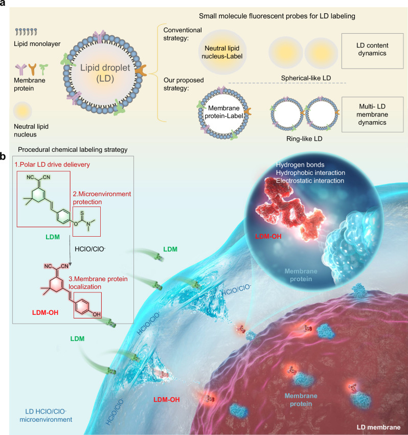
To overcome these limitations, we propose a three-pronged chemical labeling strategy that selectively labels LD membranes. This strategy integrates lipid-enhanced response, microenvironment activation, and electrostatic interactions present at and around the LD membrane. Thus, we designed a LD membrane (LDM) pro-probe (nomenclature analogous to pro-drug terminology) that is specifically delivered to the vicinity of LD membranes based on its physicochemical properties. We showed that LDM is activated by the HClO/ClO− found in the LD microenvironment to generate LDM-OH probe. LDM-OH was subsequently observed to bind to LD membrane-associated proteins through electrostatic interactions, thus selectively visualizing the LD membrane (Fig. 1). By employing LDM, we shed light on the LD membrane protein accumulation in live cells, confirming dynamic mechanism of LD membrane contacts with mitochondria and their protein accumulation parameters. Furthermore, we investigated the protein accumulation parameters at the membrane contact, and this relationship is related to the membrane protein per unit area. Taken together, LDM overcomes the limitations of current LD membrane labeling techniques, and enables real-time investigation of LD membrane dynamics under physiological conditions.
Fig. 1. Schematic diagram of chemical strategy to selectively label LD membranes.
a Schematic diagram of the LD chemical labeling strategy. Traditionally, LD labeling strategies using small molecule fluorescent probes mainly focused on labeling neutral lipid nuclei, resulting in spherical-like LD to check the dynamics of content. To achieve a dynamic description of multi-LD membrane dynamics, we propose a approach that involves labeling membrane proteins with a small molecule fluorescent probe, specifically yielding ring-like LD structures. b Schematic diagram of the chemical probe labeling mechanism for LD membrane structure. The three-pronged molecular probe design strategy employs a pro-probe called LDM, which combines lipid enhanced response, microenvironment activation, and electrostatic interaction. This comprehensive approach enables the specific delivery of LDM to the vicinity of LD membranes. Upon activation by the HClO/ClO− microenvironment, LDM-OH becomes exposed and binds to LD membranes through electrostatic interaction, facilitating labeling of LD membrane structure. Image created with Photoshop and Microsoft PowerPoint.
Results
LD membrane-specific fluorescent molecular probe concept and design components
To develop a fluorescent molecular probe suitable for selective visualization of the LD membrane, we employed a three-pronged probe design strategy that uses: (1) lipophilicity to maximize localization in the less polar regions of LD within cells12, thus delivering the probe to its subcellular target; (2) microenvironment responsive activation trigger that takes advantage of the fact that LD microenvironment exhibits a localized increase of HClO/ClO− levels11 to transform the pro-probe into (3) a probe capable of electrostatic anchoring with LD membrane proteins (see Fig. 1 for overview of the design strategy). To ensure low polarity and take advantage of the principles of similar solubility13–15, we selected (E)−2-(3-(4-hydroxystyryl)−5,5-dimethylcyclohex-2-en-1-ylidene) malononitrile as the starting point for the LDM pro-probe due to its low polarity and good fluorescence properties under neutral/weakly alkaline conditions within LDs, effectively eliminating interference from other regions. The LDM pro-probe also incorporated the N, N-dimethylthiocarbamate group as the HClO/ClO− responsive element, as it reacts with HClO/ClO− found in the LD microenvironment11 to expose the phenolic hydroxyl group in LDM-OH. The phenolic -OH in LDM-OH can form electrostatic interactions with amino groups in LD membrane proteins, similarly to what is seen in drug design16,17, thus effectively labeling the LD membrane. Additionally, the HClO/ClO−-triggered reaction transforms the green fluorescent pro-probe LDM to a red fluorescent LDM-OH, allowing for accurate visualization (Fig. 1). Taken together, our pro-probe design of LDM (green color) ensures directed delivery to LD based on lipophilicity of the molecule, rapid response to the LD microenvironment (HClO/ClO−) through formation of the red fluorescent LDM-OH, that subsequently binds LD membrane proteins, completing the labeling process of the LD membranes. Below, we describe results of detailed characterization and validation of LDM and LDM-OH, and illustrate its use for imaging LD membrane in live cells.
Optical selectively, physical properties characterization of LDM in vitro
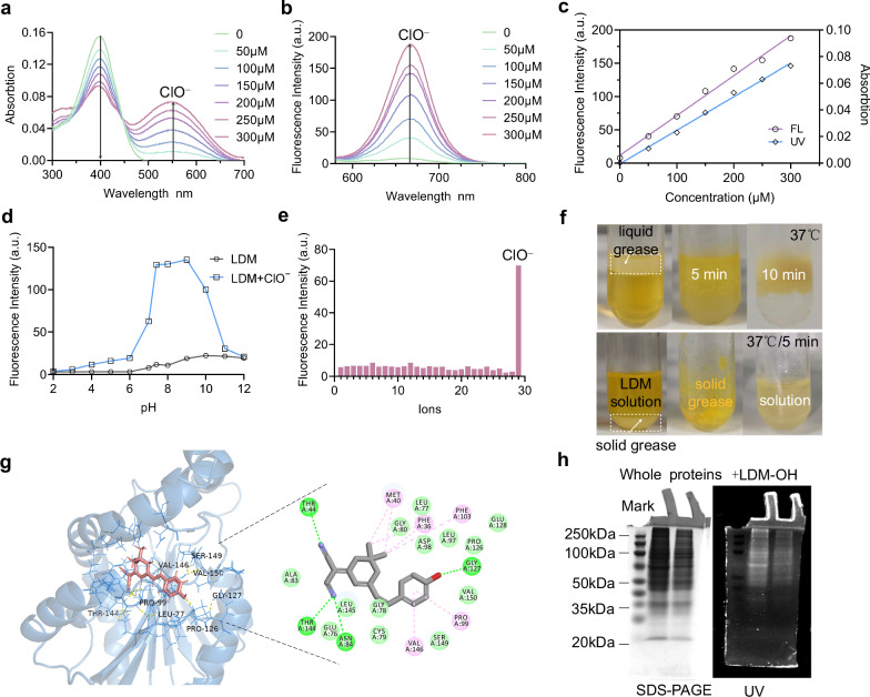
We synthesized the pro-probe molecule through a Knoevenagel condensation reaction18 (Supplementary Fig. 1–2) and confirmed the structure by standard methods (Supplementary Fig. 3–11). To confirm that HClO/ClO− triggers activation of LDM, we added NaClO (HClO/ClO− donor19), which led to an increase in fluorescence intensity at 561 nm, showing a linear correlation with R2 = 0.98933, and a detection limit of 6.8 µM (Fig. 2a–c, Supplementary Fig. 13), which depends on the environment of in vitro simulation testing20–22. The LDM to ClO− reaction was completed in 40 min (Supplementary Fig. 14), with a fluorescence quantum yield of 0.37 (Supplementary Fig. 15). Additionally, we observed a decrease in absorption peak at 405 nm, suggesting that LDM responds to HClO/ClO− to generate LDM-OH in vitro. (Supplementary Fig. 16). We further verified that the product was indeed LDM-OH using high-resolution mass spectrometry (Supplementary Fig. 12). Simultaneously investigated the effects of viscosity and polarity on LDM and LDM-OH. Results indicate that an increase in viscosity has a non-linear strengthening effect on the fluorescence intensity of LDM and LDM-OH. The effect of polarity on LDM is not significant. When the content of dioxane is 40%-50%, the polarity of the solution enhances the fluorescence intensity of LDM-OH (Supplementary Fig. 17). Since the probe LDM label the LDs membrane imaging rather than the contents, according to our experimental results (Fig. 3a and Fig. 4e), there was no significant difference in the lipid droplet membrane imaging between the normal and drug-induced treatment groups.
Fig. 2. Optical selectively, physical properties characterization of LDM in vitro.
a, b The absorption and fluorescence spectra of LDM (10.0 μM) in different NaClO (0-300.0 μM) solution (DMSO-PBS, 1:99, v/v, pH = 7.4), λex = 561 nm, slit: 5 nm/5 nm/700 V. c Linear relationship between UV absorption and fluorescence intensity and HClO/ClO− concentration in a and b, the linear regression equation is determined as Y = 59658.07143X + 11.58132, with a linear correlation R2 = 0.98933, where X represents the concentration of ClO− and Y represents the fluorescence intensity of LDM at 665 nm (d) The fluorescence intensity change of LDM (10.0 μM) in different pH (2-12) solutions with (red) or without (black) ClO− (100 μM) (λex = 561 nm), e The selectivity of LDM towards ClO−, and various reactive oxygen species in DMSO-PBS solution (100.0 μM, 1:99, v/v, pH = 7.40). 1. Blank; 2.NO2−; 3.t-Buoo−; 4.PO43-; 5.•OH; 6.HPO42-; 7.CH3COO−; 8.F−; 9.Cl−; 10.Ag+; 11.Al3+; 12.Ca2+; 13.Cr3+; 14.Co2+; 15.Fe2+; 16.Fe3+; 17.Mn2+; 18.Ni2+; 19.Pb2+; 20.Zn2+; 21.Cu2+; 22.Hg2+; 23.Cd2+; 24.H2O2; 25.NO; 26.SO42−; 27.ONOO−; 28.1O2; 29.ClO−. f Lipophilic assay for testing the distribution of the LDM in phosphate buffer. LDM molecules dissolved in the lipids, causing the phosphate buffer to change from yellow to colorless, while the lipids transformed from white to yellow, suggest. LDM with a lipid response behavior. g Macromolecular docking of LDM-OH and representative lipoprotein (Q6UX53). h The whole protein SDS-PAGE electrophoresis (left) and UV image for the protein gel incubation with LDM-OH (37 °C, 2 h) (right), indicating the LDM-OH could bind to the whole proteins in HepG2 cells. Three independent biological replicates were performed, and the results were similar. Source data are provided as a source data file.
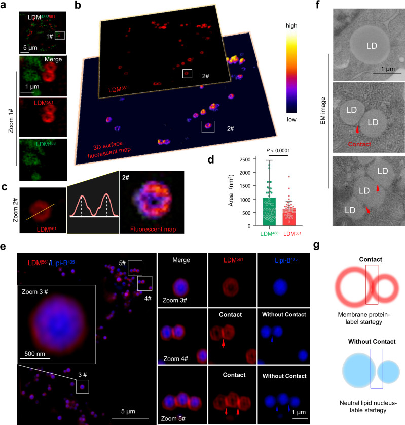
Fig. 3. Super-resolution imaging for ring-like LD membrane tracking with LDM pro-probe.
a HepG2 cells incubated with 10.0 μM LDM under SIM 488 laser and 561 laser, Zoomed-in images are of white rectangle #1. b 3D surface fluorescent map of HepG2 cells incubated with LDM under SIM 561 laser, Zoomed-in images are of white rectangle #2. c SIM imaging and 3D surface fluorescent map of representative single particles stained with LDM561, yellow dotted line in zoomed-in image #2 indicate region for fluorescence analysis. d Quantitative analysis of the area for fluorescent punctate labeled with LDM488 and LDM561. Data are mean ± SD (n = 50 areas from 10 cells). e SIM images of HepG2 cells co-stained with commercial Lipi-B405 and LDM561 channel, Zoomed-in images are of white rectangles #3, #4, #5. Three independent imaging replicates were performed, and the results were similar. f Electron microscopy imaging of single LD and multi-LD contact event. Three independent imaging replicates were performed, and the results were similar. g Schematic comparison between membrane protein ring-like LD labeling strategy and content spherical-like LD labeling strategy, suggests the traditional spherical-like labeling strategy cannot well characterize the membrane interaction event of multiple LD. LDM488 channel: λex = 488 nm, Em max 525 nm (500–550 nm); LDM561 channel: λex = 561 nm, Em max 665 nm (600–700 nm); Lipi-B405 channel: λex = 405 nm, Em max 447 nm (417–476 nm). Statistical analysis was performed using two-tailed unpaired Student’s t-test, and the data were presented as mean ± SD. P < 0.05 was considered statistically significant. Source data are provided as a source data file.
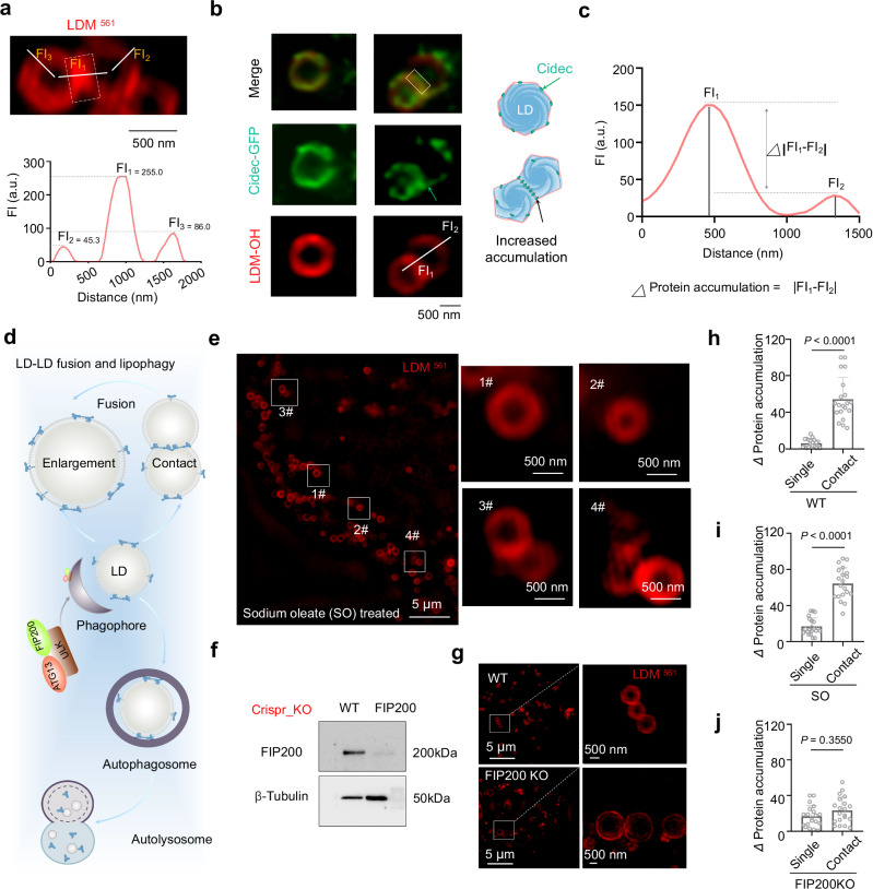
Fig. 4. LDM fluorescence aggregation as an indicator of membrane contact protein accumulation parameters.
a Representative SIM image of LD membrane dynamic stained with LDM and Lipi-B405, and simulation for single LD dynamic. b Representative SIM image of LD co-stained with Cidec-GFP in LDM-561 channel, and the schematic diagram of Cidec-GFP location in LD protein accumulation formation. Image created with Microsoft PowerPoint. c According to the LDM-OH imaging analysis of b, assuming that the protein accumulation is positively correlated with fluorescence intensity, Δprotein accumulation = |FI1-FI2 | , the data is obtained through imageJ. d Schematic diagram of LD-LD fusion and lipophilicity process, Image created with Microsoft PowerPoint. e SIM image of HepG2 cells treated with 100 μM sodium oleate, for 24 h and then stained with LDM, Zoomed-in images are of white rectangles #1-#4. white dotted line in zoomed-in images #1-#4 indicates the region for fluorescence analysis. f Western blot for detecting FIP200 protein expression in HepG2 gene-edited cells. g Representative SIM image of LD stained with LDM in HepG2 WT cells and FIP200KO cells, Zoomed-in images are of white rectangle. The white dotted line in zoomed-in images #1-#4 indicates the region for fluorescence analysis. h–j Quantitative analysis of protein accumulation changes in WT, sodium oleate and FIP200KO HepG2 cells. Data are mean ± SD (n = 20 areas from 10 cells). Cidec-GFP channel: λex = 488 nm, Em max 525 nm (500-550 nm); LDM-OH channel: λex = 561 nm, Em max 665 nm (600–700 nm). Three independent imaging or biological replicates were performed, and the results were similar. Statistical analysis was performed using two-tailed unpaired Student’s t-test, and the data were presented as mean ± SD. P < 0.05 was considered statistically significant. Source data are provided as a source data file.
To demonstrate LDM’s selectivity towards HClO/ClO−, various reactive oxygen species and nitrogen species were tested. At 10.0 µM, only HClO/ClO− triggered a fluorescence signal response centered at 665 nm, while no significant changes were observed in the presence of other ROS or biologically related species, such as H2O2, ⋅OH, ⋅O2−, metal ions (Fig. 2e, Supplementary Figs. 18, 19), or pH changes (Fig. 2d). These results indicate that LDM is selectively responsive to HClO/ClO−.
To confirm that LDM is able to localize to lipid-rich regions, we conducted a lipophilicity experiment23 to assess the probe’s distribution in phosphate buffer. We dissolved LDM in the phosphate buffer and mixed the buffer with a lipid layer (Fig. 2f). After 10 min, a substantial amount of LDM molecules dissolved in the lipids, causing the phosphate buffer to change from yellow to colorless, while the lipid layer transformed from white to yellow. This highlights that LDM has the capacity to distribute into the lipid environment, suggesting its ability to target LD.
To investigate whether LDM-OH is able to form electrostatic interactions with LD membrane proteins, we selected seven representative LD membrane proteins24–27 and performed docking assays with LDM-OH (Fig. 2g, for details, see Supplementary Fig. 20). These assays confirmed the strong binding of LDM-OH to the membrane proteins. Furthermore, we employed an SDS-PAGE gel electrophoresis experiment to validate LDM-OH’s effective binding to the entire protein in the cell, resulting in fluorescent labeling of the proteins (Fig. 2h). This result substantiates that the phenolic hydroxyl group of LDM-OH can serve to anchor the probe to LD membrane proteins.
Collectively, the results of our in vitro and docking studies provide strong support for LDM as the LD-localizing pro-probe that’s activated by HClO/ClO− into LDM-OH probe that labels LD membrane proteins to enable selective imaging of LD membranes.
Characterization of LDM in live cells
We first evaluated the cytotoxicity of LDM and LDM-OH on HeLa/HepG2 cell lines and found that the toxic effect of LDM on cell activity was negligible (Supplementary Figs. 21-22). Additionally, we established that LDM entered cells via an energy-dependent mechanism (Supplementary Figs. 23–25). In recent years, multiple excellent studies have been reported in the field of LD fusion28, fission29, cytosolic and nuclear LDs movement30, and the interaction between organelles31. Using structured illumination microscopy (SIM)32,33, we captured LDM-stained cells under SIM-488 laser (LDM488) and SIM-561 laser (LDM561) (Fig. 3a). We observed the presence of green fluorescent particles of random sizes (Zoom 1#, Fig. 3a) and a regular, red fluorescent ring-like structure (Zoom 1#, Fig. 3a). To further characterize the ring-like structure, we constructed a 3D-surface fluorescent map which demonstrated a uniform distribution of fluorescence within a single ring-like structure (Fig. 3b). The height bimodal fluorescence intensity peaks were found to be equal (Fig. 3c, Supplementary Fig. 26). However, at the contact site with multiple ring-like structures, red fluorescence showed an uneven distribution, resulting in multiple peak shapes of unequal heights (Supplementary Fig. 27). These results suggest that LDM reached LDs, underwent chemical transformation to LDM-OH and labeled the LD membrane (ring-like structures). The uneven distribution might, therefore, be due to different protein distribution patterns within different red-like ring structures, particularly at the contact site.
To further support that the Plin2 and Plin5 proteins34–36, known as uniform markers for LD membranes, were used as fluorescent markers for labeling LDs under SIM. Our analysis revealed no significant fluorescence enrichment of Plin2 or Plin5 when LD membranes approached each other (Supplementary Fig. 28a, b). To explore the possibility of specific protein aggregation, we next test Cidec37, a protein involved in LD fusion, which distributes uniformly on single LD membranes but enriches at membrane contact sites upon LD interactions. Remarkably, our LDM probe and labeled LD membranes accurately reflected this Cidec aggregation, with significant enrichment at membrane contact sites upon LD membrane proximity (Supplementary Fig. 29a, b). This validates that the fluorescence enhancement observed with LDM-labeled LD membranes is not mere fluorescence overlap but an indicator of protein dynamics at membrane contact sites.
Importantly, the size distribution of the red ring-like structures labeled by LDM561 matched the reported LD size range38. This suggests that the red ring-like structures labeled by LDM using SIM may close to represent the LD membrane (Supplementary Fig. 30). To test this hypothesis, we performed localization experiments using a commercial LD probe that labels the core neutral lipids39, Lipi-B405, and co-incubated cells with Lipi-B and LDM. As expected, the LDM561-labeled red ring-like structure effectively surrounded the Lipi-B405-labeled area indicative of LD core (Fig. 3e, enlargement Zoom 3#-5#, Supplementary Figs. 31-32), confirming that LDM labels the area immediately surrounding the LD core, most likely the LD membrane. However, we also noted that the area labeled by Lipi-B405 was somewhat smaller than the ring labeled by LDM561 (Supplementary Figs. 33-34, which can be explained by the known phenomenon that lipid dyes aggregate in the center of LD39. Therefore, we believe that the size of the LDM-based ring under SIM is a more accurate representation of the LD size, compare to traditional microscopy. Importantly, we observed that unlike Lipi-B that can’t visualize the actual physical contact between LD (Fig. 3e, Zoom 4#-5#, Supplementary Fig. 35), the LDM561-labeled ring-like LD structure provided valuable information for the multiple LD contacts (Supplementary Fig. 36), consistent with those seen by the electron microscopy (EM) (Fig. 3f). Consequently, we propose that our probe captures the more precise contact sites between multiple LD interactions in living cells (Fig. 3g, Supplementary Fig. 37), compared to probe labeling technology located in the LD core (Supplementary Fig. 30 and Supplementary Fig. 33). Furthermore, the LDM alone does not trigger the formation of LD membrane contact sites (Supplementary Fig. 38).
LDM fluorescence aggregation as an indicator of membrane contact protein accumulation parameters
It is widely accepted that LD membrane proteins move within the membrane and this mobility plays critical roles in physiological processes within living cells5. In agreement with this, we observed that the membrane protein labeled with LDM561 exhibited dynamic fluorescent coverage patterns in the inner shell of a single representative LD (Fig. 4a). We also noticed that the fluorescence intensity of the LDM561 marker at the contact site is more than twice that of the non-contact site (Supplementary Fig. 39), suggesting that the high fluorescence intensity might be due to accumulation of labeled proteins at specific. To verify this hypothesis, we co-labeled an LD membrane-related protein, Cidec-GFP, which is known to mediate adhesion between membranes37. Results demonstrated a matching trend between the positioning of Cidec-GFP and the LDM-OH-labeled membrane protein in the LD membrane contact area (Fig. 4b, Supplementary Fig. 40), further confirming LDM’s capability to accurately report on increased migration and co-localization of membrane proteins in the contact area, as indicated by increased fluorescence intensity at the contact sites (Fig. 4b, green arrow). This result further validates LDM’s utility as a marker for protein accumulation between membrane contacts, reveals that protein accumulation may depend on increased co-localization of LD membrane proteins, and provides a tool for evaluating adhesion parameters in diverse cellular processes.
To further quantify protein co-localization between contact sites, we introduced the accumulation parameter, which is the difference between the number of LDM-labeled fluorescent protein molecules at the contact site and the diagonal (Fig. 4c). Using this system, we tested the effect of chemical drug/gene intervention on accumulation parameters. Firstly, we stimulated HepG2 cells with sodium oleate for 24 h and observed a significant increase in the number of membrane contacts, including single, multiple, and even linear membrane contacts (Fig. 4e, Supplementary Figs. 41-42). More importantly, the fluorescence intensity of LDM-labeled membrane proteins in the membrane contact area increased (Fig. 4h), indicating heightened accumulation as it is generally accepted that the protein accumulation of the membrane is related to the number of proteins per unit area40.
To further verify the applicability of our probe to characterize the protein accumulation parameters, we further investigated the relationship between membrane protein accumulation and LD size. Studies have shown that blocking the process of lipophagy can inhibit the degradation of the contents of the LDs and effectively increase the area of the LD membrane. To examine this relationship further, we knocked out (KO) a key autophagy protein FIP20041 (Fig. 4f-g), thus inhibiting lipophagy to increase the accumulation of lipid components and consequently the volume of LD42 (Fig. 4d). As anticipated, the size of LD in FIP200KO cells significantly increased (Supplementary Fig. 43), while the fluorescence intensity of the LDM-labeled membrane protein in the contact area did not increase significantly, relative to a single LD (Fig. 4j). We also knocked out ATG13, another protein with the key role in autophagy43, and observed similar effects (Supplementary Fig. 44). These results indicate that when the unit membrane volume increases, the number of membrane proteins disperses, leading to decreased protein accumulation between the membranes.
In summary, these findings demonstrate that LDM fluorescence intensity effectively reflects the accumulation parameters at the membrane contact, and this relationship is related to the membrane protein per unit area.
Using LDM to track changes in LD membrane protein accumulation during the period of liver cancer cell starvation
When tumors progress rapidly, they often enter an energy-deprived state of starvation, necessitating a series of metabolic reprogramming to cope with energy stress44. Among them, liver cancer cells will accumulate a large number of LDs in the cells to supply the energy needed during periods of starvation45,46. We used LDM to measure the changes in the protein accumulation of LD in liver cancer cells, reflecting how these cells utilize LDs during starvation.
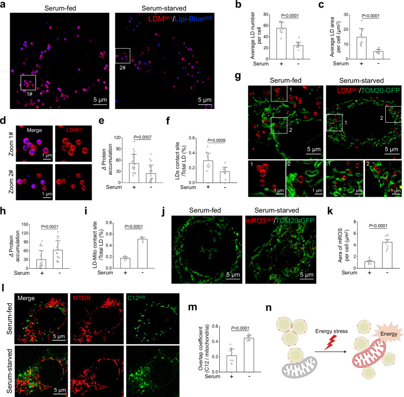
We found that the LD in liver cancer cells significantly decrease in a starving state, with the number and area of LD reduced by approximately 2-3 times (Fig. 5a–c) and the triglyceride (TG) content in LDs significantly decreases (Supplementary Fig. 45). Meanwhile, the protein accumulation between LD membranes of cancer cells decreases after starvation, and the number of contact sites between LDs significantly decreases (Fig. 5d–f). The reduction in protein accumulation contact between LDs may be associated with an increase in the utilization of LD during cancer cell starvation. This is consistent with a previous study, which indicates that when cells utilize LD, the contact and fusion between LDs decrease47.
Fig. 5. Using LDM to track changes in LD membrane protein accumulation during the period of liver cancer cell starvation.
a Representative SIM images of LD changes in HepG2 cells labeled with LDM and Lipi-B405 under serum-fed and serum-starved. b, c Quantitative analysis of the number and area of LD under serum-fed and serum-starved (n = 10 cells). d Zoomed-in images are of white rectangle in a. Quantitative analysis of intermembrane LD protein accumulation and number of contact sites of LD under serum-fed and serum-starved, e n = 20 areas from 10 cells, f n = 10 areas from 5 cells. g Representative SIM images of LD and mitochondria labeled with LDM and TOM20-GFP under serum-fed and serum-starved. Quantitative analysis of LD and mitochondria protein accumulation and number of contact sites under serum-fed and serum-starved, h n = 20 areas from 10 cells, i n = 7 areas from 5 cells. j Representative SIM images of mitochondrial ROS changes labeled with mROS561 and TOM20-GFP under serum-fed and serum-starved. k Quantitative analysis of FAs and mitochondria under serum-fed and serum-starved (n = 7 cells). l Representative SIM images of FAs and mitochondria in HepG2 cells labeled with C12488 and MTDR under serum-fed and serum-starved. m Quantitative analysis of FAs co-localization with mitochondria under serum-fed and serum-starved (n = 10 cells). n Schematic diagram of the process of LD-LD separation and LD-mitochondria interaction after starvation. Created in BioRender. Shao, S. (2024) BioRender.com/e93j405. Lipi-B405 channel: λex = 405 nm, Em max 447 nm (417–476 nm); LDM-OH channel: λex = 561 nm, Em max 665 nm (600–700 nm); TOM20-GFP channel: λex = 488 nm, Em max 509 nm (505–550 nm); mROS561 channel: λex = 561 nm, Em max 610 nm (590–610 nm); MTDR channel: λex = 644 nm, Em max 665 nm; C12488 channel: λex = 488 nm, Em max 510 nm (500–510 nm). Three independent imaging replicates were performed, and the results were similar. Statistical analysis was performed using two-tailed unpaired Student’s t-test, and the data were presented as mean ± SD. P < 0.05 was considered statistically significant. Source data are provided as a source data file.
Current research suggests that when cells are starved, they utilize LD through LD-mitochondria contact48. To examine this, we used LDM to investigate the protein accumulation status between LDs and mitochondria. Results showed that under serum-starved conditions, LDM accumulates at the contact sites between LD and mitochondrial membrane, while the number of membrane contact sites also increases (Fig. 5g–i). Next, we tested whether the enhanced protein accumulation between LD and mitochondria displayed by LDM promotes the utilization of LD by mitochondria We used mitochondrial ROS (mROS) probe561 to label mROS, thus indirectly reflecting the amount of energy produced by mitochondria. We found that mROS production increased by about fourfold in cancer cells after starvation (Fig. 5j, k). Meanwhile, we used the FA probe C12488 to indicate the migration of FAs between LD and mitochondria. The experimental results indicate that after starvation, FAs originally located on LD increase their migration towards mitochondria (Fig. 5l, m, Supplementary Fig. 46). The above results indicate that when liver cancer cells are starving, the protein accumulation between LDs decreases, and the interaction between dispersed LD and mitochondria strengthens. Subsequently, these LDs release FAs, migrate to mitochondria, and are oxidized by mitochondria, thereby providing energy for starving cancer cells (Fig. 5n). In addition to its role in cellular processes, we have explored our tool’s potential in drug evaluation. Testing known LD modulators Mos-149, CHE50, and tanshinone IIA51 revealed Mos-1 reduces LD numbers and contact, while CHE increases LD numbers but reduces contact (Supplementary Fig. 47), consistent with prior reports. Notably, our examination of tanshinone IIA with autofluorescence showed it enhances LD numbers, contact, and exhibits a membrane localization labeled by our probe (Supplementary Fig. 48). In summary, we used LDM to label LD membranes of liver cancer cells during the period of starvation, drug evaluation, and the drug location. These applications provide an attractive tool for further investigations into the specific regulatory mechanisms and drug discovery associated with LD related metabolism diseases.
Discussion
We successfully developed a fluorescent molecular probe for imaging LD membranes in live cells. Our strategy is based on the use of pro-probe LDM that localizes to LD due to its lipophilic properties. Once there, it is activated by the LD microenvironment, more specifically by increased levels of HClO/ClO− typically found around LD. This generates the probe, LDM-OH, which associates with LD membrane proteins to generate a strong fluorescent signal indicative of the LD membrane. We used this probe to visualize the LD membrane in live cells and showed that LDM treatment clearly visualizes the ring-like structure surrounding the neutral lipid core. Furthermore, we could examine LD protein accumulation, both with other LDs and with mitochondria under different conditions. As such, our probe opens new avenues for investigating the specific regulatory mechanisms and biological functions associated with LD membrane biophysics. Furthermore, our probe overcomes limitations of traditional labeling techniques based on transient fluorescent proteins or immunofluorescence52, as it does not require protein overexpression or the use of fixed cells. LDM’s ability to selectively label LD membrane structures and its compatibility with super-resolution microscopy offers a powerful tool for studying LD dynamics in live cells.
Importantly, we established LDM as a marker for protein accumulation between LD membrane contacts. By studying the aggregation of fluorescent molecules labeled by LDM-OH in the LD membrane contact area, we could accurately measure the changes in LD membrane protein co-localization and evaluate accumulation parameters in various cellular processes. Furthermore, we demonstrated that LDM fluorescence aggregation is related to the density of membrane proteins per unit area, shedding light on the relationship between membrane protein accumulation and LD size. The ability of LDM to accurately reflect membrane protein accumulation parameters is another important aspect of providing researchers with a powerful tool to investigate the dynamic mechanism of LD membrane contacts, their protein accumulation parameters, and their role in cellular physiology and pathology. By visualizing the aggregation of fluorescent molecules in the LD membrane contact area, LDM provides a direct and quantitative measure of membrane protein density, allowing for the investigation of LD-related processes in live cells. This information is critical for understanding cellular physiology and can be applied to study the effects of chemical interventions or genetic modifications on membrane protein accumulation53. In the example we use above, LDM enabled us to study the effect of starvation on the interface between LDs and mitochondria, demonstrating that liver cancer cells respond to energy stress during hunger by enhancing LD-mitochondria interactions.
More broadly, the versatility of our probe design strategy may yield similar probes for labeling other cellular structures or organelles with distinct membrane properties by tailoring the probe’s properties to target specific cellular components. Furthermore, the application of LDM in live-cell imaging allows for real-time visualization of LD membrane dynamics under physiological conditions. This capability could be harnessed to study cellular responses to external stimuli, such as changes in nutrient availability or exposure to stressors54,55, providing a deeper understanding of cellular adaptation and signaling pathways.
In conclusion, the development of the LDM probe represents a significant advancement in the field of chemical biology, enabling selective imaging of LD membrane structures and shedding light on the dynamics and functions of LD in living cells. This approach overcame the limitations of current LD membrane labeling techniques and enabled real-time investigation of LD membrane dynamics under physiological conditions. By applying LDM, we gained insights into the biophysical functions of LD membrane dynamics and its relationship with LD protein accumulation parameters, providing valuable information on LD membrane dynamics. Future use of LDM holds great potential for advancing our understanding of LD biology and other cellular processes that involve membrane dynamics and protein accumulation.
Methods
Synthesis and characterization
For synthesis and characterization of Compound 1, a solution of isophorone (3.50 mL, 23.50 mmol) and CH3COONa (1.60 g, 19.51 mmol) in ethanol (25 mL) was added malononitrile (1.30 g, 20.12 mmol). The reaction was allowed to heat and reflux for 8 h. Purification of crude product by silica gel column chromatography to obtain white powder (2.60 g, 69.01%). Melting point: 70.2-72.6oC; TLC (DCM/PE = 3:1, V/V): Rf = 0.23; 1H NMR (400 MHz, CDCl3) δ 6.54 (s, 1H), 2.52 (s, 2H), 2.22 (s, 2H), 2.03 (s, 3H), 0.94 (s, 6H). 13C NMR (101 MHz, DMSO-d6) δ 171.93, 162.99, 119.95, 113.97, 113.18, 76.60, 45.33, 42.48, 32.49, 27.80 (2 C), 25.50. HRMS: The m/z [M + H]+ was calculated as 187.1157, and the measured value was 187.1307.
For synthesis and characterization of LDM-OH, a solution of compound 1 (370.40 mg, 1.98 mmol) in ethanol (25 mL) was added 4-Hydroxybenzaldehyde (318.30 mg, 2.61 mmol) and two drops of piperidine. The reaction was allowed to heat and reflux for 12 h. Purification of crude product by silica gel column chromatography to obtain red solid (531.10 mg, 91.50%). Melting point: 200.4-201.5oC; TLC (DCM/MeOH = 20:1, V/V): Rf = 0.25; 1H NMR (400 MHz, DMSO-d6) δ 10.0 (s, 1H), 7.6 (d, J = 8.6 Hz, 2H), 7.3-7.1 (m, 2H), 6.8 (s, 1H), 6.8 (s, 2H), 2.6 (s, 2H), 2.5 (s, 2H), 1.0 (s, 6H). 13C NMR (101 MHz, DMSO) δ 170.21, 159.34, 156.65, 138.25, 129.85 (2 C), 127.10, 126.22, 121.36, 115.87 (2 C), 114.11, 113.29, 74.81, 42.29, 38.17, 31.63, 27.42 (2 C). HRMS: The m/z [M + H]+ was calculated as 291.1419, and the measured value was 291.1591.
For synthesis and characterization of LDM, a solution of LDM-OH (200.52 mg, 0.685 mmol) in CH2Cl2 (15 mL) was added dimethylaminothioformyl chloride (126.40 mg, 1.028 mmol) and 0.92 mmol (127.13 mg) of K2CO3. The reaction was allowed to heat and reflux for 4 h. Purification of crude product by silica gel column chromatography to obtain yellow powder (176.42 mg, 68.31%). TLC (DCM/EA = 30:1, V/V): Rf = 0.22; 1H NMR (400 MHz, DMSO-d6) δ 7.73 (d, J = 8.5 Hz, 2H), 7.36 (m, 2H), 7.12 (d, J = 8.4 Hz, 2H), 6.90 (s, 1H), 3.35 (d, J = 19.8 Hz, 6H), 2.59 (d, J = 27.8 Hz, 4H), 1.03 (s, 6H). 13C NMR (101 MHz, DMSO) δ 186.52, 170.81, 156.25, 155.03, 137.11, 133.99, 129.09 (2 C), 123.85 (2 C), 123.32, 114.32, 113.50, 76.90, 43.30, 42.78, 39.04, 38.65, 32.16, 27.92 (2 C). HRMS: The m/z [M + H]+ was calculated as 377.2741, and the measured value was 377.2653.
Preparation of detection solution for probe LDM
For the preparation of probe reserve solution, first, weigh the yellow solid powder LDM and dissolve it in Dimethyl sulfoxide (DMSO) to obtain LDM stock solution (20.0 μM). Then dilute the stock solution to 10.0 μM with PBS (DMSO-PBS, pH = 7.4, 1:99, v/v), and record UV and fluorescence spectra at 37 °C.
For the preparation of analytes for selective experiments, the various anionic solutions required for the experiment (F−, HPO42−, Cl− SO42−, PO43−, CH3COO−) come from the corresponding sodium salts; Various metal cation solutions (Cd2+, Ag+, Al3+, Ca2+, Cr3+, Co2+, Fe2+, Fe3+, Mn2+, Ni2+, Pb2+, Zn2+, Hg2+ and Cu2+) are prepared from corresponding chloride salts or sulfates. These solutions have a concentration of 0.01 mol/L.
LDM was dissolved in PBS buffer (0.01 M, pH = 7.4) as a reserve solution (1 mM). Various reactive oxygen species (ROS) and reactive nitrogen species (RNS), including NO2−, NO3−, ClO−, H2O2, 1O2, ∙OH, NO, t-BuOO−, ONOO−, and ⋅O2− were prepared using the following method. NO2−, NO3− and ⋅O2− were prepared with corresponding sodium salts NaNO2, NaNO3 and KO2, respectively, with a final concentration of 0.01 M. The 30% H2O2 solution was diluted and concentration was determined by light absorption at 240 nm (ε = 43.6 M−1cm−1). The commercially available Sodium hypochlorite solution was used as the source of Hypochlorite, and the concentration of Hypochlorite (ClO−) was determined at 292 nm using a molar absorption rate of 350 M−1cm−1. 1 mL of H2O2 solution (0.1 mol/L) and 1 mL of NaClO solution (0.1 mol/L) were mixed to prepare singlet oxygen (1O2), then diluted to 10 mL. (NH4)2Fe(SO4)2⋅6H2O mixed with 10 equivalents of H2O2 to prepare hydroxyl radicals (∙OH), and the concentration of ∙OH was calculated based on the Fe2+ concentration. Nitric oxide (NO) was produced from Nitroso iron Sodium cyanide (III). The t-BuOO− was generated by t-BuOOH. Add NaNO2, HCl, and H2O2 to the NaOH solution to produce ONOO−. The concentration of ONOO− was measured at 302 nm using a molar absorption rate of 1670 M−1cm−1.
Molecular docking
A theoretical simulation technique called molecular docking is used to analyze intermolecular interactions and forecast intermolecular binding affinities and patterns. Molecular docking was carried out with Discovery Studio to investigate the interactions between luteolin and the proteins in the core network. First, ChemDraw software was used to draw the LDM-OH structure and saved it as a sdf format file. Then, 7 protein crystal structures were retrieved from the UniProt (https://www.uniprot.org/), dehydrated, hydrogenated, and molecular docking using Discovery Studio Tools, and saved as pdbqt files. Next, the target proteins that were also monitored in this experiment were visualized and analyzed using Discovery Studio 2019. Finally, Pymol software identified the interacting amino acids and output the image format.
Cell culture
HepG2/HeLa cells were cultured in Dulbecco’s modified Eagle’s medium (VivaCell, Shanghai, China) supplemented with 10% fetal bovine serum, penicillin (100 units/ml), and streptomycin (100 μg/ml; 10,000 units/ml) in a 5% CO2 humidified incubator at 37 °C. Cells were obtained from Procell Life Science&Technology Co., Ltd. (Wuhan, China).
Cell treatment and imaging
A total of 2 × 10⁵ cells were seeded on a glass-bottom micro-well dish and incubated with 2 ml of DMEM supplemented with 10% FBS for 24 h, then stained with LDM (10 μM) or LDM-OH (10 μM) for 30 min, and with 100 nM Lipi-B (blue commercial LD dye, Dojindo Laboratories Kumamoto) at 37 °C for another 30 min, the cells were washed 3 times with pre-warmed free DMEM, and washed with free DMEM 3 times. Lastly, The HeLa/HepG2 cells were observed under super-resolution microscope or confocal laser scanning microscopy (LSM-980, Zeiss) and analyzed with ImageJ software. In addition, Pearson correlation coefficient (PCC, the degree of overlap between two fluorescent channels, pixel-based) was analyzed for co-localization using ImageJ software equipped a colocalization analysis plugin as previously reported56. For more information, please refer to: https://imagej.net/ij/plugins/colocalization-finder.html.
FA migration experiment
After incubating the cells in complete medium containing 1.0 µM BODIPY™ FL C12488 (Thermo Fisher Scientific) for 16 h, wash the cells twice with PBS to remove excess C12488. Then, label the mitochondria/LD with MTDR/Lipi-B405 respectively, and use confocal laser scanning microscopy for imaging. Analyze and calculate the overlap coefficient between FAs and mitochondria using ImageJ software to evaluate the migration rate of FAs from LD to mitochondria.
Data analysis
Statistical analysis was performed with Prism 8 (GraphPad). Normality and lognormality test to check the normal distribution. In the case of normal distribution, the statistical comparison of results was tested with a t-test. In the case of non-normal distribution, statistical analysis was performed using two-tailed unpaired Student’s t-test, and the data were presented as mean ± SD, P < 0.05 was considered statistically significant. Source data are provided as a source data file. Analyzed cells were obtained from three replicates. Statistical significance and sample sizes in all graphs are indicated in the corresponding Fig. legends.
Reporting summary
Further information on research design is available in the Nature Portfolio Reporting Summary linked to this article.
Supplementary information
Source data
Acknowledgements
Q.C. was funded by Young Elite Scientists Sponsorship Program by CACM (CACM-2023-QNRC1-02), Shandong Province Key R&D Program (Major Technological Innovation Project) (2021CXGC010501), National Natural Science Foundation of China (Nos. 22107059), Natural Science Foundation of Shandong Province (ZR2021QH057), Innovation Team of Shandong Higher School Youth Innovation Technology Program (2021KJ035), Taishan Scholars Program (TSQN202211221), Shandong Science Fund for Excellent Young Scholars (ZR2022YQ66), and Joint Innovation Team for Clinical & Basic Research (202408); X.C was funded by National University of Singapore (NUHSRO/2020/133/Startup/08, NUHSRO/2023/008/NUSMed/TCE/LOA, NUHSRO/2021/034/TRP/09/Nanomedicine, NUHSRO/2021/044/Kickstart/09/LOA, 23-0173-A0001), National Medical Research Council (MOH-001388-00, CG21APR1005, MOH-001500-00, MOH-001609-00), Singapore Ministry of Education (MOE-000387-00), and National Research Foundation (NRF-000352-00).
Author contributions
L.K., Q.B. and Q.W. collected all 3D-SIM super-resolution microscopy data. L.K., Q.B. and C.L. analyzed and processed the SIM data. X.S. cultured cell. L.K., C.W. (Yongchun Wei) and F.W. (Yanfeng Wang) synthesized and B.S. characterized LDM. J.S., Z.Y., and J.Y. performed confocal laser scanning microscopy. L.K. and H.F. performed Molecular docking data. X.C. and Q.C. conceived the project, designed the experiments, and wrote the manuscript with the help of all authors.
Peer review
Peer review information
Nature Communications thanks Chuan Dong, Zhaochao Xu and the other, anonymous, reviewer(s) for their contribution to the peer review of this work. A peer review file is available.
Data availability
All data are available from the corresponding author on request. Source data are provided with this paper. The original files of all images included in figures have been deposited in Zenodo with the identifier https://zenodo.org/records/13938287 Source data are provided with this paper.
Competing interests
The authors declare no competing interests.
Footnotes
Publisher’s note Springer Nature remains neutral with regard to jurisdictional claims in published maps and institutional affiliations.
These authors contributed equally: Lingxiu Kong, Qingjie Bai, Cuicui Li, Qiqin Wang.
Contributor Information
Xiaoyuan Chen, Email: chen.shawn@nus.edu.sg.
Qixin Chen, Email: chenqixin@sdfmu.edu.cn.
Supplementary information
The online version contains supplementary material available at 10.1038/s41467-024-53667-7.
References
- 1.Grabner, G. F., Xie, H., Schweiger, M. & Zechner, R. Lipolysis: cellular mechanisms for lipid mobilization from fat stores. Nat. Metab.3, 1445–1465 (2021). [DOI] [PubMed] [Google Scholar]
- 2.Roberts, M. A. & Olzmann, J. A. Protein Quality Control and Lipid Droplet Metabolism. Annu Rev. Cell Dev. Biol.36, 115–139 (2020). [DOI] [PMC free article] [PubMed] [Google Scholar]
- 3.Mece, O. et al. Lipid droplet degradation by autophagy connects mitochondria metabolism to Prox1-driven expression of lymphatic genes and lymphangiogenesis. Nat. Commun.13, 2760 (2022). [DOI] [PMC free article] [PubMed] [Google Scholar]
- 4.Talari, N. K. et al. Lipid-droplet associated mitochondria promote fatty-acid oxidation through a distinct bioenergetic pattern in male Wistar rats. Nat. Commun.14, 766 (2023). [DOI] [PMC free article] [PubMed] [Google Scholar]
- 5.Olzmann, J. A. & Carvalho, P. Dynamics and functions of lipid droplets. Nat. Rev. Mol. Cell Biol.20, 137–155 (2019). [DOI] [PMC free article] [PubMed] [Google Scholar]
- 6.Mau, K. H. T. et al. Dynamic enlargement and mobilization of lipid droplets in pluripotent cells coordinate morphogenesis during mouse peri-implantation development. Nat. Commun.13, 3861 (2022). [DOI] [PMC free article] [PubMed] [Google Scholar]
- 7.Tian, H. et al. Fluorescent probes for the imaging of lipid droplets in live cells. Coordination Chemistry Reviews427, 10.1016/j.ccr.2020.213577 (2021).
- 8.Chen, J. et al. Stable Super-Resolution Imaging of Lipid Droplet Dynamics through a Buffer Strategy with a Hydrogen-Bond Sensitive Fluorogenic Probe. Angew. Chem. Int Ed. Engl.60, 25104–25113 (2021). [DOI] [PubMed] [Google Scholar]
- 9.Liu, G. et al. Ultrabright organic fluorescent probe for quantifying the dynamics of cytosolic/nuclear lipid droplets. Biosensors and Bioelectronics241, 10.1016/j.bios.2023.115707 (2023). [DOI] [PubMed]
- 10.Song, C. W. et al. A rationally designed polarity–viscosity sensitive probe for imaging lipid droplets. Dyes and Pigments171, 10.1016/j.dyepig.2019.107718 (2019).
- 11.Ye, Z. et al. In-Sequence High-Specificity Dual-Reporter Unlocking of Fluorescent Probe Enables the Precise Identification of Atherosclerotic Plaques. Angew. Chem. Int Ed. Engl.61, e202204518 (2022). [DOI] [PubMed] [Google Scholar]
- 12.Fan, L. et al. Lipid Droplet-Specific Fluorescent Probe for In Vivo Visualization of Polarity in Fatty Liver, Inflammation, and Cancer Models. Anal. Chem.93, 8019–8026 (2021). [DOI] [PubMed] [Google Scholar]
- 13.Halaoui, R. & McCaffrey, L. Rewiring cell polarity signaling in cancer. Oncogene34, 939–950 (2015). [DOI] [PubMed] [Google Scholar]
- 14.Zhanghao, K. et al. High-dimensional super-resolution imaging reveals heterogeneity and dynamics of subcellular lipid membranes. Nat. Commun.11, 5890 (2020). [DOI] [PMC free article] [PubMed] [Google Scholar]
- 15.Peng, G. et al. Highly Efficient Red/NIR-Emissive Fluorescent Probe with Polarity-Sensitive Character for Visualizing Cellular Lipid Droplets and Determining Their Polarity. Anal. Chem.94, 12095–12102 (2022). [DOI] [PubMed] [Google Scholar]
- 16.Zhang, J. et al. Mitochondrial-Targeted Delivery of Polyphenol-Mediated Antioxidases Complexes against Pyroptosis and Inflammatory Diseases. Adv. Mater.35, e2208571 (2023). [DOI] [PubMed] [Google Scholar]
- 17.Li, Y. et al. Engineering polyphenols with biological functions via polyphenol-protein interactions as additives for functional foods. Trends Food Sci. Technol.110, 470–482 (2021). [Google Scholar]
- 18.Shi, D., Liu, W., Wang, G., Guo, Y. & Li, J. Small-molecule fluorescence-based probes for aging diagnosis %J. Acta Mater. Med.1, 4–23 (2022). [Google Scholar]
- 19.Zhang, X. et al. Ratiometric fluorescent probes for capturing endogenous hypochlorous acid in the lungs of mice. Chem. Sci.9, 8207–8212 (2018). [DOI] [PMC free article] [PubMed] [Google Scholar]
- 20.Yao, L., Song, H., Yin, C. & Huo, F. An ICT-switched fluorescent probe for visualizing lipid and HClO in lipid droplets during ferroptosis. Chem. Commun.60, 835–838 (2024). [DOI] [PubMed] [Google Scholar]
- 21.Yang, X. et al. Visualization of biothiols and HClO in cancer therapy via a multi-responsive fluorescent probe. Sensors and Actuators B: Chemical347, 10.1016/j.snb.2021.130620 (2021).
- 22.Lu, J. et al. Assessing Early Atherosclerosis by Detecting and Imaging of Hypochlorous Acid and Phosphorylation Using Fluorescence Nanoprobe. Advanced Materials35, 10.1002/adma.202307008 (2023). [DOI] [PubMed]
- 23.Zadoorian, A., Du, X. & Yang, H. Lipid droplet biogenesis and functions in health and disease. Nat. Rev. Endocrinol.19, 443–459 (2023). [DOI] [PMC free article] [PubMed] [Google Scholar]
- 24.Kitamura, T., Takagi, S., Naganuma, T. & Kihara, A. Mouse aldehyde dehydrogenase ALDH3B2 is localized to lipid droplets via two C-terminal tryptophan residues and lipid modification. Biochem J.465, 79–87 (2015). [DOI] [PubMed] [Google Scholar]
- 25.Schwefel, D. et al. Structural insights into the mechanism of GTPase activation in the GIMAP family. Structure21, 550–559 (2013). [DOI] [PubMed] [Google Scholar]
- 26.Cobbe, N. et al. The conserved metalloprotease invadolysin localizes to the surface of lipid droplets. J. Cell Sci.122, 3414–3423 (2009). [DOI] [PMC free article] [PubMed] [Google Scholar]
- 27.Yuan, M., Lin, X., Wang, D. & Dai, J. Proteins: Neglected active ingredients in edible bird’s nest. Chin. Herb. Med.15, 383–390 (2023). [DOI] [PMC free article] [PubMed] [Google Scholar]
- 28.Zhou, R. et al. Stimulated Emission Depletion (STED) Super-Resolution Imaging with an Advanced Organic Fluorescent Probe: Visualizing the Cellular Lipid Droplets at the Unprecedented Nanoscale Resolution. ACS Mater. Lett.3, 516–524 (2021). [Google Scholar]
- 29.Zhou, R. et al. A new organic molecular probe as a powerful tool for fluorescence imaging and biological study of lipid droplets. Theranostics13, 95–105 (2023). [DOI] [PMC free article] [PubMed] [Google Scholar]
- 30.Cao, M. et al. Structure Rigidification Promoted Ultrabright Solvatochromic Fluorescent Probes for Super-Resolution Imaging of Cytosolic and Nuclear Lipid Droplets. Anal. Chem.94, 10676–10684 (2022). [DOI] [PubMed] [Google Scholar]
- 31.Dai, J. et al. Super-resolution dynamic tracking of cellular lipid droplets employing with a photostable deep red fluorogenic probe. Biosensors and Bioelectronics229, 10.1016/j.bios.2023.115243 (2023). [DOI] [PubMed]
- 32.Chen, Q. et al. A dual-labeling probe to track functional mitochondria-lysosome interactions in live cells. Nat. Commun.11, 6290 (2020). [DOI] [PMC free article] [PubMed] [Google Scholar]
- 33.Wu, Y. & Shroff, H. Faster, sharper, and deeper: structured illumination microscopy for biological imaging. Nat. Methods15, 1011–1019 (2018). [DOI] [PubMed] [Google Scholar]
- 34.Bickel, P. E., Tansey, J. T. & Welte, M. A. PAT proteins, an ancient family of lipid droplet proteins that regulate cellular lipid stores. Biochimica et. Biophysica Acta (BBA) - Mol. Cell Biol. Lipids1791, 419–440 (2009). [DOI] [PMC free article] [PubMed] [Google Scholar]
- 35.Wu, Y. et al. Plin2-mediated lipid droplet mobilization accelerates exit from pluripotency by lipidomic remodeling and histone acetylation. Cell Death Differ.29, 2316–2331 (2022). [DOI] [PMC free article] [PubMed] [Google Scholar]
- 36.Kong, F. et al. Traditional Chinese medicines for non-small cell lung cancer: Therapies and mechanisms. Chin. Herb. Med.15, 509–515 (2023). [DOI] [PMC free article] [PubMed] [Google Scholar]
- 37.Lyu, X. et al. A gel-like condensation of Cidec generates lipid-permeable plates for lipid droplet fusion. Dev. Cell56, 2592–2606 e2597 (2021). [DOI] [PubMed] [Google Scholar]
- 38.Thiam, A. R., Farese, R. V. Jr. & Walther, T. C. The biophysics and cell biology of lipid droplets. Nat. Rev. Mol. Cell Biol.14, 775–786 (2013). [DOI] [PMC free article] [PubMed] [Google Scholar]
- 39.Tatenaka, Y. et al. Monitoring Lipid Droplet Dynamics in Living Cells by Using Fluorescent Probes. Biochemistry58, 499–503 (2019). [DOI] [PubMed] [Google Scholar]
- 40.Honig, B. & Shapiro, L. Adhesion Protein Structure, Molecular Affinities, and Principles of Cell-Cell Recognition. Cell181, 520–535 (2020). [DOI] [PMC free article] [PubMed] [Google Scholar]
- 41.Zhou, Z. et al. Phosphorylation regulates the binding of autophagy receptors to FIP200 Claw domain for selective autophagy initiation. Nat. Commun.12, 1570 (2021). [DOI] [PMC free article] [PubMed] [Google Scholar]
- 42.Liu, K. & Czaja, M. J. Regulation of lipid stores and metabolism by lipophagy. Cell Death Differ.20, 3–11 (2013). [DOI] [PMC free article] [PubMed] [Google Scholar]
- 43.Nakatogawa, H. Mechanisms governing autophagosome biogenesis. Nat. Rev. Mol. Cell Biol.21, 439–458 (2020). [DOI] [PubMed] [Google Scholar]
- 44.Martínez-Reyes, I. & Chandel, N. S. Cancer metabolism: looking forward. Nat. Rev. Cancer21, 669–680 (2021). [DOI] [PubMed] [Google Scholar]
- 45.Fan, T. et al. Metabolomic and transcriptomic profiling of hepatocellular carcinomas in Hras12V transgenic mice. Cancer Med.6, 2370–2384 (2017). [DOI] [PMC free article] [PubMed] [Google Scholar]
- 46.Liu, R. et al. Choline kinase alpha 2 acts as a protein kinase to promote lipolysis of lipid droplets. Mol. Cell81, 2722–2735.e2729 (2021). [DOI] [PubMed] [Google Scholar]
- 47.Zhou, L. et al. Coordination Among Lipid Droplets, Peroxisomes, and Mitochondria Regulates Energy Expenditure Through the CIDE-ATGL-PPARα Pathway in Adipocytes. Diabetes67, 1935–1948 (2018). [DOI] [PubMed] [Google Scholar]
- 48.Wang, J. et al. An ESCRT-dependent step in fatty acid transfer from lipid droplets to mitochondria through VPS13D-TSG101 interactions. Nature Communications12, 10.1038/s41467-021-21525-5 (2021). [DOI] [PMC free article] [PubMed]
- 49.Yu, Z. et al. In situ visualization of the cellular uptake and sub-cellular distribution of mussel oligosaccharides. Journal of Pharmaceutical Analysis14, 10.1016/j.jpha.2023.12.022 (2024). [DOI] [PMC free article] [PubMed]
- 50.Tang, Z.-H. et al. Induction of reactive oxygen species-stimulated distinctive autophagy by chelerythrine in non-small cell lung cancer cells. Redox Biol.12, 367–376 (2017). [DOI] [PMC free article] [PubMed] [Google Scholar]
- 51.Gao, W.-Y. et al. Tanshinone IIA Downregulates Lipogenic Gene Expression and Attenuates Lipid Accumulation through the Modulation of LXRα/SREBP1 Pathway in HepG2 Cells. Biomedicines9, 10.3390/biomedicines9030326 (2021). [DOI] [PMC free article] [PubMed]
- 52.Dean, K. M. & Palmer, A. E. Advances in fluorescence labeling strategies for dynamic cellular imaging. Nat. Chem. Biol.10, 512–523 (2014). [DOI] [PMC free article] [PubMed] [Google Scholar]
- 53.Sancho, A., Vandersmissen, I., Craps, S., Luttun, A. & Groll, J. A new strategy to measure intercellular adhesion forces in mature cell-cell contacts. Sci. Rep.7, 46152 (2017). [DOI] [PMC free article] [PubMed] [Google Scholar]
- 54.Henne, W. M., Reese, M. L. & Goodman, J. M. The assembly of lipid droplets and their roles in challenged cells. EMBO J37, 10.15252/embj.201898947 (2018). [DOI] [PMC free article] [PubMed]
- 55.Ramosaj, M. et al. Lipid droplet availability affects neural stem/progenitor cell metabolism and proliferation. Nat. Commun.12, 7362 (2021). [DOI] [PMC free article] [PubMed] [Google Scholar]
- 56.Gerst, R., Cseresnyés, Z. & Figge, M. T. JIPipe: visual batch processing for ImageJ. Nat. Methods20, 168–169 (2023). [DOI] [PubMed] [Google Scholar]
Associated Data
This section collects any data citations, data availability statements, or supplementary materials included in this article.
Supplementary Materials
Data Availability Statement
All data are available from the corresponding author on request. Source data are provided with this paper. The original files of all images included in figures have been deposited in Zenodo with the identifier https://zenodo.org/records/13938287 Source data are provided with this paper.


